báo cáo khoa học: " Lower respiratory tract infection and rapid expansion of an abdominal aortic aneurysm: a case report" pot
Bạn đang xem bản rút gọn của tài liệu. Xem và tải ngay bản đầy đủ của tài liệu tại đây (862.85 KB, 4 trang )
CAS E REP O R T Open Access
Lower respiratory tract infection and rapid
expansion of an abdominal aortic aneurysm: a
case report
Steven Naylor
1
, Zakareya Gamie
1*
, Ravinder S Vohra
1
, Sapna Puppala
2
, Patrick J Kent
1
, D Julian A Scott
1,3
Abstract
Introduction: The rate of abdominal aortic aneurysm expansion is related to multiple factors. There is some
evidence that inflammation can accelerate aneurysm expansion. However, the association between pulmonary
sepsis and rapid abdominal aortic aneurysm expansion is rarely reported.
Case presentation: Here we present a case of a rapidly expanding abdominal aortic aneurysm in a 68-year-old
Caucasian man with a concomitant lower respiratory tract infection and systemic sepsis requiring intensive
monitoring and urgent endovascular intervention. Our patient had an uncomplicated post-operative recovery and
a follow-up computed tomography scan at one month demonstrated no evidence of an endoleak.
Conclusion: This case highlights the potential association between pulmonary sepsis and rapid abdominal aortic
aneurysm expansion. In such cases, a policy of frequent monitoring should be adopted to identify those patients
requiring definitive management.
Introduction
Studies suggest t hat an abdominal aort ic aneurysm
(AAA) expands on average at 0.25 cm per annum [1].
This is proportional to the size of the AAA [2,3], and
has been linked to factors such as smoking, hyperten-
sion, advanced age and cardiac disease [4]. There are
rare reports that aneurysmal disease can expand in the
presence of lung sepsis over a few months [5,6]. The
presence of pulmonary disease may increase inflamma-
tory mediators and result in weakening of the aortic
wall [5]. Here we report the case of a sudden expansion
of an infra-renal AAA in a patient with a lower respira-
tory tract infection (LRTI) and sepsis.
Case presentation
A 68-year-old Caucasian man with a known infra-renal
AAA was admitted with shortness of breath, a presumed
community acquired LRTI and increasing back pain and
epigastric discomfort. His past medical history included
ischemic heart disease and he underwent a coronary
artery bypass graft in 1987. He smoked approximately
20 cigarettes per day for 20 years. The AAA had been
under six-monthly surveillance since 2005. At that time,
the maximum diameter was 4.9 cm and it had grown to
5.2 cm over a period of one year. The most recent ultra-
sound scan was performed two months prior to admis-
sion and at that time the maximal diameter of the AAA
was 5.4 cm. Our patient was hemodynamically stable,
but pyrexial and hypoxic. Clinical examination revealed
signs of a left bronchopneumonia and tender epigas-
trium. A leukocytosis with neutrophilia of 15.03 × 10
9
/L
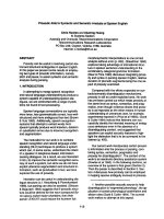
was demonstrated on his blood investigations. An
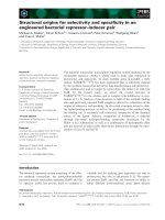
urgent computed tomography (CT) aortogram con-
firmed a non-leak ing 5.6 cm AAA (Figure 1). In addi-
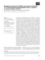
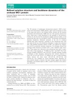
tion, extensive lower lobe consolidation and collapse
with hilar lymphadenopathy was noted (Figure 2). A
diagnosis of a lobar pneumonia was made. However, th e
tenderness in the epigastrium was a concern and could
represent either a symptomatic aneurysm or referred
pain from th e lobar pneumonia. Thus , our p atient was
closely observed and the pneumonia treated aggressively
with intravenous antibiotics, supplemental oxygen and
physiotherapy.
* Correspondence:
1
The Leeds Vascular Institute, The General Infirmary at Leeds, Great George
Street, Leeds LS1 3EX, UK
Full list of author information is available at the end of the article
Naylor et al. Journal of Medical Case Reports 2010, 4:333
/>JOURNAL OF MEDICAL
CASE REPORTS
© 2010 Naylor et al; licensee BioMed Central Ltd. This is an Open Access article distributed under the terms of the Creative Commons
Attribution License ( which permits unrestricted use, distribution, and reproduction in
any medium, provided the original work is properly cited.
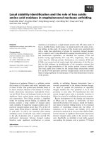
Forty-eight hours following admission our patient
reported symptoms of pre-syncope with a brief period
of hypotensio n. A repeat CT aort ogram demonstrated a
rapid increase in size o f the AAA to 7.0 cm and r etro-
peritoneal fat stranding (Figures 3 and 4). The neck of
theaneurysmdidnotshowanysignificantangulation
and its juxta-renal diameter was 22.1 mm increasing to
25.4 mm in its infra-renal segment. In addition, there
was a significant stenosis of the left common iliac artery.
Despite aggressive treatment of our patien t’spneumo-
nia, he remained hypoxic. The options were either an
emergency open bifurcated aortic graft or an endovascu-
lar aorto -uni-iliac repair with a femoral-to-femoral
cross-over procedure. Following a full discussion with
our patient, anesthe tists and endovascular radiologists,
the latter procedure was performed. Our patient had an
uncomplicated post-operative recovery. He was con tin-
ued on intravenous antibiotics for a further five days
and discharged. He was followed up clinically at four,
five and seven months post-operati vely. CT scans at one
Figure 1 CT aortogram demonstrating a 5.6 cm anteroposterior
diameter AAA.
Figure 2 CT thorax demonst rating extensive lower lobe
consolidation and collapse noted in the left lung with
extensive hilar lymphadenopathy.
Figure 3 CT aortogram demonstrating a rapid increase in the
size of the AAA measuring 7.0 cm in the anteroposterior
diameter. There is a new beak in the left lateral aortic thrombus
and signs of impending rupture.
Figure 4 Coronal CT angiogram image demonstrating the 7.0
cm aneurysm. The neck of the aneurysm was not angulated and
its diameter at the renal arteries was 22.1 mm and below the renal
arteries was 25.4 mm.
Naylor et al. Journal of Medical Case Reports 2010, 4:333
/>Page 2 of 4
and six months post-operatively showed good stent
position and patency.
Discussion
The expansion rate of AAAs varies accord ing to numer-
ous factors. The probability of rupture of a 5 cm and 7
cm AAA is less than 16% and 25% per year, respectively
[7] and guidelines in the United Kingdom recommend
three-monthly assessment with an abdominal ultrasound
for AAAs greater than 5 cm [8]. In small AAAs with a
size of 3.0 to 3 .9 cm growth, the growth rate has been
reported as an average 0.11 cm annual ly [2]. AAAs with
adiameterofbetween4.0and4.9cmhavebeenfound
to have a much larger rate of growth with an average
rate of 0.79 cm per year in those with continuous
expansion compared to 0.27 cm per year with discontin-
uous (staccato) expansion [3]. Thus the typical expan-
sionrateisabout0.25cmperannum,andifthe
aneurysm diameter increases by 0.4 to 0.8 cm per year
more frequent surveillance is recommended [9]. AAA
expansion varies individually and inflammation can
influence this process and dramatically accelerate AAA
expansion as a result of specific cellular immune
responses [10,11].
In the case presented here, the AAA had increased in
size by about 0.3 cm per annum until admission. In the
presence of concomitant sepsis it suddenly expanded. In
the wall of an AAA there is up-regulation of pro-inflam-
matory IL-1b,IL-6,IL-10andTNF-a, which have been
shown to positively correlate with aneurysm growth
[12,13]. Such cytokines, chemokines and growth factors
are known to be further potentiated during septic events
such as a LRTI [14]. One possibility is that concomitant
sepsis could increase these specific inflammatory media-
tors within the AAA wall further weakening the aortic
wall, increasing the risk of expansion and rupture.
There is a documented association between ongoing
pulmonary sepsis, expansionofaorticaneurysmsand
aortic dissection [5,15]. This has been more commonly
reported in thoracic than AAAs. However, hematogen-
ous seeding may also affect the abdominal aorta, if there
is no contiguous focus o f infection [6]. The expansion
and change in fat around the AAA found on t he repeat
CT aortogram suggested inflammation or an impending
leak. In the case presented here, it is n ot clear whe ther
this represen ted a mycotic AAA. However, the fusiform
nature of the pre-existing AAA and lack of air in the
aneurysm sac do not support a myc otic AAA. Regard-
less, there is controversy regarding the use of endovas-
cular app roaches in s uch aneurysms; however, there are
several reports which demonstrate better outcomes
when compared to conventional surgery in these high
risk cases [16]. Despite these concerns we proceeded
with an endovascular repair, which was uneventful.
Conclusions
This case h ighlights the potential association between
pulmonary sepsis and rapid AAA expansion. In these
patients there must be a hi gh index of clinica l suspicion
for rapid progre ssion, and a policy of frequent monitor-
ing may be adopted to identify those patients requiring
definitive management. Endovascular repair may be sui-
table in certain cases depending on aneurysm morphol-
ogy and local experience.
Consent
Written informed consent was obtained from the patient
for publication of this case report and any accompany-
ing images. A copy of the written c onsent is available
for review by the Editor-in-Chief of this journal.
Abbreviations
AAA: abdominal aortic aneurysm; CT: computed tomography; LRTI: lower
respiratory tract infection.
Author details
1
The Leeds Vascular Institute, The General Infirmary at Leeds, Great George
Street, Leeds LS1 3EX, UK.
2
Department of Interventional Radiology, The
General Infirmary at Leeds, Great George Street, Leeds LS1 3EX, UK.
3
Division
of Cardiovascular and Diabetes Research, Leeds Institute of Genetics, Health
and Therapeutics, University of Leeds, Clarendon Way, Leeds LS2 9JT, UK.
Authors’ contributions
SN reviewed the literature and wrote a first draft of the manuscript. ZG
reviewed the literature, corrected, finalized and submitted the manuscript.
RSV reviewed the literature and was involved in manuscript preparation and
editing. SP interpreted the radiological images and performed the
endovascular stent procedure. PJK carried out the surgical procedure and
was involved with manuscript editing and reviewing. DJAS was involved
with the conception of the report and was involved with the surgical
procedure. All authors read and approved the final manuscript.
Competing interests
The authors declare that they have no competing interests.
Received: 7 December 2009 Accepted: 21 October 2010
Published: 21 October 2010
References
1. Schlosser FJ, Tangelder MJ, Verhagen HJ, van der Heijden GJ, Muhs BE, van
der Graaf Y, Moll FL: Growth predictors and prognosis of small
abdominal aortic aneurysms. J Vasc Surg 2008, 47:1127-1133.
2. Santilli SM, Littooy FN, Cambria RA, Rapp JH, Tretinyak AS, d’Audiffret AC,
Kuskowski MA, Roethle ST, Tomczak CM, Krupski WC: Expansion rates and
outcomes for the 3.0- cm to the 3.9- cm infrarenal abdominal aortic
aneurysm. J Vasc Surg 2002, 35:666-671.
3. Vega de Ceniga M, Gomez R, Estallo L, de la Fuente N, Viviens B, Barba A:
Analysis of expansion patterns in 4-4.9 cm abdominal aortic aneurysms.
Ann Vasc Surg 2008, 22:37-44.
4. Chang JB, Stein TA, Liu JP, Dunn ME: Risk factors associated w ith rapid
growth of small abdominal aortic aneurysms. Surgery 1997, 121:117-122.
5. Monaco M, Di Tommaso L, Oliviero U, Iannelli G, Stassano P: A rapidly
expanding descending thoracic aortic aneurysm: an unusual
complication. J Card Surg 2008, 23:260-261.
6. Kandpal H, Seith A: Rapidly enlarging mediastinal mass in a middle-aged
patient with fever. Br J Radiol 2008, 81:357-359.
7. Kent KC, Zwolak RM, Jaff MR, Hollenbeck ST, Thompson RW,
Schermerhorn ML, Sicard GA, Riles TS, Cronenwett JL: Screening for
abdominal aortic aneurysm: a consensus statement. J Vasc Surg 2004,
39:267-269.
Naylor et al. Journal of Medical Case Reports 2010, 4:333
/>Page 3 of 4
8. Devaraj S, Dodds SR: Ultrasound surveillance of ectatic abdominal aortas.
Ann R Coll Surg Engl 2008, 90:477-482.
9. Dehlin JM, Upchurch GR: Management of Abdominal Aortic Aneurysms.
Curr Treat Options Cardiovasc Med 2005, 7:119-130.
10. Shimizu K, Mitchell RN, Libby P: Inflammation and cellular immune
responses in abdominal aortic aneurysms. Arterioscler Thromb Vasc Biol
2006, 26:987-994.
11. Jagadesham VP, Scott DJ, Carding SR: Abdominal aortic aneurysms: an
autoimmune disease? Trends Mol Med 2008, 14:522-529.
12. Middleton RK, Lloyd GM, Bown MJ, Cooper NJ, London NJ, Sayers RD: The
pro-inflammatory and chemotactic cytokine microenvironment of the
abdominal aortic aneurysm wall: a protein array study. J Vasc Surg 2007,
45:574-580.
13. Wallinder J, Bergqvist D, Henriksson AE: Proinflammatory and anti-
inflammatory cytokine balance in patients with abdominal aortic
aneurysm and the impact of aneurysm size. Vasc Endovascular Surg 2009,
43:258-261.
14. Gallagher PM, Lowe G, Fitzgerald T, Bella A, Greene CM, McElvaney NG,
O’Neill SJ: Association of IL-10 polymorphism with severity of illness in
community acquired pneumonia. Thorax 2003, 58:154-156.
15. Mory M, Hansmann J, Allenberg JR, Bockler D: Images in vascular
medicine. Rapid expansion of an inflammatory abdominal aortic
aneurysm. Vasc Med 2007, 12:381-382.
16. Kan CD, Lee HL, Yang YJ: Outcome after endovascular stent graft
treatment for mycotic aortic aneurysm: a systematic review. J Vasc Surg
2007, 46:906-912.
doi:10.1186/1752-1947-4-333
Cite this article as: Na ylor et al.: Lower respiratory tract infection and
rapid expansion of an abdominal aortic aneurysm: a case report. Journal
of Medical Case Reports 2010 4:333.
Submit your next manuscript to BioMed Central
and take full advantage of:
• Convenient online submission
• Thorough peer review
• No space constraints or color figure charges
• Immediate publication on acceptance
• Inclusion in PubMed, CAS, Scopus and Google Scholar
• Research which is freely available for redistribution
Submit your manuscript at
www.biomedcentral.com/submit
Naylor et al. Journal of Medical Case Reports 2010, 4:333
/>Page 4 of 4