báo cáo khoa học: " Eosinophilic infiltrate in a patient with severe Legionella pneumonia as a levofloxacin-related complication: a case report" potx
Bạn đang xem bản rút gọn của tài liệu. Xem và tải ngay bản đầy đủ của tài liệu tại đây (1.21 MB, 5 trang )
CAS E REP O R T Open Access
Eosinophilic infiltrate in a patient with severe
Legionella pneumonia as a levofloxacin-related
complication: a case report
Nicola Facciolongo
1*
, Francesco Menzella
1
, Claudia Castagnetti
1
, Alberto Cavazza
2
, Roberto Piro
1
,
Cristiano Carbonelli
1
, Luigi Zucchi
1
Abstract
Introduction: Legionella pneumonia can appear with different levels of severity and it can often present with
complications such as acute respiratory distress syndrome.
Case presentation: We report the case of a 44-year-old Caucasian man with Legionella pneumonia with successive
development of severe acute respiratory distress syndrome. During his stay in intensive care the clinical and
radiological situation of the previously observed acute respiratory distress syndrome unexpectedly worsened due
to acute pulmonary eosinophilic infiltrate of iatrogenic origin.
Conclusion: Levofloxacin treatment caused the occurrence of acute eosino philic infiltrate. Diagnosis was possible
following bronchoscopic examination using bronchoaspirate and transbronchial biopsy.
Introduction
Since the pneumonia epidemic that struck the delegates
of the American Legion Convention in Philadelphia in
1976, Legionella spp. has become a relatively frequent
cause of community acquired pneumonia [1].
Legionella may appear in different forms, from subcli-
nical presentations to Legionnaires’ disease, which has a
mortality rate as high as 30 to 50% in cases of hospital
infections and in cases of complications such as acute
respiratorydistresssyndrome(ARDS).Thefatalityrate
is 5 to 25% even in patients who are immunocompete nt
[2].
Other complications are rare, although a significant
number of drugs used in the treatment of Legionella
pneumonia can be associated with the a ppearance of
pulmonary eosinophilic infiltrates, especially non-steroi-
dal anti-inflammatory drugs (NSAIDs) and antibiotics
[3]. The diagnosis is mainly based on the temporal cor-
relation between the administration of drugs and the
appearance of the clinical condition, but it is often not
easy to determine the etiologic agent with certainty.
This report concerns the case of a man with Legio-
nella pneumonia that evolved into ARDS and then
became complicated with eosinophilic infiltration as an
effect of treatment with levofloxacin. Usually this drug
is safe, though in some cases can cause eosinophilic
pneumonia [4].
Case presentation
A 44-year-old Caucasian man presented to our hospital
for hyperpyrexia (over 39°C) for about a week, with gen-
eral weakness and strong headaches; he had been trea-
ted by his general p ractitioner with amoxicillin and
clavulanate administrated orally with no improvement.
His case history revealed that he was a smoker (20
packs/year). No other pathologies or trips abroad had
been registered in the last 6 months.
On admission, he had hyperpyrexia (38.9°C), headache,
dry cough, diarrhea, general weakness and sinus tachy-
cardia (100 beats/minute); his oxygen saturation was
95% (no oxygen supplement).
The results of a physical examination of his chest were
reduced vesicular respiration and crackling in the med-
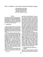
ian axillary line to the left and in front; a chest X-ray
showed extensive inconsistent parenchymal consolida-
tion at the fissure of the left upper lobe (Figure 1A).
* Correspondence:
1
Department of Pneumology, S. Maria Nuova Hospital, 42123 Reggio Emilia,
Italy
Full list of author information is available at the end of the article
Facciolongo et al. Journal of Medical Case Reports 2010, 4:360
/>JOURNAL OF MEDICAL
CASE REPORTS
© 2010 Facciolongo et al; licensee BioMed Central Ltd. This is an Open Access article dis tributed under the terms of the Creative
Commons Attribu tion License ( 2.0), which permits unrestricted use, distribution, and
reproduction in any medium, provided the original work is properly cited.
The results of initial laboratory examinations revealed
his white blood cell count was 2020 cells/mm
3
,total
bilirubin level was (1.6 mg/dL), he had reduced albumi-
nemia (2.7 g/dL), increased alkaline phosphatase (382
U/L), g-glutamyl transferase (69 U/L) and creatine phos-
phokinase (422 U/L). His serology test results w ere
negative for Hepatitis B virus, Hepatitis C virus and
HIV. His initial blood culture test results were negative
for aerobic and anaerobic germs and mycetes.
Our patient began treatment with intravenous pipera-
cillin and tazobactam (13.5 g/day) and clarithromycin
orally (1 g/day). On the third day the results of his urin-
ary antigen test were found to be positive for Legio nella
serogroup 1, so clarithromycin was suspended and sub-
stituted with intravenous levofloxacin (750 mg/day). We
maintain ed the piperacillin and tazobactam treatment to
help prevent secondary infection from other Gram-posi-
tive and Gram-negative bacteria.
On the sixth day, his clinical condition worsened.
After consultation with an infectious disease specialist,
we added rifampicin (900 mg/day) to support the
levo-
floxacin action against Legionella pneumonia.
On the ninth day he showed respiratory distress (40
breaths/minute). An Arterial Blood Gas analysis in room
air gave the following results: partial O
2
pressure (pO
2
)
of 50 mmHg, partial CO
2
pressure (pCO
2
) of 30 mmHg,
pH 7.50 and oxygen saturation (SaO
2
) of 86%.
A computed tomography (CT) scan of his chest
rev ealed multiple areas of parenchymal consolidation in
the entire upper left pulmonary lob e, mixed with
ground-glass areas and abundant pleural effusion. In the
right lung, in the dorsal and basal regions, there were
ground-glass areas mixed with consolidation areas (Fig-
ure 1B).
On the 10th day PaO
2
/fraction of inspired O
2
(FiO
2
)
ratio was 101 and he was moved to our intensive care
A
C
D
B
R L
R L
R L
R L
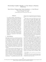
Figure 1 Chest X-ray and computed tomography (CT) images. A) A chest X-ray taken on admission: extensive pulmonary consolidation can
be seen in the upper left lobe (arrow). There was an absence of pleural effusion and no cardiomegaly. B) A chest CT scan taken on the ninth
day: consolidation areas can be seen on the whole superior left lobe, mixed with ground-glass areas and air bronchogram. There was an
absence of pleural effusion. C, D) A CT scan taken on the 21st day: on the left there is parenchymal consolidation with air bronchogram and
pneumothorax, and several areas of parenchymal consolidation on the right superior lobe. There was an absence of pleural effusion.
Facciolongo et al. Journal of Medical Case Reports 2010, 4:360
/>Page 2 of 5
unit. Here he was placed on a ventilator on continuous
positive airway pressure modality, with noticeable
improvement of t he respiratory parameters (PaO
2
/FiO
2
ratio of 254). On the 17th day, levofloxacin was sus-
pended in order to allow wash-out and taking of further
blood cultures. On the 19th day levofloxacin was
resumed; after advice from an infectious diseases specia-
list intravenous levofloxacin 1500 mg p er day together
with intravenous fluconazole 800 mg per day were given
On the 21st day, after an initial improvement, he
showed respiratory distress. A CT scan showed
increased parenchy mal consolidation with left pneu-
mothorax (Figure 1C, D).
On the 22nd day, because of the unexpected occur-
rence of muscular exhaustion, orotracheal intubation
was perform ed and he was placed on a mechanical ven-
tilator in synchronized intermittent mandatory ventila-
tion mode associated with appropriate kinetic therapy
on a reclining bed. A fibrobronchoscopy study, carried
out with bronchoalveolar lavage (BAL) for bacteriologi-
cal reasons and in order to define the cytological profile,
revealed the presence of numerous macrophages (32%),
lymphocytes (26%; CD4/CD8 ratio 0.8), neutrophilic
granulocytes (40%) and some eosinophilic granulocytes
(2%). Protozoa, fungus and neoplastic cells were absent.
On the 23rd day, methylprednisolone (120 mg/day
intravenously) was added to the therapy.
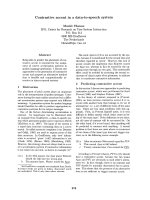
On the 26th day, he underwent another bronchoscopy,
with BAL and transbronchial biopsy in the basal seg-
ments of the lower right lobe, which revealed a histolo-
gical condition compatible with acute eosinophilic
pneumonia (Figures 2 and 3). The BAL confirmed the
presence of eosinophils 28%, macrophages 57%, lympho-
cytes 15%, neutrophilic granulocytes 2% and a CD4/CD8
ratio of 1. Incidental findings showed masses of finely
pigmented macrophages (due to our patient ’ssmoking
habit). Serum levels of total IgE were within normal lim-
its, and the specific IgE antibody results for allergens
(food, pollen, fungal) were also negative. Fecal and sero-
logical test results were negative for parasites.
On the 27th day, his steroid therapy was increased
(methylprednisolone 1 g/day) while levofloxacin was
suspended. His response to steroid therapy was rapid,
with a general improvement starting from the fifth day
of treatment (the 32nd day overall), associated with
accompanying improvement of respiratory exchange and
subsequent return to spon taneous breathing on the 41st
day (PaO
2
/FiO
2
ratio of 357).
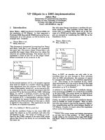
On the 51st day, a chest X-ray showed that the pneu-
monia bilateral consolidation had completely resolved
(Figure 4).
Discussion
ARDS is a common medical emergency and is usually a
complication of a previous illness, which is the etiological
cause [5]. In our patient, the unusual fact was the over-
lapping of acute eosinophilic infiltrate in legionellosis.
Eosinophilic pneumonias include a wide range of pul-
monary pathologies, characterized by alveolar and per-
ipheral blood eosinophilia. Peripheral eosinophilia may
be absent, in particular in the early stages of acute idio-
pathic eosinophilia pneumonia or in patients taking sys-
temic corticosteroids. It may occur with extremely
variable forms of seriousness, from asymptomatic pul-
monary infiltrates to acute respirator y distress syndrome
associated with respiratory insufficiency. The possible
causes, such as drugs or parasitic infections, have been
widely studied, but are, in most cases, idiopathic [6]. In
our opinion, in accordance with the findings of other
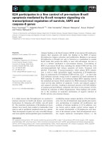
Figure 2 Histological images. Transbronchial biopsies showed
several eosinophils associated with fibrin (hematoxylin and eosin
stain, 200×).
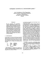
Figure 3 Histological images.Focally,hyalinemembraneswere
present (hematoxylin and eosin stain, 200×).
Facciolongo et al. Journal of Medical Case Reports 2010, 4:360
/>Page 3 of 5
authors [7], early low-dose steroid therapy leads to a
better outcome of pneumonia with severe respiratory
distress; however it could determine a delayed onset of
eosinophilic pneumonia.
In our patient, we are inclined to consider it as having
an iatrogenic etiopathogenesis. O ther causes were
excluded by laboratory tests for differential diagnosis
options (serum total and specific IgE, fecal and serologic
examinations for parasite infections).
Eosinophilic pneumonia has been linked to more than
80 drugs , alth ough only 20 of these (for the mo st part
NSAIDs and antibiotics) can be considered as common
causes of this pathology [6]. All the drugs administered in
the weeks prior to the appearance of eosinophilic infiltrate
should be suspected as a p ossible cause of the pathology.
Iatrogenic eosinophilic infiltrates usually develop progres-
sively, with dyspnea, cough and fever in subjects who have
taken certain drugs for weeks or months.
The diagnosis of drug-induced eosinophilic pneumo-
nia is mainly based on a detailed history of drug expo-
sure, evidence of eosinophil accumulation in the lung
and exclusion of other causes. Numerous methods have
been studied in order to demonstrate sensitivity to one
or more drugs. One of the most commonly applied
methods is the lymphocyte stimulation test (LST),
which measures the p roliferation of T lymphocytes in
response to a drug in vitro , in order to di agnose a pre-
vious reaction in vivo.Thisconceptwasconfirmedby
the finding of drug-specific T lymphocyte clones that
can interact with cellular receptors without being meta-
bolized and without bonding to protein carriers [8].
We did not consider it necessary to carry out the LST
with our patient because this method is not specific and
sensitive, and it has the major drawback of being diffi-
cult to interpret [8].
With regard to the challenge test in vivo, this was not
performed because of the serious clinical condition of
our patient, who in any case did not give his consent.
However, voluntary challenge may cause life-threatening
adverse reactions and it should be limited to rare situa-
tions [9].
Among the possible causes we considered, the first
was levofloxacin. There are some reports in the litera-
ture regarding the possibility of development of eosino-
philic pneumonia during the course of levofloxacin
therapy [4]; moreover, it was the drug administered to
our patient for the greatest number of days (21 in total).
Other points can be taken into account: (1) the drug
was suspended for four days in order to allow for wash-
out and subsequent blood culture; afterward s, the same
drug was resumed. At t he same time, the clinical radi-
ological findings became worse, with an unintentional
challenge effect. (2) The BAL on the 22nd day, as some
other authors have reported, still showed compatibility
with ARDS Legionella, [10] while the following BAL
showed eosinophilia (28%) compatible with an acute
eosinophilic pneumonia [6], which histological exams
confirmed (Figure 3).
With regard to the other drugs administered, there are
reports of isolated cases of eosinophilia associated with
parenchymal infiltrates as a consequence of rifampicin
therapy [11]. There is only one reported case where
clarithromycin may have led to eosinophilic pneumonia
[12], but our patient was only treated with this drug for
two days. Moreover, it is possible that eosinophilic
pneumonia could be an adverse reaction to smoking in
predisposed subjects: this sometimes happens to patients
who have recently started smoking or who have modi-
fied their ‘way’ of smoking (for example, increasing or
changing type of smoking). Our patient, however, did
not report any changes, either in quantity or in quality,
in his smoking habits, so this would seem to exclude
any relation to smoking [13].
However, it is plausible that smoking could have acted
as a cofactor (together with the drugs) in triggering the
clinical condition, be cause it is a known fact that acute
eosinophilic infiltrates are often frequent in smokers
[14].
Conclusion
In conclusion, levofloxacin may be the most probable
cause of the occurrence of acute eosinophilic infiltrate
in this patient. It is important to emphasize that we
decided to change t he diagnostic and therapeutic
approach only when the presence of eosinophilic infil-
trate was proven by transbronchial biopsy. Published
studies dealing with risks of invasive endoscopic
Figure 4 Chest X-ray. Thickening areas and parenchymal distortion
can be seen on the left upper lobe. Diffuse thickening can be seen
on medial and lower lobes (arrow).
Facciolongo et al. Journal of Medical Case Reports 2010, 4:360
/>Page 4 of 5
procedures in a patient who was critically ill on
mechanical ventilatio n showed a higher incidence of
complications such as hemorrhage and pneumothorax.
Correlating the endoscopic risk to the percentage o f
correctly carried out diagnoses, which varies from 33%
to 76%, with consequent change in therapeutic strategy,
it may be stated that the risk/benefit ratio of the endo-
scopic procedure in terms of therapeutic response is
surely in its favor and it is, therefore, recommended
[15].
Consent
Written informed consent was obtained from the patient
for publication of this case report and any accompany-
ing images. A copy of the written consent is available
for review by the Editor-in-Chief of this journal.
Author details
1
Department of Pneumology, S. Maria Nuova Hospital, 42123 Reggio Emilia,
Italy.
2
Department of Pathology, S. Maria Nuova Hospital, 42123 Reggio
Emilia, Italy.
Authors’ contributions
NF coordinated diagnostic and therapeutic stages and was one of the
principal contributors in writing the manuscript. MF contributed to the
clinical approach, analyzed and interpreted the data and was a major
contributor in writing the manuscript. CC was a contributor in writing the
manuscript. AC performed the histological examination of the lung and was
a contributor in writing the manuscript. CC was a contributor in writing the
manuscript. RP was a contributor in writing the manuscript. LZ was a
contributor in writing the manuscript and he gave final approval of the
version to be published. All authors read and approved the final manuscript.
Competing interests
The authors declare that they have no competing interests.
Received: 24 October 2009 Accepted: 11 November 2010
Published: 11 November 2010
References
1. Diederen BM: Legionella spp. and Legionnaires’ disease. J Infect 2008,
56:1-12.
2. DeMello D, Kierol-Andrews L, Scalise PJ: Severe sepsis and acute
respiratory distress syndrome from community-acquired Legionella
pneumonia: case report. Am J Crit Care 2007, 16:317-319.
3. Camus PH, Foucher P, Bonniaud PH, Ask K: Drug-induced infiltratives lung
disease. Eur Respir J 2001, 18:93s-100s.
4. Fujimori K, Shimatsu Y, Suzuki E, Arakawa M, Gejyo F: Levofloxacin-induced
eosinophilic pneumonia complicated by bronchial asthma. Nihon Kokyuki
Gakkai Zasshi 2000, 38:385-390.
5. Bauer TT, Ewig S, Rodloff AC, Müller EE: Acute respiratory distress
syndrome and pneumonia: a comprehensive review of clinical data. Clin
Infect Dis 2006, 43:748-756.
6. Katz U, Shoenfeld Y: Pulmonary eosinophilia. Clin Rev Allerg Immunol 2008,
34:367-371.
7. Confalonieri M, Urbino R, Potena A: Hydrocortisone infusion for severe
community-acquired pneumonia. A preliminary randomized study. Am J
Respir Crit Care Med 2005, 171:242-248.
8. Pichler WJ, Tilch J: The lymphocyte transformation test in the diagnosis
of drug hypersensitivity. Allergy 2004, 59 :809-820.
9. Choquet-Kastylevsky G, Vial T, Descotes J: Drug allergy diagnosis in
humans: possibilities and pitfalls. Toxicology 2001, 158:1-10.
10. Trisolini R, Agli LL, Cancellieri A, Procaccio L, Candoli P, Alifano M, Patelli M:
Bronchoalveolar lavage findings in severe community-acquired
pneumonia due to Legionella pneumophila serogroup 1. Respir Med 2004,
98:1222-1226.
11. Lee M, Berger HW: Eosinophilia caused by rifampicin. Chest 1980, 77:579.
12. Ohnishi H, Abe M, Yokoyama A, Hamada H, Ito R, Hirayama T, Nishimura K,
Higaki J: Clarithromycin-induced eosinophilic pneumonia. Intern Med
2004, 43:3.
13. Uchiyama H, Suda T, Nakamura Y, Shirai M, Gemma H, Shirai T: Alterations
of smoking habits are associated with acute eosinophilic pneumonia.
Chest 2008, 133:1174-1180.
14. Miki K, Miki M, Okano Y, Nakamura Y, Ogushi F, Ohtsuki Y, Nakayama T:
Cigarette smoke-induced acute eosinophilic pneumonia accompanied
with neutrofilia in the blood. Intern Med
2002, 41:11.
15. Bulpa PA, Dive AM, Mertens L, Delos MA, Jamart J, Evrard PA, Gonzalez MR,
Installé EJ: Combined bronchoalveolar lavage and transbronchial lung
biopsy: safety and yield in ventilated patients. Eur Respir J 2003,
21:489-494.
doi:10.1186/1752-1947-4-360
Cite this article as: Facciolongo et al.: Eosinophilic infiltrate in a patient
with severe Legionella pneumonia as a levofloxacin-related
complication: a case report. Journal of Medical Case Reports 2010 4:360.
Submit your next manuscript to BioMed Central
and take full advantage of:
• Convenient online submission
• Thorough peer review
• No space constraints or color figure charges
• Immediate publication on acceptance
• Inclusion in PubMed, CAS, Scopus and Google Scholar
• Research which is freely available for redistribution
Submit your manuscript at
www.biomedcentral.com/submit
Facciolongo et al. Journal of Medical Case Reports 2010, 4:360
/>Page 5 of 5