báo cáo khoa học: "Development of a duodenal gallstone ileus with gastric outlet obstruction (Bouveret syndrome) four months after successful treatment of symptomatic gallstone disease with cholecystitis and cholangitis: a case report" pps
Bạn đang xem bản rút gọn của tài liệu. Xem và tải ngay bản đầy đủ của tài liệu tại đây (502.51 KB, 5 trang )
CASE REPO R T Open Access
Development of a duodenal gallstone ileus with
gastric outlet obstruction (Bouveret syndrome)
four months after successful treatment of
symptomatic gallstone disease with cholecystitis
and cholangitis: a case report
Arnd Giese
1
, Jürgen Zieren
2
, Guido Winnekendonk
3
, Bernhard F Henning
1*
Abstract
Introduction: Cases of gallstone ileus account for 1% to 4% of all instances of mechanical bowel obstruction. The
majority of obstr ucting gallstones are located in the terminal ileum. Less than 10% of impacted gallstones are
located in the duodenum. A gastric outlet obstruction secondary to a gallstone ileus is known as Bouveret
syndrome. Gallstones usually enter the bowel through a biliary enteral fistula. Little is known about the formation
of such fistulae in the course of gallstone disease.
Case presentation: We report the case of a 72-year-old Caucasian woman born in Germany with a gastric outlet
obstruction due to a gallstone ileus (Bouveret syndrome), with a large gallstone impacted in the third part of the
duodenum. Diagnostic investigations of our patient included plain abdominal films, gastroscopy and abdominal
computed tomography, which showed a biliary enteric fistula between the gallbladder and the duodenal bulb. Our
patient was successfully treated by laparotomy, duodenotomy, extraction of the stone, cholecystectomy, and
resection of the fistula in a one-stage surgical ap proach. Histopathological examination showed chronic and acute
cholecystitis, with perforated ulceration of the duodenal wall and acute purulent inflammation of the surrounding
fatty tissue. Four months prior to developing a gallstone ileus our patient had been hospitalized for cholecystitis, a
large gallstone in the gallbladder, cholangitis and a small obstructing gallstone in the common biliary duct. She
had been treated with endoscopic retrograde cholangiopancreatography, endoscopic biliary sphincterotomy,
balloon extraction of the common biliary duct gallstone, and intravenous antibiotics. At the time of her first
presentation, abdominal ultrasound and endoscopic examination (including esophagogastroduodenoscopy and
endoscopic retrograde cholangiopancreatography) had not shown any evidence of a biliary enteral fistula. In the
four months preceding the gallstone ileus our patient had been asymptomatic.
Conclusion: In patients known to have gallstone disease presenting with symptoms of ileus, the differential
diagnosis of a gallstone ileus should be considered even in the absence of preceding symptoms related to the
gallbladder disease. Gallstones large enough to cause intestinal obstruction usually enter the bowel by a biliary
enteral fistula. During the formation of such a fistula, patients can be asymptomatic.
* Correspondence:
1
Department of Internal Medicine, Gastroenterology Unit, Marienhospital,
Ruhr-University Bochum, Hölkeskampring 40, 44625 Herne, Germany
Full list of author information is available at the end of the article
Giese et al. Journal of Medical Case Reports 2010, 4:376
/>JOURNAL OF MEDICAL
CASE REPORTS
© 2010 Giese et al; licensee BioMed Central Ltd. This is an Open Access article distributed under the terms of the Creative Commons
Attribution License ( nses/by/2.0), which permits unrestricted use, distribution, and reproduction in
any mediu m, provided the original work is properly cited.
Introduction
Gallstone ileus accounts for approximately 1% to 4% of
all cases of mechanical bowel obstruction. H owever, in
the population over the age of 65 it is the cause of 25%
of non-strangulated small bowel obstructions. Diagnosis
is often delayed and morta lity is high, ranging at 15% to
18%, which may also reflect the age and comorbidity of
affected patients [1]. Gallstones usually enter t he bowel
through a biliary enteric fistula, which complicates 2%
to 3% of cases of cholecystolithi asis with associated epi-
sodes of cholecystitis [2]. Due to the sedimentation of
intestinal content, gallstones increase in diameter as
they pass the bowel. The majority of obstructing gall-
stones are located in the terminal ileum (50% to 75%),
followed by the proximal i leum and jejunum (20% to
40%). Gallstones impacted in the duodenum account for
less than 10% [3]. A gast ric outlet obs truction secondary
to an impact ed gallstone in the duodenum or pylorus is
calle d Bouveret syndrome. It was first described in 1896
by the French internist Leon Bouveret, and up to 1999
only 175 cases had bee n described in the medical litera-
ture [4]. Our case is a rare description of Bouveret syn-
drome developing four months after successful
treatment of sym ptomatic gallstone disease and after a
four-month period with no symptoms.
Case report
A 7 2-year-old Caucasian woman born in Germany was
admitted to our hospital with acute onset of nausea,
vomiting and diffuse abdominal pain. Her only medica-
tions were metoprolol tartate and ra mipril for arterial
hypertension and chronic compensated heart failure.
Physical examination was normal apart from diffuse
pain on abdominal palpation. There were no signs of
peritonitis. Laborator y findings (Table 1) included
a white blood count of 14.3 cells/nL, an elevated
C-reactive protein (CRP) level of 25.9 mg/dL, mildly ele-
vated plasma aspartate aminotransferase and alanine
aminotransferase (AST and ALT) levels of 51 U/L and
83 U/L, a moderate elevation of the g glutamyl trans-
peptidase (GGT) level of 487 U/ L and an al kaline phos-
phatase (AP) level of 368 U/L. Her total bilirubin level
was elevated to 1 .17 mg/dL and h er serum creatinine
level was 1.84 mg/dL. An abdominal ultrasonography
scan showed thickening and edema of the gallbladder
(GB) wall (12 mm), double wall sign, the presence of a
large gallstone and a local hypoechogenic mass in the
GB adheri ng to the GB wall with no signs of vasculari-
zation on color flow imaging. The common b iliary duct
(CBD) was dilated to 10 mm. Endoscopic retrograde
cholangiopancreatography (ERCP) performed on the
day of admission revealed a normal pancreatic duct
and a small pigmented gallstone of the CBD that was
extracted with an extraction balloon after endoscopic
biliary sphincterotomy. Esophagogastroduodenoscopy
(EGD) findings were normal without any signs of
perforation or fistula. Under antibiotic treatment
(ceftriaxon 2 g intravenously a day and metronidazole
400 mg intra venously four times a day for 10 d ays), our
patient recovered completely. Her white blood count
normalized and CRP and GGT levels fell (CRP 1.6 mg/
dL, GGT 284 U/L t wo days bef ore discharge). She was
discharged after 11 days. After discharge our patient
continued her antibiotic treatment (cefuroxim 500 mg
orally twice a day and metronidazole 500 mg orally
three times a day) for another four days.
As she remained asymptomatic, our patient did not
attend the cholecystectomy scheduled two months after
hospital discharge. Instead, four months afte r her initial
discharge, she re-presented to our hospital with abdominal
Table 1 Laboratory data for blood at admission
Normal range First
admission
Second
admission
WBC 4.0 to 10.0 cells/nL 14.3 cells/nL 11.7 cells/nL
Segmented cells 85% NA
Lymphocytes 25% to 40% 6% NA
Monocytes 2% to 6% 8% NA
Eosinophils 2% to 7% 0% NA
Basophils 0% to 1% 1% NA
ESR after 1 hour 6 to 11 mm 104 mm 44 mm
RBC 4.1 to 5.1 cells/pL 4.53 cells/pL 5.16 cells/pL
Hemoglobin 12 to 16 g/dL 13.5 g/dL 14.2 g/dL
Hematocrit 35% to 45% 39.7% 42.2%
Platelets 140 to 440 cells/nL 303 cells/nL 360 cells/nL
Bilirubin (total) <1.2 mg/dL 1.68 mg/dL 0.97 mg/dL
Bilirubin
(conjugated)
<0.5 mg/dL 1.17 mg/dL NA
Creatinine 0.5 to 0.9 mg/dL 1.84 mg/dL 1.12 mg/dL
AP 40 to 150 U/L 368 U/L 97 U/L
GGT 9 to 39 U/L 487 U/L 84 U/L
AST 5 to 31 U/L 51 U/L 29 U/L
ALT 0 to 34 U/L 83 U/L 12 U/L
LDH <243 208 U/L 252 U/L
Potassium 3.5 to 5.1 mmol/L 3.60 mmol/L 3.93 mmol/L
Sodium 136 to 145 mmol/
L
137 mmol/L 144 mmol/L
Lipase 8 to 78 U/L 32 U/L 43 U/L
CRP <0.5 mg/dL 25.93 mg/dL 0.83 mg/dL
INR 0.85 to 1.17 0.87 0.99
pTT 25 to 40 seconds 37 seconds 32 seconds
ALT = alanine aminotran sferase; AP = alkaline phosphatase; AST = aspartate
aminotransferase; CRP = C-reactive protein; ESR = erythrocyte sedimentation
rate; GGT = g glutamyl transpeptidase; INR = international normalized ratio;
LDH = lactate dehydrogenase; NA = not applicable; pTT = partial
thromboplastin time; RBC = red blood cell count; WBC = white blood
cell count.
Giese et al. Journal of Medical Case Reports 2010, 4:376
/>Page 2 of 5
right upper quadrant (RUQ) pain and repeated post-pran-
dial vomiting.
Physical examination at this time showed RUQ pain
with no local tenderness or other signs of peritonitis. At
admission her blood pressure was 140/90 mmHg and
her body temperature was 37°C. The laboratory findings
at the t ime of her second admission revealed a white
blood count of 11.7 cells/nL, a slightly elevated CRP
level of 0.83 mg/dL and normal liver test results apart
from an elevated GGT level of 84 U/L.
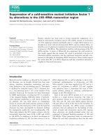
EGD was performed, during which 1.5 L of gastric con-
tent was removed by endoscopic suction. A fistula lead-
ing into a cavity of 2 cm diameter was detected just distal
of the pyloric sphincter on the dorsal wall of the duode-
nal bulb, as well as some small fibrin-covered erosions on
the anterior wall of the duodenal bulb (Figure 1).
Chest radiography results revealed an absence of pul-
monary infiltrate. On plain abdominal film no signs of
ileus, pneumobilia or free air could be detected. A CT
scan of the abdomen with oral and intr avenous contrast
(Figure 2) revealed a gallstone ileus with a 4 cm × 3 cm
gallstone in the third part of the duodenum associated
with a fistula between the GB and the duodenal bulb, as
well as minimal pneumobilia. The impacted gallstone
was surgically removed by laparotomy and duodenot-
omy. It measured 5 cm × 3 cm. Cholecystectomy and
excision o f the fistula was performed. A histopathologic
examination revealed a gallbladder with chronic and
acute cholecystitis, high-grade c hronic granulating
xanthom atous and puru lent pericholecystiti s with a for-
eign body granuloma. The duodenal wall excision
showed high-grade chronic fibrosing and acute ulcerat-
ing inflammation with perforated ulceration as well as
chronic and acute purulent inflammation of the
surrounding fatty tissue . Postoperative duoden al leakage
or persistence of duodenal obstruction was ruled out by
a contrast swallow. Our patient’s recovery was unevent-
ful. At seven weeks after discharge (eight weeks after
surgery) she was doing well, and was able to continue
her usual daily activities immediately after discharge.
Discussion
This case report is the first published observation of this
particular course of gallstone disease. In our patient, a
duodenal gallstone ileus developed f our months after a
cholecystitis associated with a large gallstone in the GB,
a small obstructing gallstone in the CBD, and cholangi-
tis. It is s triking that the formation of the biliary enteral
fistula must have taken place in an asymptomatic period
of four months. Fistula formation and dislocation of a
gallstone from the GB into the duodenum happened
even after sufficient biliary drainage and antibiotic treat-
ment during our patient’s first hospitalization.
It is generally believed that pericholecystic inflamma-
tion after cholecystitis, as well as pressure necrosis by
the gallst one against the biliary wall, may lead to fo rma-
tion of a b iliary enteric fistula. Fistula formation is a
complication of 2% to 3% of all cases of cholelithiasis
with associated episodes of cholecystitis [2]. Obstruction
of the biliary systems is known to promote cholecystitis.
It also seems to play a role in the formation of a biliary
enteric fistula. In a large series reported by B eltran et
al., 89.5% of patients with cholecystoenteric fistulae
were also found to have a CBD obstruction caused by
Figure 1 Endoscopic view of the duodenal bulb. Arr ow A: view
into the descending duodenum. Arrow B: biliary enteral fistula.
Figure 2 Abdomin al computed tomograp hy (CT) sca n with
intravenous and oral contrast enhancement. Labels on the
figure are as follows. 1: Contrast material in the lumen of the
stomach. 2: Pyloric sphincter. 3: Contrast material in the lumen of
the duodenal bulb. 4: Gallstone impacted in the lumen of the third
part of the duodenum. *: Contrast material in the lumen of the
gallbladder (notice thickening of the wall of the gallbladder and
communication with the duodenal bulb). #: Contrast material in the
lumen of the descending duodenum.
Giese et al. Journal of Medical Case Reports 2010, 4:376
/>Page 3 of 5
an extrinsic compression from an impacted stone in the
cystic duct, known as Mirizzi syndrome [5]. A biliary
enteric fistula provides a pas sage for large gallstones to
enter the bowel and eventually cause gallstone ileus.
Bili ary enter ic fistulae are compr ised of 60% cholecysto-
duode nal fistulae, but cholecystocolonic and cholecysto-
gastric fistulae can also lead to a gallstone ileus [6].
Although a gallstone ileus is usually preceded by the
formation of a biliary e nteric fistula, there also exists a
description in the literature of a gallstone ileus after
endoscopic biliary sphincterotomy [7] with a large
extracted stone causing gallstone ileus. We do not
believe that this was the pathomechanism in our patient
since the migration of the large stone through the fistula
between the GB and the duodenum seems to be more
likely than a passage through the CBD.
ERCP performed during our patient’s first hospitaliza-
tion revealed only a small gallstone in the CBD. The his-
topathologic findings from our patient also support the
theory of pericholecystitis leading to fistula formation.
The hypoechogenic mass in the GB found at ab dominal
ultrasonography during our patient’s first visit may have
been a sign of granuloma format ion. It could also corre-
spond to GB sludge. Although early cholecystectomy
seems to yield equivalent outcomes as delayed cholecys-
tectomy [8], we decided to opt for a del ayed cholecys-
tectomy as our patient prese nted with cholangitis,
severe inflammation, signs of serious local inflammation
and e levated creatinine at the time of her first visit. As
the situation corresponded to a moderate to severe
(grade I to II) acute cholecystitis according to the Tokyo
guidelines, this approach seems reasonable [9].
In our patient, p lain abdominal films did not show
pneumobilia or a gall stone. The diagnosis was made on
the basis of the results of an abdominal CT scan and
gastroscopy. However, a biliary enteral fistula and a gall-
stone ileus may also be seen by ultrasound imaging [10].
The therapeutic approach to our patient having gall-
stone ileus remains a subject of debate, most ly due to a
lack of large prospective studies. Our patient recovered
well after a laparotomy with simultaneous extraction o f
the gallstone, cholecystectomy and resection of the fis-
tula. However, in the recent literature a high periopera-
tive mortality rate of up to 35% is described. The high
mortalityismainlyattributedtothedelayoftime
between first symptoms and admission, with an average
of three to five days.
Possible strategies are a one-stage approach with enter-
otomy, cholecystectomy and resection of the fistula at
once, or a two-stage approach with an emergency enter-
otomy to remove the obstructing gallstone and cholecys-
tectomy after a period of recuperation. It seems
reasonable to restrict the one-stage approach to clinically
stable patients and to c hoose a two-stage approach in
patients with severe cholecystitis and a high perioperative
risk as a result of concomitant comorbidities [11]. For
Bouveret syndrome, endoscopic extraction of the gall-
stone [12] has been described, as well as extracorporeal
shockwave lithotripsy and argon plasma coagulation [13]
or duodenotomy. As with more distal gallstone ileus the
primary therapeutic goal should be to relieve the gall-
stone obstruction. In principle, laparoscopic treatment of
gallstone ileus is possible and was initially considered for
our patient. However, the location of gallstones along the
entire length of the bowel, especially in the presence o f
obstruction, and a probably longer operation time may
be problematic [14]. Also, laparoscopic extraction of large
gallstones may cause problems. With the gallstone of
our patient measuring 3 cm × 4 cm on a CT scan and
because we were planning a one-stage s urgery , we per-
formed a laparotomy instead of choosing a laparoscopic
approach.
Conclusion
In a patient with gallstone disease with abdominal pain,
nausea and vomiting, the possibility of a gallstone ileus
leading to gastric outlet obstruction (Bouveret syndrome)
should be considered. A CT scan of the abdomen can be
helpful in making the diagnosis. Gastroscopy should be
performed and may in some cases offer non-invasive
treatment optio ns. If the patient is not heavily compro-
mised by the gallstone ileus itself or by comorbidities , a
one-stage surgical approach with simultaneous enterot-
omy, cholecystectomy and fistula resection is feasible.
The formation of a biliary enteric fistula can be preceded
by an asymptomatic period.
Consent
Written informed consent was obtained from the patient
for publicatio n of this case report and any accompany-
ing images. A copy of the written c onsent is available
for review by the Editor-in-Chief of this journal.
Author details
1
Department of Internal Medicine, Gastroenterology Unit, Marienhospital,
Ruhr-University Bochum, Hölkeskampring 40, 44625 Herne, Germany.
2
Department of Surgery, Marienhospital, Ruhr-University Bochum,
Hölkeskampring 40, 44625 Herne, Germany.
3
Department of Radiology,
Marienhospital, Ruhr-University Bochum, Hölkeskampring 40, 44625 Herne,
Germany.
Authors’ contributions
AG conceived the case report, drafted and revised the manuscript and the
relevant literature. He also was responsible for our patient’s
gastroenterological management. JZ was responsible for our patient’s
surgical management and for editing the manuscript. GW was responsible
for the radiological findings and provided the CT scan figure. BH was
responsible for the coordination and supervision of our patient’s
gastroenterological management and manuscript editing. All authors read
and approved the final manuscript.
Giese et al. Journal of Medical Case Reports 2010, 4:376
/>Page 4 of 5
Competing interests
The authors declare that they have no competing interests.
Received: 7 February 2010 Accepted: 23 November 2010
Published: 23 November 2010
References
1. Reisner R, Cohen J: Gallstone ileus: a review of 1001 reported cases. Am
Surg 1994, 60:441-446.
2. Roslyn J, Thompson JJ, Darvin H, DenBesten L: Risk factors for gallbladder
perforation. Am J Gastroenterol 1987, 82:636-640.
3. Clavien P, Richon J, Burgan S, Rohner A: Gallstone ileus. Br J Surg 1990,
77:737-742.
4. Ariche A, Czeiger D, Gortzak Y, Shaked G, Shelef I, Levy I: Gastric outlet
obstruction by gallstone: Bouveret syndrome. Scand J Gastroenterol 1999,
35:781-783.
5. Beltran M, Csendes A, Cruces K: The relationship of Mirizzi syndrome and
cholecystoenteric fistula: validation of a modified classification. World J
Surg 2008, 32:2237-2243.
6. van Hillo M, van der Vliet J, Wiggers T, Obertop H, Terpstra O, Greep J:
Gallstone obstruction of the intestine: an analysis of ten patients and a
review of the literature. Surgery 1987, 101:273-276.
7. Despland M, Clavien P, Mentha G, Rohner A: Gallstone ileus and bowel
perforation after endoscopic sphincterotomy. Am J Gastroenterol 1992,
87:886-888.
8. Gurusamy K, Samraj K: Early versus delayed laparoscopic cholecystectomy
for acute cholecystitis. Cochrane Database Syst Rev 2006, CD005440.
9. Mayumi T, Takada T, Kawarada Y, Nimura Y, Yoshida M, Sekimoto M,
Miura F, Wada K, Hirota M, Yamashita Y, Nagino M, Tsuyuguchi T, Tanaka A,
Gomi H, Pitt HA: Results of the Tokyo Consensus Meeting Tokyo
Guidelines. J Hepatobiliary Pancreat Surg 2007, 14:114-121.
10. Rauh P, Neye H, Ensberg D, Bönicke P, Georgiew E, Rickes S:
Ultrasonographic diagnosis of a biliary-digestive fistula with gallstone
ileus. Dtsch Med Wochenschr 2010, 135:287-289.
11. Kirchmayr W, Mühlmann G, Zitt M, Bodner J, Weiss H, Klaus A: Gallstone
ileus: rare and still controversial. ANZ J Surg 2005, 75:234-238.
12. Lubbers H, Mahlke R, Lankisch P: Gallstone ileus: endoscopic removal of a
gallstone obstructing the upper jejunum. J Intern Med 1999, 246:593-597.
13. Gemmel C, Weickert U, Eickhoff A, Schilling D, Riemann J: Successful
treatment of gallstone ileus (Bouveret’s syndrome) by using
extracorporal shock wave lithotripsy and argon plasma coagulation.
Gastrointest Endosc 2007, 65:173-175.
14. Ayantunde A, Agrawal A: Gallstone ileus: diagnosis and management.
World J Surg 2007, 31:1292-1297.
doi:10.1186/1752-1947-4-376
Cite this article as: Giese et al.: Development of a duodenal gallstone
ileus with gastric outlet obstruction (Bouveret syndrome) four months
after successful treatment of symptomatic gallstone disease with
cholecystitis and cholangitis: a case report. Journal of Medical Case
Reports 2010 4:376.
Submit your next manuscript to BioMed Central
and take full advantage of:
• Convenient online submission
• Thorough peer review
• No space constraints or color figure charges
• Immediate publication on acceptance
• Inclusion in PubMed, CAS, Scopus and Google Scholar
• Research which is freely available for redistribution
Submit your manuscript at
www.biomedcentral.com/submit
Giese et al. Journal of Medical Case Reports 2010, 4:376
/>Page 5 of 5