báo cáo khoa học: "A new predisposing factor for trigemino-cardiac reflex during subdural empyema drainage: a case report" pot
Bạn đang xem bản rút gọn của tài liệu. Xem và tải ngay bản đầy đủ của tài liệu tại đây (874.5 KB, 4 trang )
CAS E REP O R T Open Access
A new predisposing factor for trigemino-cardiac
reflex during subdural empyema drainage:
a case report
Toma Spiriev
1,2*
, Nora Sandu
2,3
, Belachew Arasho
2,4
, Slavomir Kondoff
1
, Christo Tzekov
1
, Bernhard Schaller
2,4
,
Trigemino-Cardiac Reflex Examination Group (TCREG)
1
Abstract
Introduction: The trigemino-cardiac reflex is defined as the sudden onset of parasympathetic dysrhythmia,
sympathetic hypotension, apnea, or gastric hypermotility during stimulation of any of the sensory branches of the
trigeminal nerve. Clinically, trigemino-cardiac reflex has been reported to occur during neurosurgical skull-base
surgery. Apart from the few clinical reports, the physiological function of this brainstem reflex has not yet been
fully explored. Little is known regarding any predisposing factors related to the intraoperative occurrence of this
reflex.
Case presentation: We report the c ase of a 70-year-old Caucasian man who demonstrated a clearly expressed
form of trigemino-cardiac refl ex with severe bradycardia requiring intervention that was recorded during surgical
removal of a large subdural empyema.
Conclusion: To the best of our knowledge, this is the first report of an intracranial infection leading to
perioperative trigemino-cardiac reflex. We therefore add a new predisposing factor for trigemino-cardiac reflex to
the existing literature. Possible mechanisms are discussed in the light of the relevant literature.
Introduction
For more than a century, it has been well known that
electrical, chemical, or mechanical stimulation of the tri-
geminal nerve leads to trigemino-respiratory reflexes fol-
lowed by cardiac arrhythmias [1]. In the early 20th
century, this phenome non gained increased clinical
attention i n the form of the oculocardiac reflex (OCR),
which represents the cardiac response associated with
stimulation of the ophthalmic division of the trigeminal
nerve during ocular surgery [2]. In 1999, Schaller [3]
demonstrated for the first time that a similar reflex
occurs with stimulation of the intracranial (central) por-
tion of the trigeminal nerve dur ing skull-base surgery
and subsummarized all these trigemino-depressor
responses under the term “ trigemino-cardiac reflex
(TCR)” [4]. He also defined the TCR in a way that is
now generally accepted. Later, his group also described
the TCR for intraoperative stimulation of the peripheral
portion [5].
Since then, ther e has been increasing discussion about
the TCR itself, its provoking factors, and its treatment
during intra cranial or extracranial neurosurgical proce-
dures. Several predisposing factors for intraoperative
occurrence of TCR have been described [6-8], but until
now no case o f intracranial infection in combination
with intraoperative TCR has been reported.
Case presentation
Preoperative history
A 70-year-old Caucasian man was admitted for the sec-
ond time to the Department of Neurosurgery at our
hospital. His personal history included symptomatic epi-
lepsy and chronic anemia after nephrectomy because of
kidney carcinoma two years before admission to our
clinic.
Two m onths before the current admission, he under-
went surgery for a giant left frontotemporal meningioma
which was removed “gross totally.” One month after this
* Correspondence:
1
Department of Neurosurgery, Tokuda Hospital, Sofia, Bulgaria
Full list of author information is available at the end of the article
Spiriev et al. Journal of Medical Case Reports 2010, 4:391
/>JOURNAL OF MEDICAL
CASE REPORTS
© 2010 Spiriev et al; licensee BioMed Central Ltd. This is an Open Access article distributed under the terms of the Creative Commons
Attribution License ( censes/by/2.0), which permits unrestricted use, distribution, and reproduction in
any medium, provide d the original work is properly cited.
intervention, there was seen a fistula with emission of
pus in the middle third o f the operative scar. After
another neurosurgical consultation, he was admitted to
our department for surgery. At this occasion, the patient
presented afebrile, with a blood pressure (BP) of 150/70
mmHg and a heart rate (HR) of 82 beats per minute
(beats/minute), complaining of headache as well as
vomiting. In the neurolo gical examination, there was
seen a right-side horizontal nys tagmus, a right -side
hemiparesis (MRC grade 3) and complete motor apha-
sia. The only medic ation that he was taking was carba-
mazepine 2× 200 mg for epileptic prophylaxis. On the
cranial computed tomography (CT) scan without con-
trast performed in our hospital, a partial osteolysis of
the frontotemporal bone flap was demonstrated, the sur-
rounding tissues (including the dura) were seen as
thick er (due to the associated inflammation), an d a sub-
dural collection with capsule organization and peri-
lesional brain edema on the side of t he previous tumor
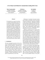
was described (see Figures 1, 2, and 3). On cranial CT
bonereconstruction,theosteolyticfociandfistulawere
clearly visible. The laboratory examination showed,
besides t he chronic a nemia, normal C-reactive protein
but a monocytosis of 1.04 10
-9
/L (normal value, 0.1 to
0.8). The patient was diagnosed with a subdural
empyema and an indication for the operative treatment
was set.
Anesthetic technique
The patient underwent surgery several days af ter this
second hospitalization. No pre-operative antibiotics were
given. The patient fasted f or eight hours prior to sur-
gery. Routine monitoring during surgery included elec-
trocardiography (ECG), end-tidal (ET) concentration of
CO
2
and sevoflurane, and pulse oximetry. All hemody-
namic parameters were monitored continuously and
recorded throughout the neurosurgical procedure.
Anesthesia was induced with midazolam (1 mg total
dosage) and propofol (2 mg/kg) followed by suxametho-
nium chloride (1.1 mg/kg), atracurium (0.6 mg/kg), and
fentanyl (100 μg total dosage). After the trachea was
intubated, the lungs were mechanically ventilated (S/5
Aespire Config; Datex-Ohmeda Ins., Madison, WI, USA)
with a mixture of air and O
2
. Anesthesia was maintained
with sevo flurane (1%). An additional 50 mg of prop ofol
and 1 mg of midazolam were applied during t he inter-
vention when necessary.
Surgical technique and postoperative management
A frontotemporal skin incision was made using the same
method used in the first intervention. Between the bone
flap and galea aponeurotica in the left frontotemporal
region, a large quantity (approximately 7-12 ml) of pus
was removed. Intraoperatively, the bone flap was f ound
to be changed by the osteomyelitic process. It was eroded
by the inflammation, with multiple pus-filled channels
connecting the inner and outer bone tables. After open-
ing the dura, a gray-white thick pus was removed. During
the whole intervention, the patient’s baseline mean arter-
ial blood pressure (MABP) was 91.0 mmHg (range, 76.7-
98.7 mmHg), and baseline mean heart rate (HR) was 82.5
bpm (range, 80-89 bpm). One hour and 20 minutes aft er
skin incision during the removal of subdural pus and
working around the dura, the patient’s blood pressure
dropped to 37/0 m mHg (MABP, 12.3 mmHg; a 86.49%
drop from baseline) and concomitantly HR dropped to
61 bpm (a 26.07% drop from baseline). There was no sig-
nificant blood loss at the time of the incident. The surgi-
cal procedure was discontinued, and the patient was
given ephedrin (20 mg), atropin (0.5 mg), and methyl-
prednisolone (60 mg) (s ee Figure 4). T wo to three min-
utes aft er the administrati on of these drugs, the patient’s
Figure 1 Preoperative computed tomography (CT) scan.
Subdural collection with capsule organization and collateral brain
edema on the side of the previous tumor is clearly visible.
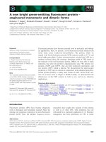
Figure 2 Preoperative CT scan. The surrounding tissues (including
the dura) are thicker, related to the associated inflammation.
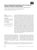
Figure 3 Preoperati ve CT s can. CT bone window shows partial
osteolysis of the bone flap, due to osteomyelitic process.
Spiriev et al. Journal of Medical Case Reports 2010, 4:391
/>Page 2 of 4
hemodynamic parameters returned to normal, and the
surgical intervention was continued. This phenomenon
was reproducible. The skin fistulae were excised, and two
subgaleal drainage systems (Dainobag Lock 300 V;
B. Braun, Melsungen, Germa ny) with a d iameter of 12
mm were left. The patient’s postoperative period was
uneventful, and he presented with no additional neurolo-
gical deficit. On microbiologi cal examination, actinomy-
cosis was reported as the cause of the empyema that
was treated with cefoperazone 2× 1 g for 12 days. The
patient’ s C-reactive protein and leucocyte count
remained normal. The postoperative period was unevent-
ful. The patient was discharged from our hospital 13 days
after the intervention.
Discussion
The presented case report is unique and adds a new and
important risk factor for the intraoperative occurrence of
TCR to the existing literature. It seems that infected intra-
cranial tissue may be a new predisposing factor in combi-
nation with surgical manipulation on the meninges, a
routine surgi cal operative techni que that has never been
described before to be associated with TCR occurrence.
It has already been shown that mechanical stimulation
of the cerebral falx results in hypera ctivity of trigeminal
ganglion, thereby triggering the TCR [9]. The neural sup-
ply of the cranial dura mater involves mainly the three
divisions of the trigeminal nerve, the first three cervical
spinal nerves, and the cervical sympathetic trunk. A case
of immediate, reproducible, and re flexive response of
asystole upon stimulation of t he cerebral falx during
operative resection of a parafalcine meningioma was pre-
viously reported [9], being most likely related to bilater al
trigeminal stimulation of the falx. According to the studies
of Penfield and McNaughton [10], the nervus tentorii, a
recurrent branch of the ophthalmic branch of the trigem-
inal nerve bilaterally innervates the tentorium cerebelli,
the dura of the parieto-occipital region, the posterior third
of the falx, and the adjacent sinuses. In our present case,
however, the subdural empyema was located in the middle
cranial fossa that is predominantly innervated by the V2
and V3 branches of trigeminal nerve [11]. However, it has
been previously shown by us and others that surgical pro-
cedures at the anterior, middle, and posterior skull base
(any branch of the central part of trigeminal nerve) may
elicit the TCR.
In this special case, one may suggest that the patient
had simply a (physiological) Cushing reflex with consecu-
tive elevated MABP before operation that only normal-
ized after elevation of the mass lesion. But the Cushing
reflex is not a possible explanation of the MABP and HF
drop as seen in our case. In our case, the intraoperative
phenomenon was reproducible, which would be not the
case if there were a Cushing reflex. Our case show s,
therefore, a clear cause-and-effect rel ationship necessary
for the TCR and as described earlier in detail [3].
Different retrospective studies have shown an incidence
of TCR ranging from 8% [12] to 18% [13] using all the
same inc lusi on criteria as defined earlier by us [3]. How-
ever, it seems that TCR is often unrecognized intraopera-
tively, so the identification of possible provoking factors
is important but often elusive. There are several reports
for the provoking factor for the peripheral initiation and
central initiatio n of the TCR. To date, several risk factors
for the intraoperative occurrence of TCR have been iden-
tified, such as light general anesthesia, childhood, and the
nature of the provoking stimulus (strength and duration
of stimulus) [3,8]. In addition, there are several known
prov oking drugs such as potent narcotic agents (sufenta-
nil a nd alfentanil), b-blockers, and calcium channel
blockers [3,8]. Until now, no report for intracranial infec-
tions as a provoking factor for intraoperative TCR occur-
rence has been identified.
Intracranial infections, as in the current case of sub-
dural empyema, could lead to a pathological process
called sensitization of trigeminal afferents in the dura
mater [14]. It was demonstrated that chemical stimula-
tion of dur al receptive fields with i nflammatory media-
tors such as prostaglandin E
2
,bradykinin,orhistamine
directly excite the neu rons and enhance their mecha nical
sensitivity [1,5], such that they can be easily activated by
mechanical stimuli that initiallyhadevokedlittleorno
response [14,15]. It seems that meningeal sensory inner-
vation is not known to subserve multiple sensory
Figure 4 Anesthesiology chart.Beforetheoccurrenceof
trigemino-cardiac reflex (TCR), mean arterial blood pressure (MABP)
was 91.0 mmHg and heart rate (HR) was 82.5 beats/minute. At the
time of the TCR record, the patient’s blood pressure dropped to
37/0 mmHg (MABP, 12.3 mmHg; 86.49% drop from baseline), and
concomitantly HR dropped to 61 beats/minute (26.07% drop from
baseline). No significant blood loss at the time of the incident was
recorded. The applied medications were ephedrin (20 mg), atropin
(0.5 mg) and methylprednisolone (60 mg). After drug administration,
the patient’s hemodynamic parameters returned to normal and the
intervention was reinitiated.
Spiriev et al. Journal of Medical Case Reports 2010, 4:391
/>Page 3 of 4
modalities [10,14]. Meningeal afferents are thought to
become activated only under potentially harmful or
pathological conditions [10]. However, although the
dural afferent population does not appear to mediate dis-
tinct sensory modalities, it shows a pattern of variation in
mechanosens itivity as a function of conduction velocities
[10,16]. Mechanical response properties of dura are
attributed to A and C primary afferent neurons. Such
exaggerated mechanical sensitivity and manipulation of
theduramatercouldplayaroleintheinitiationofTCR
in our case.
Conclusion
To the best of our knowledge, this is the first report of an
intracranial infection with the intra-operative occurrence
of TCR during a routine neurosurgical maneuver.
Infected (intracranial) tissue may be a new and important
predisposing factor for the occurrence of TCR, a phe-
nomenon that is different from the falcine TCR caused
by bilateral stimulation of tentorial nerve that was
described earlier. Further laboratory and clinical investi-
gations are needed to clarify this new information about
TCR.
Consent
Written informed consent was obtained form the patient
for publication of this case report and accompanying
images. A copy of the written consent is available for
review by the Editor-in-chief of this journal.
Author details
1
Department of Neurosurgery, Tokuda Hospital, Sofia, Bulgaria.
2
Department
of Neurosurgery, University Hospital Lariboisiere, Paris, France.
3
Department
of Neurosurgery, University of Lausanne, Switzerland.
4
Department of
Neurology, University Addis Ababa, Ethiopia.
Authors’ contributions
TS and BS wrote the article. TS collected the data. BS interpreted and
analyzed the data. SK and CK performed the operation and the patient’s
treatment and provided substantial information regarding the patient’s case
and were therefore major contributors to writing the manuscripts. NS and
BA provided some specific and general ideas that initiated the work and
helped to finish the work. Without both contributions, this report would not
have been possible. NS made substantial corrections to the manuscript. All
authors read and approved the final manuscript.
Competing interests
The authors declare that they have no competing interests.
Received: 21 June 2010 Accepted: 30 November 2010
Published: 30 November 2010
References
1. Angell-James JE, Daly MB: Nasal reflexes. Proc R Soc Med 1969,
62:1287-1293.
2. Ashner B: Über einen bisher noch nicht beschriebenen Reflex, vom Auge
auf Kreislauf und Atmung. Verschwinden des Radialispulses bei Druck
auf das Auge. Wien Klin Wochenschr 1908, 21:1529-1530.
3. Schaller B, Probst R, Strebel S, Gratzl O: Trigeminocardiac reflex during
surgery in the cerebellopontine angle. J Neurosurg 1999, 90:215-220.
4. Schaller B: Trigeminocardiac reflex: a clinical phenomenon or a new
physiological entity? J Neurol 2004, 251:658-665.
5. Schaller BJ, Filis A, Buchfelder M: Trigemino-cardiac reflex in humans
initiated by peripheral stimulation during neurosurgical skull-base
operations: its first description. Acta Neurochir (Wien) 2008, 150:715-717.
6. Blanc VF, Hardy JF, Milot J, Jacob JL: The oculocardiac reflex: a graphic
and statistical analysis in infants and children. Can Anaesthet Soc J 1983,
30:360-369.
7. Schaller B, Cornelius JF, Prabhakar H, Koerbel A, Gnanalingham K, Sandu N,
Ottaviani G, Filis A, Buchfelder M, Trigemino-Cardiac Reflex Examination
Group (TCREG): The trigemino-cardiac reflex: An update of the current
knowledge. J Neurosurg Anesthesiol 2009, 21:187-195.
8. Bauer DF, Youkilis A, Schenck C, Turner CR, Thompson BG: The falcine
trigeminocardiac reflex: case report and review of the literature. Surg
Neurol 2005, 63:143-148.
9. Penfield W, McNaughton F: Dural headache and innervation of the dura
mater. Arch Neurol Psychiatr 1940, 44:43-75.
10. Strassman AM, Raymond SA, Burstein R: Sensitization of meningeal
sensory neurons and the origin of headaches. Nature 1996, 384:560-564.
11. Jeker A, Martins C, Rhoton AL Jr: Meningeal Anatomy. In Meningiomas.
Edited by: Pamir MN, Black MP, Fahlbusch R. Amsterdam: Elsevier; 2010.
12. Koerbel A, Gharabaghi A, Samii A, Gerganov V, von Gösseln H, Tatagiba M,
Samii M: Trigeminocardiac reflex during skull base surgery: mechanism
and management. Acta Neurochir (Wien) 2005, 147:727-733.
13. Schaller B: Trigemino-cardiac reflex during microvascular trigeminal
decompression in cases of trigeminal neuralgia. J Neurosurg Anesthesiol
2005, 17:45-48.
14. Strassman AM, Levy D: Response properties of dural nociceptors in
relation to headache. J Neurophysiol 2006, 95:1298-1306.
15. Harriott AM, Gold MS: Electrophysiological properties of dural afferents in
the absence and presence of inflammatory mediators.
J Neurophysiol
2009, 101:3126-3134.
16. Strassman AM, Levy D: Mechanical response properties of A and C
primary afferent neurons innervating the rat intracranial dura. J
Neurophysiol 2002, 88:3021-3031.
doi:10.1186/1752-1947-4-391
Cite this article as: Spiriev et al.: A new predisposing factor for
trigemino-cardiac reflex during subdural empyema drainage: a case
report. Journal of Medical Case Reports 2010 4:391.
Submit your next manuscript to BioMed Central
and take full advantage of:
• Convenient online submission
• Thorough peer review
• No space constraints or color figure charges
• Immediate publication on acceptance
• Inclusion in PubMed, CAS, Scopus and Google Scholar
• Research which is freely available for redistribution
Submit your manuscript at
www.biomedcentral.com/submit
Spiriev et al. Journal of Medical Case Reports 2010, 4:391
/>Page 4 of 4