báo cáo khoa học: " A case of limbic encephalitis presenting as a paraneoplastic manifestation of limited stage small cell lung cancer: a case report" ppsx
Bạn đang xem bản rút gọn của tài liệu. Xem và tải ngay bản đầy đủ của tài liệu tại đây (1.04 MB, 4 trang )
CAS E REP O R T Open Access
A case of limbic encephalitis presenting as a
paraneoplastic manifestation of limited stage
small cell lung cancer: a case report
Ahmed Fahim
1*
, Mohammad Butt
2
, Damian V McGivern
3
Abstract
Introduction: The differential diagnosis of altered mental status and behavioral change is very extensive.
Paraneoplastic limbic encephalitis is a rare cause of cognitive impairment, which should be considered in the
differential diagnosis.
Case presentation: A 64-year-old British Caucasian woman presented to our hospital with a 12-week history of
confusion and short-term memory loss. She was hyponatremic wi th a serum sodium level of 128mmol/L.
Moreover, there was evidence of left hilar prominence on the chest radiograp h. A thoracic computed tomo graphy
scan sho wed left hilar opacity with confluent lymphadenopathy. A percutaneous biopsy confirmed a diagnosis of
small cell lung cancer. There was no radiological evidence of brain metastasis on the computed tomography scan.
In view of continued cognitive impairment, which was felt to be disproportionate to hyponatremia, a magnetic
resonance imaging scan of the brain was undertaken. It showed hyperintense signals from both hippocampi,
highly suggestive of limbic encephalitis presenting as a paraneoplastic manifestation of small cell lung cancer. She
had a significant radiological and clinical response following chemotherapy and radiotherapy.
Conclusion: This case highlights the importance of considering paraneoplastic syndromes in patients with
neurological symptoms in the context of lung malignancy. If initial investigations fail to reveal the cause of
cognitive impairment in a patient with malignancy, magneti c resonance imaging may be invaluable in the
diagnosis of limbic encephalitis. The clinical presentation, diagnostic techniques and management of
paraneoplastic limbic encephalitis are discussed in this case report.
Introduction
The differential diagnosis of cognitive impairment in a
patient with lung malignancy is extensive. Paraneoplastic
neurological syndromes, including limbic encephalitis,
should be suspected as a c ause of altered behavior and
short-term memory loss, if the more common causes
(brain metastasis, biochemical derangement, infection or
drug related delir ium) have been excluded. We report a
case of paraneoplastic limbic encephalitis (PLE) asso-
ciated with limited stage small cell lung cancer, which
highlights the importance of considering this entity as a
cause of cognitive dysfunction in a patient with lung
carcinoma.
Case presentation
A 64-year-old British Caucasian woman with a medical
history of fibromy algia, hypertension and asthma pre-
sented to our hospital with collapse and brief loss of
consciousness. Our patient had no recollection of the
event, and she did not have a history of witnessed
seizures. According to her family, she ha d experienced
progressively worsening short-term memory for the pre-
vious three months. She was a lifelong smoker with a
50-pack-year history. Her medications included citalo-
pram, co-amilozide, salbutamol and beclomethasone
inhalers.
On examination, she was hemodynamically stable with
pulserateof60beats/minuteandbloodpressureof
107/75. Oxygen saturations were 95% on air. Her abbre-
viated mental test score was 7/10. On neurological
examination there was no evidence of nystagmus,
* Correspondence:
1
Department of Cardiovascular and Respiratory Studies, Castle Hill Hospital,
Cottingham, UK
Full list of author information is available at the end of the article
Fahim et al . Journal of Medical Case Reports 2010, 4:408
/>JOURNAL OF MEDICAL
CASE REPORTS
© 2010 Fahim et al; licensee BioMed Central Ltd. This i s an Open Access article distributed under the terms of the Creative Commons
Attribution License ( which permits unrestricted us e, distr ibution, and reprod uction in
any medium, provided the original work is properly cited.
impaired coordination, sensory loss or muscle wasting.
The rest of her systemic examination results were within
normal limits. There was evidence of significant postural
hypotension contributing to the clinical presentation of
collapse a nd brief loss of consciousness. An electrocar-
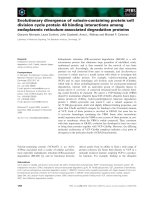
diogram showed a normal sinus rhythm. H owever, a
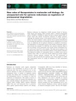
chest radio graph (Figure 1) was abnormal with l eft hilar
sha dowing. A biochemical profile showed hyponatremia
with a serum sodium level of 128mmol/L. In view of
her significant smoking history the most likely diagnosis
was broncho genic carcino ma with brain metastasis, and
so a computed tomography (CT) scan of the thorax and
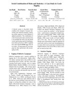
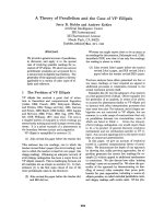
head was arranged. The thoracic CT scan (Figure 2)
revealed confluent left hilar lymphadenopathy encasing
the left lower lobe pulmonary artery, and a parenchymal
opacity in the left lower lobe was highly suggestive of
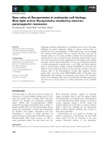
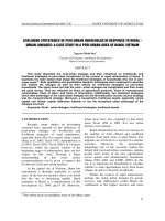
bronchogenic carcinoma. The contrast-enhanced CT
scan of her head (Figure 3) did not show any significant
abnormality. Flexible fiber-optic bronchoscopy results
were normal and bronchial washings were negative for
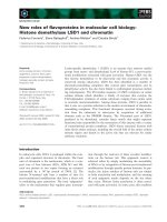
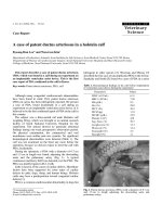
malignant cells. We therefore performed a CT-guided
biopsy (Figure 4) of the left lower lobe lesion. It was
suggestive of neoplastic infiltration of lung parenchyma
by small cell carcinoma. Furthermore, immunohisto-
chemistry showed positive staining with CD56 (Figure 5),
pan cytokeratin, chromograni n and thyroid transcription
factor 1 (TTF1), consistent with a diagnosis of small cell
lung cancer. As there was no improvement in her cogni-
tive function, a lumbar puncture was performed. The
results of a cerebrospinal fluid (CSF) examination were
unremarkable. In view of her persistent neurol ogical
symptoms, an MRI scan of the brain was performed
that showed numerous small foci of increased T2-
weighted signals scattered throughout the cerebral white
matter, particularly in the frontal and parietal areas.
Axial fluid attenuation inversion recovery (FLAIR)
sequences showed hyperint ense signals from the medial
temporal lobe on the left (F igure 6) and right side
(Figure 7), consistent with the radiological diagnosis of
limbic encephalitis. There was no evidence of metastatic
disease. A diagnosis of paraneoplastic limbic encephalitis
associated with limited stage small cell lung cancer was
made based on the clinical and radiological results. Our
patient was treated with prophylactic cranial irradiation
followed by platinum-based chemotherapy. Her
Figure 1 Chest radiograph showing left hilar abnormality
(arrow) and opacity in the left mid zone.
Figure 2 Computed tomography (CT) scan showing
parenchymal opacity in the apical segment of the left lower
lobe (arrow), highly suggestive of lung malignancy.
Figure 3 Computed tomography (CT) image of our patient’ s
brain on presentation, without significant acute pathology.
Fahim et al . Journal of Medical Case Reports 2010, 4:408
/>Page 2 of 4
cognitive function improved considerab ly over the
course of next few months and her condition remained
stable 18 months after presentation.
Discussion
Paraneoplastic limbic encep halitis, first described as a
clinical entity in 1968 [1] is characterized by short-term
memory deficits, mood and behavioral changes and rela-
tive preservation of other cognitive functions. There
may be seizures, which are most often partial complex
in nature. Moreover, hypothalamic involvement can
manifest with hyperthermia, hyperphagia or pituitary
hormonal deficits [2]. Patients with PLE often present
with symptoms of neurological involvement distant
from the limbic system (commonly brainstem and
cerebellum). Bakheit and colleagues [3] found that only
32% of patients had isolated limbic encephalitis. The
neurological symptoms often precede identification of
the tumor by weeks or months and the non-specific nat-
ure and diversity of symptoms add to the difficulty in
diagnosing this rare clinical entity.
Figure 5 Immunohistochemistry of a lung biopsy specimen
showing positive staining with CD56, suggestive of small cell
carcinoma of lung.
Figure 4 Histology from a computed tomography (CT)-guided
lung biopsy showing infiltration of the left lower lobe with
neoplastic cells, consistent with small cell lung cancer.
Figure 6 Axial fluid attenuation inversion recovery (FLAIR) MRI
of the brain showing a bright signal from the medial temporal
lobe on the left side (arrow) consistent with limbic
encephalitis.
Figure 7 Hyperintense signal from the hippocampus/medial
temporal lobe on the right (arrow).
Fahim et al . Journal of Medical Case Reports 2010, 4:408
/>Page 3 of 4
The most frequent neoplasms associated with PLE are
small cell lung cancer, testicular tumors, thymoma,
Hodgkin’s lymphoma and breast cancer. I n an analysis
of 50 patients with PLE, Gultekin and colleagues [4]
found that lung cancer was the most common neoplasm
identified in 50% of cases, followed by testicular and
breast carcinoma in 20% and 8%, respectively. The neu-
rological symptoms preceded the diagnosis of cancer in
approximately two-thirdsofpatientswithamedian
duration of three and a half months.
Paraneoplastic limbic encephalitis can pose a diagnos-
tic challenge in patients with cognitive impairment,
especially when there is no evidence of malignant dis-
ease, and can be easily mistaken for viral encephalomye-
litis or rapidly progressive neurodegenerative disease.
Eveninthepresenceofmalignancy,theneurological
symptoms can be easily attributed to cranial metastases.
An MRI scan of the brain is the most sensitive radi-
ological investigation to diagnose limbic encephalitis.
Typically, it shows hyperintense lesions in the medial
temporal lobes and these are best visualized in T2 and
axial FLAIR sequences without significant contrast
enhancement. A CSF examination is seldom diagnostic
of this condition, and the most common findings are
consistent with inflammatory changes (pleocytosis,
increased protein, oligoclonal bands and increased
immunoglobulin content). Anti-neuronal antibodies are
frequently found in the serum or CSF of patients with
PLE, but the absence of these antibodies does not
exclude the diagnosis. The most common is the anti-Hu
antibody, which is present in about 50% of patients with
small cell lung cancer presenting with limbi c encephali-
tis and the anti-Ta antibody associated with testicular
cancer [4]. Moreover, the presence of antibodies can be
predictive for a good response to immunosuppressive
therapy [5].
Electroencephalography can be a useful tool in order
to support the diagnosis of limbic encepha litis, as it can
demonstrate focal or generalized slowing and/or sharp
wave epileptiform activity p redominantly in the tem-
poral regions [6]. The treatment should be directed at
the associated malignancy, which frequently improves
the neurolo gical symptoms and is superior to immuno-
modulatory therapy.
Conclusion
Our patient ’ s case highlights the importan ce of consid-
ering PLE in the differential diagnosis of altered mental
status in patients with lung malignancy, if there is no
readily identifiable cause of c ognitive impairment on
initial investigations. Prompt diagnosis and early treat-
ment of malignancy provides the best chance of clinical
improvement in patients with this rare disorder.
Consent
Written informed consent was obtained from the patien t
for publication of this case report and any accompanying
images. A copy of the written consent is available for
review by the Editor-in-Chief of this journal.
Acknowledgements
We thank Dr A Campbell for providing Figures 4 and 5 and Dr R Hill, Dr G
Avery and Dr R Bartlett for their advice on the radiological images presented
in this case report.
Author details
1
Department of Cardiovascular and Respiratory Studies, Castle Hill Hospital,
Cottingham, UK.
2
Department of Oncology, Castle Hill Hospital, Cottingham,
UK.
3
Respiratory Medicine, Castle Hill Hospital, Cottingham, UK.
Authors’ contributions
AF, DVM and MB contributed to the writing of the manuscript. All authors
read and approved the manuscript.
Competing interests
The authors declare that they have no competing interests.
Received: 18 January 2010 Accepted: 17 December 2010
Published: 17 December 2010
References
1. Corsellis JA, Goldberg GJ, Norton AR: ’Limbic encephalitis’ and its
association with carcinoma. Brain 1968, 91:481-496.
2. Rosenfield MR, Dalmau J: Paraneoplastic limbic encephalitis associated
with small cell lung cancer. Comm Oncol 2007, 4:491-494.
3. Bakheit AM, Kennedy PG, Behan PO: Paraneoplastic limbic encephalitis:
clinico-pathological correlations. J Neurol Neurosurg Psychiatry 1990,
53:1084-1088.
4. Gultekin SH, Rosenfield MR, Voltz R, Eichen J, Posner JB, Dalmau J:
Paraneoplastic limbic encephalitis: neurological symptoms,
immunological findings and tumour association in 50 patients. Brain
2000, 7:1481-1494.
5. Storstein A, Bru A, Vedeler CA: Limbic encephalitis-a diagnostic challenge.
Tidsskr Nor Laegeforen 2007, 127:3077-3080.
6. Lawn ND, Westmoreland BF, Kiely MJ, Lennon VA, Vernino S: Clinical,
magnetic resonance imaging, and electroencephalographic findings in
paraneoplastic limbic encephalitis. Mayo Clin Proc 2003, 78:1363-1368.
doi:10.1186/1752-1947-4-408
Cite this article as: Fahim et al .: A case of limbic encephalitis presenting
as a paraneoplastic manifestation of limited stage small cell lung
cancer: a case report. Journal of Medical Case Reports 2010 4:408.
Submit your next manuscript to BioMed Central
and take full advantage of:
• Convenient online submission
• Thorough peer review
• No space constraints or color figure charges
• Immediate publication on acceptance
• Inclusion in PubMed, CAS, Scopus and Google Scholar
• Research which is freely available for redistribution
Submit your manuscript at
www.biomedcentral.com/submit
Fahim et al . Journal of Medical Case Reports 2010, 4:408
/>Page 4 of 4