báo cáo khoa học: " Epstein Barr Virus-positive large T-cell lymphoma presenting as acute appendicitis 17 years after cadaveric renal transplant: a case report" docx
Bạn đang xem bản rút gọn của tài liệu. Xem và tải ngay bản đầy đủ của tài liệu tại đây (6.68 MB, 8 trang )
CAS E REP O R T Open Access
Epstein Barr Virus-positive large T-cell lymphoma
presenting as acute appendicitis 17 years after
cadaveric renal transplant: a case report
Shiva K Ratuapli
1*
, Shishir Murarka
1
, Karen A Miller
2
, James C Ferraro
1
, Haider Zafar
1
Abstract
Introduction: The majority of post-transplant lymphoproliferative disorders in renal transplant patients are of the
B-cell phenotype, while the T-cell phenotype is rare. We report a case of Epstein Barr Virus-positive, T-cell
lymphoma in a renal transplant patient, presenting unusually as acute appendicitis.
Case presentation: A 45-year-old Hispanic male renal transplant patient presented with right-side abdominal pain
17 years after transplant. The laboratory studies were unremarkable. Laparoscopic exploration showed an inflamed
appendix so a laparoscopic appendectomy was performed. Pathology of the appendix showed large cells positive
for CD3, CD56 and Epstein Barr Virus-encoded RNA staining, and negative for CD20 and CD30. The tissue tested
positive for T-cell receptor gene rearrangement by polymerase chain reaction analysis. Treatment management
involved reduction of immunosuppression and initiation of chemotherapy with cisplatin, etoposide, gemcitabine,
and solumedrol followed by cyclophosphamide, hydroxydaunorubicin, vincristine and prednisone). He recovered
and the allo-grafted kidney is fully functional.
Conclusion: We report a rare case of post-renal transplant large T-cell lymph oma, with an unusual presentation of
acute appendicitis and Epstein Barr Virus-positivity, which responded well to chemotherapy.
Introduction
Solid organ transplantation has been increasingly per-
formed in recent years with the use of highly potent
immunosuppressive agents to avoid rejection by the
host. Post-transplant lymphoproliferative disorders
(PTLD) are well known malignanci es found in trans-
plant patients, with an incidence reportedly 28 to 49
times greater than in the general population [1]. PTLD
in renal transplant patients is reported to be h igher in
the paediatric population (10.1%) than the adult popula-
tion (1.2%) [2]. PTLD in renal transplant patients was
first described as a complication with azathioprine-based
therapy [3], but was later described after therapy with
multiple more novel immunosuppressive agents.
While the majority of PTLD in renal transplant
patients are of the B-cell phenotype, a few exhibit the
T-cell phenotype [4]. We report a case of Epstein Barr
Virus (EBV)-positive T-cell lymphoma in a patient, who
underwent cadaveric renal transplant 17 years ago and
was on chronic multi-drug immunosuppression. Our
patient presented unusually with abdominal pain and
acute appendicitis.
Case presentation
A 45-year-old Hispani c male who underwent c adaveri c
renal transplant in t he right l ower quadrant 17 years
earlier, presented to the hospital with a t hree-month
history of generalized abdominal pain with localization
to the right side for two weeks. He was on chronic
immunosuppression with tacrolimus, a zathioprine, siro-
limus and prednisone. The pain was more pronounced
in the right upper quadrant, and the ultrasound ima ging
of the abdomen was suggestive of cholecystitis. Labora-
tory studies did not reveal any abnormalities. He could
not confirm if he had had any problems or surgeries on
his gall bladder. Hence, he underwent laparoscopic
exploration of the gall bladder fossa. During surgery,
adhesions of the omentum were found in the gall
* Correspondence:
1
Department of Medicine, Banner Estrella Medical Center, 9201 W. Thomas
Road, Phoenix, AZ 85037, USA
Full list of author information is available at the end of the article
Ratuapli et al. Journal of Medical Case Reports 2011, 5:5
/>JOURNAL OF MEDICAL
CASE REPORTS
© 2011 Ratuapli et al; licensee BioMed Central Ltd. This is an Open Access article distributed under the te rms of the Creative Co mmons
Attribution License ( which permits unrestricted use, distribution, and reproduction in
any medium, provided the original work is properly cited.
bladder fossa in the absence of the gall bladder, and an
inflamed appendix was found elevated due to the trans-
planted kidney in the right lower quadrant. Laparoscopic
appendectomy was performed and the tissue underwent
pathological examination. He was discharged after an
uneventful post-operative course.
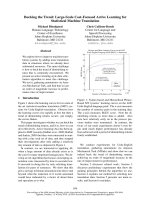
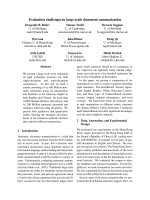
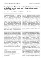
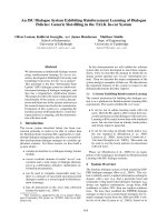
Pathology of his appendix by immunostaining revealed
anaplastic cells strongly positive for CD3, CD56 together
with strong focal EBV-encoded RNA (EBER) staining
(Figures 1, 2, 3 and 4). The malignant cells were nega-
tive for CD20, CD30, CD45, CD5, Alk-1 and TCK. The
tissue was found to be positive for the T-cell receptor
(TCR) by gamma gene rearrangement studies by PCR
analysis (Figure 5). Immunoglobulin heavy chain rear-
rangement (IgH) by PCR analys is did not detect a clonal
B-cell population, thereby confirming T-cell lymphoma.
A bone marrow examination revealed no involvement
with negative flow cytometry and showed normal male
karyotype (46, XY). A staging positron emission tomo-
graphy (PET) scan showed increased r adiotracer uptake
in the r ight cervical and left groin lymph nodes along
with the 3.3 cm liver mass. Non-specific uptake in the
stomach was also observed.
The patient was re-admitted to the hospital 10 days
later, with increasing abdominal pain, symptoms of gas-
tric outlet obstruction, weight loss, headaches and fever.
A lumbar puncture was negative for infection or lym-
phoma. Cranial imaging with a computed tomography
(CT) scan was also negative. An esophagogastroduode-
noscopy (EGD) was performed revealing multiple ulcer-
ated nodular masses in the stomach and duodenum
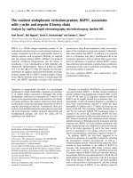
(Figures 6 and 7). A stomach biopsy gave similar results
as the appe ndix with large anaplastic cells with i rregular
nuclei. Immunostaining of the gastric specimen con-
firmed T-cell lymphoma as well as positive EBER
staining.
Initial treatment ma nagement involved reducing the
dose of the patient’s immunosuppressive agents and
starting chemotherapy. Administration of azathioprine,
prednisone and tacrolimus was stopped and low dose
sirolimus at 1 mg was given daily. The first cycle of
chemotherapy (PEGS) included cisplatin 25 mg/m
2
,
Figure 1 Appendix biopsy showing large, pleomorphic lymphocytes with irregular nuclear contours and large nucleoli. (400 X).
Ratuapli et al. Journal of Medical Case Reports 2011, 5 :5
/>Page 2 of 8
Figure 2 Positive staining of lymphoid infiltrate for CD3 (400 X).
Figure 3 Gastric biopsy showing atypical lymphoid infiltrate (200 X).
Ratuapli et al. Journal of Medical Case Reports 2011, 5 :5
/>Page 3 of 8
etoposide 40 mg/m
2
and solumedrol 250 mg adminis-
tered on days one, two and three, and gemcitabine
(Gemzar) 1000 mg/m
2
on day one (ongoing Phase II
trial SWOG 0350). Our patient had a positive and rap id
clinical response to this regimen. Thus, the chemother-
apy was changed to a standard CHOP regimen (c yclo-
phosphamide, doxorubicin [Adriamycin ], vincristine,
prednisone). His gastric ou tlet obstruction was sup-
ported with total parenteral nutrition (TPN) for a few
weeks, after which the patient was able to eat well. A
repeat PET scan after the second cycle of CHOP
showed a significant response.
The main complications during the ther apy were pan-
cytopenia, febrile neutropenia and pneumonia. These
were managed successfully. He recovered well and is
presently receiving treatment as an outpatient. His allo-
grafted kidney is also fully functional. Restaging is
planned after a total of six cycles of CHOP with a PET
scan and EGD.
Discussion
Our case is interesting due to the latency of PTLD
development, the lack of hematological abnormalities
and the EBV-positivity, even though the lymphoma was
of T-cell origin. The diagnosis became apparent when
an appendectomy w as performed for abdominal pain.
The cytopathology of our patient shows all the typical
features of a peripheral T-cell variant of PTLD, where
the T-cell lineage of the lymphoma was confirmed by
TCR gene rearrangement studies.
While presenting symptoms in the majority of patients
are non-specific such as fever and w eight loss, approxi-
mately 15% of cases present as an emergency with intest-
inal perforation [5]. Similar to other reports on T-cell
type PTLD, which generally occurred more than five
years after transplant, this case occurred 17 years after
the renal t ransplant. The high levels of immunosuppres-
sion were due to two episodes of graft rejection in the
preceding four years.
Figure 4 Appendiceal infiltrate showing scattered Epstein Barr Virus-positive cells (100 X).
Ratuapli et al. Journal of Medical Case Reports 2011, 5 :5
/>Page 4 of 8
PTLD can be categorized into three distinct groups
basedontheWorldHealthOrganization classification
of lymphoid tissue neoplasms [6] (Table 1). The first
group has diffuse B-cell hyperplasia, w hich is relatively
benign and responds well to a reduction in immunosup-
pression. The second group consists of polymorphic
PTLD, which is the most common group in both the
adult and pediatric populations. The third group con-
sists of high-grade invasive lymphomas of either T- or
B-cell monoclonality. Monomorphic T-cell PTLD is
further subdivided into large cell, anaplastic or an
unspecified type. While the incidence of T-cell PTLD in
renal transplant patients is approximately 15%, a recent
study of 21 cas es of post-transplant hepatosplenic T-cell
lymphoma by Tey et al. [7] reported 19 patients who
underwent prior renal transplant. This and several other
reports [5,7,8] appear to show increased incidence,
which might be due to heightened awareness along with
use of increasingly potent immunosuppressants.
The etiopathogenesis of T-cell PTLD is not entirely
known, although it may be similar to non-Hodgkins
lymphoma (NHL) seen in the general population. While
Figure 5 T cell receptor gene rearrangement by PCR analysis showing monoclonal spike.
Ratuapli et al. Journal of Medical Case Reports 2011, 5 :5
/>Page 5 of 8
aputativeroleofEBVhasbeensuggestedin89%of
cases of B-cell PTLD [9], no direct role for this lympho-
tropic virus has been confirmed in T-cell PTLD. EBV
infects and immortalizes B cells causing unchecked pro-
liferation of EBV-infected cells, as the critical T-cell
control of B-cells is lacking in immunosuppressed
patients [10]. Human T-cell Lymphotropic Virus 1
(HTLV1) has been reported to cause post renal trans-
plant T-cell lymphomas in Japan [11], due t o a higher
prevalence of the virus among hemodialysis patients.
Our case was unusual in that the lymphoma tested posi-
tive for EBV, even though it was of T-cell origin, which
is only seen in a small minority of patients [12].
The initial step in treating PTLD is reduction
in immunosuppression, and the response is usu ally seen
in three to four weeks, resulting in long term remission
in 25% to 63% [13] of adults. Early polyclonal PTLD
responds well to a reduction in immunosuppression,
whereas monoclonal PTLD generally does not respond
to the redu ction and has a high mortality rate of 50% to
90% [14]. Early use of anthracycline-based chemotherapy
results in long term disease free survival rates of 50% to
60% in monoclonal B-cell lymphomas [15], whereas
T-cell PTLD responds very poorly. There are no stan-
dardized chemotherapy regimens for PTLD in general
and for the T-cell phenotype in particular. Multiple
treatment regimens such as CHOP, VAPEC-B (Adria-
mycin, etoposide, cyclophosphamide, methotrexate,
bleomycin and vincristine), anti-IL-6 mAb, bexarotene
and antivira l agents have been used with varied results.
Other salvage regimens for high-grade lymphomas have
also been suggested in the literature [14,15].
Figure 6 EGD showing large ulcerated gastric nodule together with large nodules in the duodenum.
Ratuapli et al. Journal of Medical Case Reports 2011, 5 :5
/>Page 6 of 8
Conclusion
In summary we report the case of a patient with post-renal
transplant large T-cell lymphoma, with an unusual presen-
tation of acute appendicitis and EBV positivity. We report
successful treatment with chemotherapy and stress the
need for heightened awareness of this malignancy in
patients with prolonged immunosuppression. Monoclonal
T-cell PTLD remains a high mortality disease, and further
large multi-centre studies are required to understand the
pathogenesis and develop better treatment regimens.
Consent
Written informed consent was obtained from the patient
for publication of this case report and accompanying
images. A copy of the written consent is available for
review by the Editor-in-Chief of this journal.
Figure 7 EGD showing multiple large gastric nodules with central ulceration.
Table 1 The World Health Organization classification of PTLD
Early Lesions Polymorphic PTLD Monomorphic PTLD
B-Cell Lymphomas T-Cell Lymphomas Other Types
Reactive Plasmacytic
Hyperplasia
Polyclonal
Monoclonal
• Diffuse large B cell
lymphoma
• Burkitt/Burkitt-like
lymphoma
• Plasma cell myeloma
• Peripheral T cell lymphoma
• Large CellAnaplastic
• Unspecified
• Raretypes (gammadelta, Hepatosplenic,
T/NKcell)
1. Hodgkin’s disease-
like
2. Plasmacytoma-like
Ratuapli et al. Journal of Medical Case Reports 2011, 5 :5
/>Page 7 of 8
Author details
1
Department of Medicine, Banner Estrella Medical Center, 9201 W. Thomas
Road, Phoenix, AZ 85037, USA.
2
Department of Pathology, Banner Estrella
Medical Center, 9201 W. Thomas Road, Phoenix, AZ 85037, USA.
Authors’ contributions
SKR and HZ participated in the conception and literature search. KAM
provided and reviewed the pathology slides. SKR, SM, JF and HZ helped to
draft the manuscript. All authors read and approved the final manuscript.
Competing interests
The authors declare that they have no competing interests.
Received: 20 February 2010 Accepted: 12 January 2011
Published: 12 January 2011
References
1. Boubenider S, Hiesse C, Goupy C, Kriaa F, Marchand S, Charpentier B:
Incidence and consequences of post-transplantation lymphoproliferative
disorders. J Nephrol 1997, 10:136-145.
2. Shapiro R, Nalesnik M, McCauley J, Fedorek S, Jordan ML, Scantlebury VP,
Jain A, Vivas C, Ellis D, Lombardozzi-Lane S, Randhawa P, Johnston J,
Hakala TR, Simmons RL, Fung JJ, Starzl TE: Posttransplant
lymphoproliferative disorders in adult and pediatric renal transplant
patients receiving tacrolimus-based immunosuppression. Transplantation
1999, 68:1851-1854.
3. Penn I, Hammond W, Brettschneider L, Starzl TE: Malignant lymphomas in
transplantation patients. Transplant Proc 1969, 1:106-112.
4. Leblond V, Sutton L, Dorent R, Davi F, Bitker MO, Gabarre J, Charlotte F,
Ghoussoub JJ, Fourcade C, Fischer A, et al: Lymphoproliferative disorders
after organ transplantation: a report of 24 cases observed in a single
center. J Clin Oncol 1995, 13:961-968.
5. Taylor AL, Marcus R, Bradley JA: Post-transplant lymphoproliferative
disorders (PTLD) after solid organ transplantation. Crit Rev Oncol Hematol
2005, 56:155-167.
6. Harris NL, Jaffe ES, Diebold J, Flandrin G, Muller-Hermelink HK, Vardiman J,
Lister TA, Bloomfield CD: The World Health Organization classification of
hematological malignancies report of the Clinical Advisory Committee
Meeting, Airlie House, Virginia, November 1997. Mod Pathol 2000, 13:193-207.
7. Tey SK, Marlton PV, Hawley CM, Norris D, Gill DS: Post-transplant
hepatosplenic T-cell lymphoma successfully treated with HyperCVAD
regimen. Am J Hematol 2008, 83:330-333.
8. Balachandran I, Walker JW Jr, Broman J: Fine needle aspiration cytology of
ALK 1(-), CD 30(+) anaplastic large cell lymphoma post renal
transplantation: A case report and literature review. Diagn Cytopathol
2010, 38:213-216.
9. Pasquale MA, Weppler D, Smith J, Icardi M, Amador A, Gonzalez M, Kato T,
Tzakis A, Ruiz P: Burkitt’s lymphoma variant of post-transplant
lymphoproliferative disease (PTLD). Pathol Oncol Res 2002, 8:105-108.
10. Cohen JI: Epstein-Barr virus infection. N Engl J Med 2000, 343:481-492.
11. Hoshida Y, Li T, Dong Z, Tomita Y, Yamauchi A, Hanai J, Aozasa K:
Lymphoproliferative disorders in renal transplant patients in Japan. Int J
Cancer 2001, 91:869-875.
12. Magro CM, Weinerman DJ, Porcu PL, Morrison CD: Post-transplant EBV-
negative anaplastic large-cell lymphoma with dual rearrangement: a
propos of two cases and review of the literature. J Cutan Pathol 2007, 34:1-8.
13. Gottschalk S, Rooney CM, Heslop HE: Post-transplant lymphoproliferative
disorders. Annu Rev Med 2005, 56:29-44.
14. Orjuela M, Gross TF, Cheung YK, Alobeid B, Morris E, Cairo MS:
A pilot
study of chemoimmunotherapy (cyclophosphamide, prednisone, and
rituximab) in patients with post-transplant lymphoproliferative disorder
following solid organ transplantation. Clin Cancer Res 2003, 9:3945S-3952S.
15. Taylor AL, Bowles KM, Callaghan CJ, Wimperis JZ, Grant JW, Marcus RE,
Bradley JA: Anthracycline-based chemotherapy as first-line treatment in
adults with malignant posttransplant lymphoproliferative disorder after
solid organ transplantation. Transplantation 2006, 82:375-381.
doi:10.1186/1752-1947-5-5
Cite this article as: Ratuapli et al.: Epstein Barr Virus-positive large T-cell
lymphoma presenting as acute appendicitis 17 years after cadaveric
renal transplant: a case report. Journal of Medical Case Reports 2011 5:5.
Submit your next manuscript to BioMed Central
and take full advantage of:
• Convenient online submission
• Thorough peer review
• No space constraints or color figure charges
• Immediate publication on acceptance
• Inclusion in PubMed, CAS, Scopus and Google Scholar
• Research which is freely available for redistribution
Submit your manuscript at
www.biomedcentral.com/submit
Ratuapli et al. Journal of Medical Case Reports 2011, 5 :5
/>Page 8 of 8