báo cáo khoa học: " Peritoneal mesothelioma in a woman who has survived for seven years: a case report" pptx
Bạn đang xem bản rút gọn của tài liệu. Xem và tải ngay bản đầy đủ của tài liệu tại đây (317.56 KB, 4 trang )
CASE REP O R T Open Access
Peritoneal mesothelioma in a woman who has
survived for seven years: a case report
Krishna Pillai, Javed Akhter, Mohammad H Pourgholami
*
, David L Morris
Abstract
Introduction: Malignant peritoneal mesothelioma is a rare cancer with poor patient survival. Fem ale gender has
been identified as a positive prognostic factor. Recently, it has been suggested that the expression of estrogen
receptor b in malignant mesothelioma leads to tumor suppression and a better prognosis.
Case presentation: We report the c ase of a 48-year -old Caucasian woman who is alive and disease-free seven
years after the initial diagnosis and treatment of malignant peritoneal mesothelioma.
Conclusion: This patient’s long survival may be attributable to a combination of factors, including minimal disease,
complete cytoreductive surgery and hyperthermic intraperitoneal chemotherapy plus the estrogen receptor b
positivity of the tumor.
Introduction
Peritoneal mesothelioma is a rare but fatal disease; the
incidence is approximately one per million, and perito-
neal mesothelioma accounts for about 20% to 30% of all
cases of mesothelioma [1]. Although asbestos has been
implicated as the main carcinogen [2,3], other factors
such as radiation, peritonitis and SV40 have all been
implicated [4].
Peritoneal mesothelioma progresses with unspecific
symptoms, and when presented, it is commonly in the
form of increased abdominal girth, pain and weight loss;
hence, diagnosis is late, with a poor prognosis. A num-
ber of therapeutic regimens have been used to improve
prognosis [5], and currently debulking surgery is fol-
lowed by hyperthermic intraperitoneal chemotherapy
(HIPEC). This has led to marked improvement in
patients who were once classified as preterminal [6].
The current median survival is around 10 months, and
relative five-year survival is in approximately 16% [7].
Hence, more information on the disease and more effec-
tive therapies are needed to improve prognosis.
Case presentation
A Caucasian women, now aged 48 years, presented her-
self at the age of 40 years with abdominal pain (four to
five days), a bad taste in her mouth an d tiredness. She
had epigastric discomfort caused by abdominal disten-
sion for the past four to five years and had multiple
upper and lower gastrointestinal endoscopic examina-
tions. Her medical history involved obesity, treatment
for a blocked salivary duct, hypertension, endometriosis,
appendectomy, Bell’s palsy and hormone replacement
therapy. Recent laproscopic cholecystectomy showed
areas of abnormality, and a biop sy revealed the presence
of malignant mesothelioma of epi thelial histology.
A c omputed tomography (CT) scan showed peritoneal
tumor, not widely spread with no parenchymal liver dis-
ease. There were no pleural nodules or fluid collection
along with the absence of metastasis. Disease volume as
determined by peritoneal cancer index was low. The
patient denied any exposure to asbestos.
Tumor markers such as CA125 appeared to be normal
and ranged from 7 to 11, which fell within the reference
range (0-35 IU/mL). Blood analysis revealed that the
patient had mild to moderate anemia with moderate
thrombocytosis.
Laparotomy with peritonectomy performed one month
later revealed the accumulation of ascitic fluid (four
litres) with no liver disease but some disease affecting
the diaphragm, small bowel, colon and uterus. Complete
cytoreduction was carried out, with preservation of the
spleen (minor diseased part removed). Similarly, disease
affecting the small bowel, mesentery and co lon was also
* Correspondence:
Department of Surgery, St. George Hospital, University of New South Wales,
Kogarah, NSW, Australia
Pillai et al. Journal of Medical Case Reports 2011, 5:36
/>JOURNAL OF MEDICAL
CASE REPORTS
© 2011 Pillai et al; licensee BioMed Central Ltd. This is an Open Access article distributed under the terms of the Creative Commons
Attribution License ( which permits unres tricted use, distribution, and reproduction in
any medium, provided the original work is properly cited.
removed. Disease in the uterus was diathermised, and
HIPEC was carried out with 50 mg/m
2
of cisplatin and
15 mg/m
2
of Adriamycin for 90 minutes at 41.5°C and
20 mg/m
2
(5 cycles) of paclitaxel, with insertion of a
peritoneal catheter and port. The patient refused post-
operative chemotherapy.
Macroscopic findings showed a peritoneal tumor
(multiple pieces of omentum 400 × 200 × 50 mm in
aggre gates ), and microscopic inves tigat ion showed some
areas of prominent papillary tumor on mesothelial
surface. Nuclear atypia varied from minimal to focally
moderate with nuclear membrane irregularities and an i-
sonucleosis. Mitosis was rare (<1/10 high-power field).
Further focal stromal invasion of small groups of cells
and single cells was seen in underlying fat with an
absence of desmoplastic response. Very rare psammoma
bodies were seen, and necrosis was absent in this sec-
tion. Atyp ical mesothelial proliferation was also seen in
all sections examined. Chronic inflammation was also
seen in the subserosal connective tissues. Although the
tumor was WDPM entering into differential diagnosis,
theextentofthediseaseandthepresenceofinvasion
mitigated against this diagnosis. The immunohistochem-
ical (ICH) findings are shown in Table 1.
Two years later, the patient presented herself with epi-
gastric discomfort, gastric reflux, abdominal pain, consti-
pation and diarrhea. A CT scan was normal, but t he
patient underwent laparotomy and a second peritonect-
omy. The findings were adhesions (significant at term-
inal small bowel and right colon), few nodules (bowels
and mesentery) and a thin membranous septum on the
small bowel. Macroscopic examination showed occa-
sional atypical cells with minor peritoneal disease
(epitheloid cells) consisten t with mesothelioma. There
was absence of tumor in lymph nodes, the lesser omen-
tum and the hepatic artery. Microscopic examination
results are shown in Table 2. Hence, it was concluded
that very low-volume disease was present, and pain was
mainly attributable to adhesions.
Treatment with HIPEC (cisplatin 200 mg + mitomycin
C 25 mg/90 min/41.5°C), extensive division of adhe-
sions, peritoneal biopsy and intraoperativ e ultrasonogra-
phy w ere carried out. The membranous septum on the
smal l bowel along with two nodul es were also removed.
The patient refused to have postoperative chemotherapy.
Seven months later, the patient presented with a para-
spinal mass, and microscopic examination showed no
tumor; the patient was negative for CK5/6, cytokeratin
5.2, epithelial membrane antigen and mesothelial cell
membrane protein. Therefore, diagnosis was made in
favor of fibrosis and mild chronic inflammation.
Another 11 months later, the patient underwent laparot-
omy during which division of adhesions and repair of a
hernia was carried out. No evidence of mesothelioma
was found at this stage, and two months later, laparot-
omy was repeated for division of adhesions and debride-
ment of a large abscess owing to infection. The wound
did not heal, so the patient was prescribed antibiotic
therapy with an open wound-hea ling regimen. Finally,
the wound resolved, and currently the patient is well.
Discussion
Although the tumor was WDPM entering into differen-
tial diagnosis, the extent of the disease and the presence
of invasio n mitigated against this diagnosis. Hence, the
patient underwent cytoreductive surgery with HIPEC,
which is most effective for patients with ma lignant peri-
toneal mesothelioma (MPM). Only two peritonectomies
with HIPEC were ca rried out in this patient, and she
did not receive postoperative chemotherapy. Although
the patient developed some complications as a result of
adhesions, this was rectified during recovery with subse-
quent laparotomy. CT scans and ICH findings showed
the absence of any malignancy after the second HIPEC.
Unlike many patients, who succumb to the disease
soon after treatment, this patient is alive and well past
seven years. Close scrutiny of the case seems to reveal
three s alient features that may have contributed to this
person’s favorable prognosis. First, it appears from diag-
nosis (macroscopic a nd microscopic) that the patient
Table 1 Protein markers that have been identified by
immuno histochemistry in the patient tumor samples
Protein Markers Positive (+) Negative (-)
CAM5.2 +
Cytokeratin +
HBME-1 + (thick or membranous)
CD 15 -
BER-EPA -
CEA -
Human epithelial antigen -
EMA + (focal staining)
Table 2 Diagnostic findings from patient specimens
(formalin fixed and paraffin embedded)
Section Result of Examination
Formalin fixed Atypical epithelioid cells present
Higher nuclear:cytoplasmic ratio
Paraffin
embedded
Presence of nodules of cellular tumor
Positive for HBME-1 and EMA
Absence of tumor in lymph nodes
Adipose tissue, omentum: low-grade epithelioid
mesothelioma
Pillai et al. Journal of Medical Case Reports 2011, 5:36
/>Page 2 of 4
mayhavealessaggressiveformofMPM(lowdisease
volume with no metastasis) that with complete cytore-
duction is probably more amenable to HIPEC. This con-
clusion has been derived because the patient responded
well to HIPEC treatment in the first three years, after
which her examination revealed abatement of the dis-
ease. Indeed, the last laparotomy performed did not
reveal any disease.
Second, the pathology report indica ted that the patient
had an epithelioid type of MPM with abundant cellular
cytoplasm that is less aggressive and more amenable to
HIPEC compared with the sarcomatous or mixed type [8].
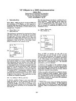
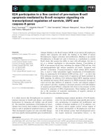
Finally, immunohistochemical examination of the
tumor tissues revealed that the patient has a high
expression of estrogen receptor b (ER
b
) (Figure 1). Very
recent studies by Pinton et al. [9] have indicated that
ER
b
expression in pleural mesothelioma has prognostic
significance and that high expression of these receptors
has endowed marked longevity in these patients. These
authors have also suggested that manipulation of ER
b
receptors may offer a new mode of therapy for this type
of cancer. Our studies have also shown that the expres-
sion of ER
b
in peritoneal mesothelioma offers a better
prognosis (unpublished data).
Noticeab ly, this patient’ s plasma estradiol was mea-
sured to be 483 rmol/L, which is comparable t o levels
found in women during the follicular phase of the ovar-
ian cycle. Estradiol is a universal ligand for both ER
a
and ER
b
.ER
b
is endowed with modulatory function on
ER
a
-dep endant cell proliferation [10], and when present
by itself, it is able to control cell replication through th e
G2-M phase cell arrest in a ligand-dependant and -inde-
pendent manner [9]. Hence, it may be suggested that
the high level of estradiol together with th e high expres-
sion of ER
b
could have led to better disease control and
hence longer survival.
Conclusion
Taken as whole, cytoreduction with HIPEC has con-
ferred good prognosis on this patient owing to the mild
nature of the disease of epitheloid histology with ER
b
expression and high plasma estradiol level.
Consent
Written informed consent was obtained from the patient
for publicatio n of this case report and any accompany-
ing images. A copy of the written consent is available
for review from the Editor-in Chief of the journal.
Abbreviations
CT: computed tomography; ER
β
: estrogen receptor β; HIPEC: hyperthermic
intraperitoneal chemotherapy; ICH: immunohistochemical; MPM: malignant
peritoneal mesothelioma; WDPM well-differentiated papillary mesothelioma.
Authors’ contributions
KP, MHP, JA and DLM collected, analyzed and interpreted patient data. KP
was the major contributor in writing the manuscript. All authors read and
approved the final manuscript.
Competing interests
The authors declare that they have no competing interests.
Received: 7 May 2010 Accepted: 26 January 2011
Published: 26 January 2011
References
1. Ahmed I, Koulaouzidis A, Iqbal J, Tan WC: Malignant peritoneal
mesothelioma as a are cause of ascites: a case report. J Med Case Reports
2008, 2:121-124.
2. Lee AM, Raz DJ, He B, Jablons DM: Update of the molecular biology of
malignant mesothelioma. Cancer 2007, 109:1454-1461.
3. Boffetta P: Epidemiology of peritoneal mesothelioma: a review. Ann
Oncol 2007, 18:985-990.
4. Cutrone R, Lidnisk J, Dunn G, Rizzo P, Bocchetta M, Chumakov K, Minor P,
Carbone M: Some oral poliovirus vaccine were contaminated with
infectious SV40 after 1961. Cancer Res 2005, 65:10273-10279.
5. Mohammad F, Sugarbaker PH: Peritoneal mesothelioma. Curr Treat Options
Oncol 2002, 3:375-386.
6. Chua TC, Yan TD, Morris DL: Peritoneal mesothelioma: current
understanding and management. Can J Surg 2009, 52:59-64.
Figure 1 Immunohistochemical staining of paraffin-embedded slides (3 μminthickness). A-Negative for estrogen receptor b (ER
b
)
(breast tissue) stained blue. B-Patient slide with heavy staining for ER
b,
stained brown.
Pillai et al. Journal of Medical Case Reports 2011, 5:36
/>Page 3 of 4
7. Feldman AL, Libutti SK, PingPank JF, Bartlett DL, Beresnev TH,
Mavroukakis SM, Steinberg SM, Liewehr DJ, Kleiner DE, Alexander HR:
Analysis of factors associated with outcome in patients with malignant
peritoneal mesothelioma undergoing surgical debulking and
intraperitoneal chemotherapy. J Clin Oncol 2003, 21:4560-4566.
8. Bridda A, Padoan I, Mencarelli R, Frego M: Peritoneal mesothelioma: a
review. Med Gen Med 2007, 9:32.
9. Pinton G, Brunelli E, Murer B, Puntoni R, Puntoni M, Fennell DA, Gaudino G,
Mutti L, Moro L: Estrogen receptor β affects the prognosis of human
malignant mesothelioma. Cancer Res 2009, 11:4598-4604.
10. Osborne CK, Schiff R: Estrogen receptor biology: continuing progress and
therapeutic implications. J Clin Oncol 2005, 23:1616-1622.
doi:10.1186/1752-1947-5-36
Cite this article as: Pillai et al.: Peritoneal mesothelioma in a woman
who has survived for seven years: a case report. Journal of Medical Case
Reports 2011 5:36.
Submit your next manuscript to BioMed Central
and take full advantage of:
• Convenient online submission
• Thorough peer review
• No space constraints or color figure charges
• Immediate publication on acceptance
• Inclusion in PubMed, CAS, Scopus and Google Scholar
• Research which is freely available for redistribution
Submit your manuscript at
www.biomedcentral.com/submit
Pillai et al. Journal of Medical Case Reports 2011, 5:36
/>Page 4 of 4