Báo cáo y học: "Extracorporeal immune therapy with immobilized agonistic anti-Fas antibodies leads to transient reduction of circulating neutrophil numbers and limits tissue damage after hemorrhagic shock/resuscitation in a porcine model" doc
Bạn đang xem bản rút gọn của tài liệu. Xem và tải ngay bản đầy đủ của tài liệu tại đây (1.37 MB, 13 trang )
Lögters et al. Journal of Inflammation 2010, 7:18
/>Open Access
RESEARCH
BioMed Central
© 2010 Lögters et al; licensee BioMed Central Ltd. This is an Open Access article distributed under the terms of the Creative Commons
Attribution License ( which permits unrestricted use, distribution, and reproduction in
any medium, provided the original work is properly cited.
Research
Extracorporeal immune therapy with immobilized
agonistic anti-Fas antibodies leads to transient
reduction of circulating neutrophil numbers and
limits tissue damage after hemorrhagic
shock/resuscitation in a porcine model
Tim T Lögters*
1,2
, Jens Altrichter
1
, Adnana Paunel-Görgülü
1
, Martin Sager
1
, Ingo Witte
1
, Annina Ott
1
, Sarah Sadek
1
,
Jessica Baltes
1
, José Bitu-Moreno
3
, Alberto Schek
1
, Wolfram Müller
4
, Teresa Jeri
1
, Joachim Windolf
1
and Martin Scholz
1
Abstract
Background: Hemorrhagic shock/resuscitation is associated with aberrant neutrophil activation and organ failure. This
experimental porcine study was done to evaluate the effects of Fas-directed extracorporeal immune therapy with a
leukocyte inhibition module (LIM) on hemodynamics, neutrophil tissue infiltration, and tissue damage after
hemorrhagic shock/resuscitation.
Methods: In a prospective controlled double-armed animal trial 24 Munich Mini Pigs (30.3 ± 3.3 kg) were rapidly
haemorrhaged to reach a mean arterial pressure (MAP) of 35 ± 5 mmHg, maintained hypotensive for 45 minutes, and
then were resuscitated with Ringer' solution to baseline MAP. With beginning of resuscitation 12 pigs underwent
extracorporeal immune therapy for 3 hours (LIM group) and 12 pigs were resuscitated according to standard medical
care (SMC). Haemodynamics, haematologic, metabolic, and organ specific damage parameters were monitored.
Neutrophil infiltration was analyzed histologically after 48 and 72 hours. Lipid peroxidation and apoptosis were
specifically determined in lung, bowel, and liver.
Results: In the LIM group, neutrophil counts were reduced versus SMC during extracorporeal immune therapy. After
72 hours, the haemodynamic parameters MAP and cardiac output (CO) were significantly better in the LIM group.
Histological analyses showed reduction of shock-related neutrophil tissue infiltration in the LIM group, especially in the
lungs. Lower amounts of apoptotic cells and lipid peroxidation were found in organs after LIM treatment.
Conclusions: Transient Fas-directed extracorporeal immune therapy may protect from posthemorrhagic neutrophil
tissue infiltration and tissue damage.
Background
Hemorrhagic shock is a leading cause of complications
and death in combat casualties and civilian trauma [1]. It
has been shown to cause systemic inflammatory response
syndrome (SIRS), multiple organ dysfunction syndrome
(MODS), and multiple organ failure (MOF) [2]. Despite
intensive investigations, the pathophysiology of posthem-
orrhagic multiple organ failure remains incompletely
understood. Recently, it has been reported that neutro-
phils recruited by mitochondrial products (formyl pep-
tides and mitochondrial DNA) released from damaged
tissues and cells are responsible for the inflammation
seen in SIRS [3]. However, tissue infiltration with acti-
vated polymorphonuclear neutrophils is associated with
collateral tissue damage elicited by excessive amounts of
neutrophil-derived proteases and oxygen radicals which
may affect all major organs and largely contribute to
MODS [4-17].
* Correspondence:
1
Department of Trauma and Hand Surgery, University Hospital, Düsseldorf,
Germany
Full list of author information is available at the end of the article
Lögters et al. Journal of Inflammation 2010, 7:18
/>Page 2 of 13
One major reason for the collateral damage mediated
by hyperactivated neutrophils is the prolonged neutro-
phil survival time in conjunction with resistance against
apoptosis [18]. There is increasing evidence that pro-
longed neutrophil survival is due to reduced susceptibil-
ity to proapoptotic mediators as a result of
proinflammatory cytokines [19] and cytokines [20].
Moreover, intracellular inhibitors of apoptosis proteins
(IAPs) are important regulators of neutrophil survival
time under inflammatory conditions [21]. Unfortunately,
the role of modified neutrophil susceptibility against
proapoptotic signaling in the posttraumatic/posthemor-
rhagic situation and its potential for therapeutic targeting
is largely unknown.
Recently, we developed an extracorporeal immune
therapy approach to inactivate circulating neutrophils by
targeting neutrophil Fas [22-25]. It is known that ade-
quate cross-linking of Fas (APO-1, CD95) on the neutro-
phil surface membrane stimulates proapoptotic signaling
pathways [26,27] but probably may also lead to cellular
changes independent from apoptosis [28]. In this regard,
we could show earlier that neutrophils rapidly become
inactive following contact with membrane bound FasL
[29] or with immobilized agonistic anti-Fas IgM antibody
[24]. Moreover, evidence has been obtained that the tran-
sient contact of technetium-labelled neutrophils with
immobilized anti-Fas IgM leads to their rapid sequestra-
tion in the spleen [22]. This proposed mechanism might
efficiently reduce the number of preapoptotic circulating
neutrophils within the circulation. In addition, we
recently showed that apoptosis resistance of hyperacti-
vated neutrophils from patients with major trauma may
be overcome by agonistic Fas stimulation [30] which may
also lead to a shorter life time of activated circulating
neutrophils.
This experimental study was done to find out whether
neutrophil Fas-directed extracorporeal immune therapy
may limit posthemorrhagic inflammation and MODS.
Therefore, an extracorporeal mini circuit was developed
for the use in a porcine hemorrhagic shock model. As the
functional unit, a down-scaled adaptation of the anti-Fas
containing leukocyte inhibition module (LIM) as it was
used previously for the integration in heart-lung
machines [24] was connected to the circuit. The module
allows Fas specific inactivation of circulating neutrophils
at a flow of 300 ml/min. At this flow neutrophils adhere
to and roll over biofunctionally modified three dimen-
sional polyurethane surfaces that carry covalently immo-
bilized anti-Fas (anti-CD95) monoclonal IgM antibodies.
Upon contact with the biofunctional surface, inactivated
neutrophils rapidly lose their ability to adhere and to
migrate towards chemotactic signals [12,29]. Conse-
quently, neutrophils detach from the artificial surface and
may be efficiently cleared from the blood probably by
phagocytic engulfment [31] and degradation in the spleen
[22].
To define whether this specific extracorporeal immune
therapy is superior over standard medical care, one group
of animals was hemorrhaged/resuscitated without any
further treatment whereas the verum group underwent
posthemorrhagic extracorporeal immune therapy with
the mini-circuit.
Methods
Animals and groups
The animal experiments were performed according to the
National Institutes of Health Guidelines for the use of
experimental animals. This study was approved by the
regional government of Düsseldorf and supervised by the
animal health officer of the University of Düsseldorf.
Twenty-four pigs (Munich mini pigs; 30.3 ± 3.3 kg) were
allocated to 2 groups (each n = 12). All animals were
fasted 24 hours before surgery and only received water ad
libitum. For histological control samples five additional
untreated healthy animals were sacrificed.
Premedication and anesthesia
The animals were premedicated with ketamine and azap-
eron. Pigs were anesthetized with analgosedation (Thio-
pental), relaxed, and intubated endotracheally.
Ventilation was performed with Isoflurane (1%) and
nitrous oxide:oxygen (3:1) mixture with a tidal volume
adjusted to maintain PaCO
2
values between 36 and 44
Torr [4.8 and 5.9 kPa] and PaO
2
between 100 and 150
Torr [13.3 and 20 kPa].
Surgical preparation
All invasive procedures were accomplished using aseptic
technique. Several catheters were inserted for hemody-
namic monitoring, blood sampling and connection of the
circuits for LIM. A median cut at the ventral neck was
accomplished to allow insertion of a 5-Fr catheter into the
left carotid artery for continuous arterial pressure moni-
toring. An 8-Fr Sheldon catheter was placed into the left
external jugular vein. This catheter was used for con-
trolled hemorrhage, extracorporeal circulation, and inter-
mittent blood sampling. In addition an 8-Fr introducer
sheath was placed into the right external jugular vein fol-
lowed by a Swan-Ganz catheter (Edwards Lifesciences,
Irvine, California, USA) insertion. After verifying proper
calibration of arterial and Swan-Ganz-catheter all cathe-
ters were fixed subcutaneously.
Extracorporeal Fas-targeted immune therapy with the
Leukocyte inhibition module (LIM)
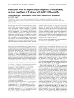
The extracorporeal immune therapy circuit (Figure 1)
consists of a Sheldon catheter, a tubing set, and a func-
tional unit with a total volume of 70 ml housing an open
Lögters et al. Journal of Inflammation 2010, 7:18
/>Page 3 of 13
porous polyurethane foam with specific 3-dimensional
characteristics that allows blood flow of 300 ml/min. The
foam is coated with anti-Fas (CD95/APO-1) directed
agonistic antibodies (clone CH11). The circuit was
primed with 70 ml Ringer' solution. After anticoagulation
by means of systemic administration of 200 IU/kg hepa-
rin (Liquemin; Roche, Grenzach-Wyhlen, Germany) the
housing was connected with both lines to the Sheldon
catheter (Fig. 1A). To rule out a possible bias, pigs under-
going hemorrhagic shock/resuscitation without extracor-
poreal immune therapy (standard medical care; SMC)
received the same amounts of heparin.
Experimental protocol
All animals were allowed to equilibrate for 15 minutes
before baseline measurements (time point 0; Figure 1B).
After two additional baseline measurements within 10
minutes, each animal was hemorrhaged rapidly through
the Sheldon catheter over 15 minutes in order to reach a
mean arterial pressure (MAP) of 35 ± 5 mmHg. Average
volume of withdrawn blood was 586 ± 22 ml (SMC: 555 ±
34 ml; LIM: 616 ± 26 ml, n.s.). All animals were kept
hypotensive for the next 30 minutes at an MAP of 35 ± 5
mmHg and for further 15 minutes at 40 ± 5 mmHg.
Subsequently, resuscitation was carried out by transfu-
sion of 961 ± 28 ml crystalloid (Ringer') solution back to
about 90% of the baseline MAP level (SMC: 916 ± 50 ml;
LIM: 1005 ± 18 ml, n.s.). Fifteen minutes after resuscita-
tion extracorporeal circuits were connected to the Shel-
don catheter and extracorporeal circulation was initiated
with a flow rate of 300 ml/min (LIM group, n = 12). After
3 hours the circuit was flushed with Ringer's solution and
disconnected. All animals were then allowed to recover
and observed for 48 hours (n = 12, 6 of each group) or 72
hours (n = 12, 6 of each group). Then animals underwent
anesthesia, intubation and ventilation again. Catheters
were reconnected and after a steady-state stabilization
period of 30 minutes hemodynamic parameters were
examined for 15 minutes. Finally, pigs were sacrificed and
autopsy was performed.
Figure 1 Scheme (A) of the Fas-directed extracorporeal immune therapy (LIM) in the porcine model and (B) schematic depiction of exper-
imental procedures over time.
Open porous polyurethane
foam with covalently
immobilized anti-Fas rapidly
inactivates neutrophils
LIM
Mini-pumpSheldon catheter
The total volume of the
circuit is <70 ml
Flow
Munich Mini-Pig
[30.3 ± 3.3 kg]
Neutrophils
15 min 15 min 45 min
Equilibration Baseline 1-3 Hemorrhage Hypotensive shock Resuscitation
180 min10 min
LIM on LIM off
Begin
data/sample
collection
Begin
Bleeding
MAP 35 ± 5
mmHg
Reperfusion End of
extracorporeal
therapy
Begin
experiment
15 min
Begin of
extracorporeal
therapy
Therapy
0min
10min
25min 70min-15min 85min 265min
A
B
Lögters et al. Journal of Inflammation 2010, 7:18
/>Page 4 of 13
Hemodynamics
During anesthesia following hemodynamic variables
were continuously measured with Swan-Ganz and arte-
rial catheter: mean arterial pressure (MAP), heart rate
(HR), cardiac output (CO), central venous pressure
(CVP), pulmonary capillary wedge pressure (PCWP),
mean pulmonary arterial pressure (MPAP), and central
venous oxygen saturation (svO2). Blood gas samples were
collected every 10 minutes throughout the experimental
procedure and measured with a blood gas analysis system
(ABL800 Flex, Radiometer GmbH, Willich, Germany).
From beginning of baseline measurements venous blood
samples were collected at time points 10, 25, 70, 85, 95,
115, 145, 205, 265 minutes as well 12, 24, 48, 72 h after
surgery and were analyzed with standardized methods of
clinical chemistry. Red blood count, leukocyte count and
differential, erythrocyte parameters and platelets were
analyzed from EDTA blood (scil animal care company
GmbH, Viernheim, Germany).
Histology and staining procedures
All animals included in this study as well as five healthy
control animals without any treatment have been
euthanised in order to harvest organs for histological
evaluation. Tissue samples were fixed in 4% formaldehyde
and embedded in paraffin according to standard proce-
dures. Sections (5 μm) were stained with hematoxylin-
eosin for pathological examination. In addition, chlorace-
tatesterase staining was performed for specific detection
and quantification of tissue infiltration by neutrophils.
Neutrophils were counted in a blinded and standardized
fashion by microscopy (Axiovert 40, Zeiss, Jena, Ger-
many). Briefly, an ocular micrometer (x10) was used to
count neutrophils in 10 different high power fields (HPF)
of each section. Mean values from each organ and animal
were allocated to predefined ranges of countings/0.09
mm
2
(0-5, 6-10, 11-20, 21-50, 51-100, 101-500).
Quantification of apoptotic cells in tissue sections by
TUNEL - Assay
For histological evaluation of apoptotic cells in the por-
cine tissues, tissue samples of lung, liver, and bowel were
frozen directly after removal in liquid nitrogen and stored
at -80°C before further utilization. For Tdt-mediated
dUTP Nick-End Labeling (TUNEL)-Assay, samples were
first embedded in paraffin and 5 μm - sections were pre-
pared according to standard protocols. All following steps
were done according to instructions of DeadEnd™ Fluoro-
metric TUNEL System kit (Promega GmbH, Mannheim,
Germany). Microscopic examination of DAPI (4'-6-
Diamidin-2'-phenylindol-dihydrochlorid) stained nuclei
and apoptotic domains was carried out with a fluores-
cence microscope (Axioskop 40, Zeiss, Jena, Germany) in
400 fold magnification. Different visual fields were
selected for each tissue type to count up to 1000 DAPI
positive cells. The percentage of apoptotic cells was cal-
culated as the number of TUNEL positive cells from all
DAPI positive cells counted. As a positive control for the
staining procedure some slides were incubated with
DNase before TUNEL staining, resulting in 100% TUNEL
positive cells in each field.
Polymerase chain reaction
Total RNA from tissue was extracted using TRI
REAGENT (Sigma, Munich, Germany) according to the
manufacturer's instructions. 10 μl of total RNA was
reverse transcribed using oligo (dT) 15 primer (Sigma,
Munich, Germany), employing Omniscript Reverse Tran-
scriptase (Qiagen, Hilden, Germany) and following the
manufacturer's instructions. PCR was carried out using
gene specific primer sequences for heme oxygenase-1
(HO-1; pHO-1-R: 5'-CGTAGCGCTTGGTGGCCT-
GCG-3'; -F: 5'-CAGCCCAACAGCATGCCCCAG-3',
Genosys-Sigma, Munich, Germany). Primers for glyceral-
dehyde 3-phosphate dehydrogenase (GAPDH)
(hGAPDH-R: 5'-GAAGTCAGAGGAGACCACCA-3'; -F:
5'-CACCACCATGGAGAAGGCTG-3', Genosys-Sigma,
Munich, Germany) were used as controls. 2.5 μl of cDNA
were amplified using Taq PCR Core Kit (Qiagen, Hilden,
Germany) and products were separated on 1.8% agarose
gel and visualized under UV after Sybr Gold (Invitrogen,
Karlsruhe, Germany) staining.
Western blot analysis
Tissue samples were suspended in RIPA buffer (1% Non-
idet-P40 (NP40), 0.5 mM sodium deoxycholate, 0.1%
sodium dodecyl sulfate (SDS) in PBS) supplemented with
the Complete Protease Inhibitor Cocktail (Roche, Man-
nheim, Germany). Samples were sonicated and incubated
at 4°C for 15 min. After centrifugation at 8,000 × g for 10
min and 4°C, protein concentration was assayed using the
Dc Protein Assay kit from Bio-Rad. Protein (30 μg/sam-
ple) was separated on SDS-polyacrylamide gel electro-
phoresis and transferred to nitrocellulose membrane.
Membranes were saturated in Tris-buffered saline (TBS)
containing 0.1% Tween-20 and 5% w/v nonfat dry milk
(blocking buffer) for 60 min at room temperature and
then incubated with mouse HO-1 monoclonal primary
antibodies specific against pig HO-1 (Stressgen, Victoria,
Canada) diluted in TBS containing 0.1% Tween-20 and
5% w/v nonfat dry milk. After three washes in TBS con-
taining 0.1% Tween-20, the membranes were incubated
for 60 min at room temperature with the horseradish per-
oxidase-labelled polyclonal goat anti-mouse secondary
antibody for HO-1 (Dako Cytomation, Glostrup, Den-
mark), diluted 1:1,000 in TBS, 0.1% Tween-20 and
washed as described above. Bands were visualized by the
enhanced chemiluminescence method (SuperSignal West
Lögters et al. Journal of Inflammation 2010, 7:18
/>Page 5 of 13
pico Chemiluminescent Substrate, Pierce, Bonn, Ger-
many). Equal loading of gels was confirmed both by Pon-
ceau S staining of membranes and by re-incubation of the
filters with a polyclonal antibody for beta-Actin (Santa
Cruz, Heidelberg, Germany). The amount of specific pro-
tein was quantified by densitometry (Quantity One, Bio-
Rad, Munich, Germany).
Lipid peroxidation assay
The determination of lipid peroxidation in tissue homo-
genates was done by quantification of thiobarbituric acid
reactive substances (TBARS; Cayman Chemical Com-
pany, Ann Arbor, MI). Lipid peroxides, derived from
polyunsaturated fatty acids, are unstable and decompose
to form a complex series of compounds, which include
reactive carbonyl compounds, such as malondialdehyde
(MDA). The assay is based on the reaction of MDA with
thiobarbituric acid (TBA) which is added to the sample.
MDA-TBA adducts formed by the reaction of MDA and
TBA under high temperature (90-100°C) and acidic con-
ditions is measured colorimetrically at 530-540 nm (Vic-
tor 3, Perkin Elmer). Briefly, 25 mg of frozen tissue (-
80°C) were mixed with RIPA buffer (1% Nonidet-P40
(NP40), 0.5 mM sodium deoxycholate, 0.1% sodium
dodecyl sulfate (SDS) in PBS) with protease inhibitors
(Complete Mini, Roche). The mixture was homogenized
with a pestle and sonicated (Ultrasonic processor UP50H,
Hielscher) for 15 seconds on ice. The tubes were then
centrifuged at 1600 × g for 10 minutes at 4°C. The super-
natant was used for protein concentration analysis (Dc
Protein Assay, Biorad), standarized at 1 mg protein/ml
solution and utilized for TBARS-assay immediately. The
assay was done in duplicates in 96 well plates. Data were
compared with standards provided by the manufacturer.
The obtained MDA values were calculated using the for-
mula provided by the manufacturer. The dynamic range
of the kit is 0-50 μM MDA equivalents.
Statistical analysis
Statistical analysis was carried out using the SAS/Stat for
Windows software (SAS Institute, Inc, Cary, NC, version
8) and SPSS (SPSS, Inc, Chicago, IL, version 15). Non-
parametric tests of the raw data were used to analyze spe-
cific inter-group and over-time differences. Data was
considered to be statistically significant at p < 0.05. Wil-
coxon two-sample test was used for specific inter-group
(LIM versus SMC groups) difference and Wilcoxon
paired test for over time differences (time point versus
start value).
Results
Effects of LIM on leukocyte counts
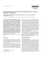
Time kinetics of leukocyte counts was determined
throughout the entire experiments (Figure 2). As shown
in Figure 2A, after beginning of resuscitation with LIM
leukocyte counts were found to be depressed until the
end of extracorporeal immune therapy in the LIM group
compared with SMC. This was due to the depression of
neutrophil numbers (Figure 2B) and monocyte numbers
(Figure 2C), whereas lymphocyte numbers were not sig-
nificantly modified (Figure 2D). Three hours after reper-
fusion, neutrophil counts increased in both groups.
Furthermore, 72 hours after beginning of resuscitation
neutrophil counts were significantly reduced in the LIM
group compared to SMC (p < 0.05). However, 24 and 48
hours after beginning of resuscitation no intergroup dif-
ferences were evident for neutrophil counts (data not
shown).
Effects of LIM on hemodynamics
MAP in both groups was equivalent at baseline (SMC:
75.7 ± 2.57 mmHg; LIM: 75.2 ± 3.11 mmHg) and
decreased in a similar pattern during hemorrhage (Figure
3). During resuscitation MAP reached 89% of the base-
line levels. However, it was found to be significantly (p <
0.05) decreased in the post resuscitation period in both
groups (Figure 3, Table 1). After 72 h MAP values were
significantly higher in the LIM group compared with
SMC (p < 0.05, Table 1). Heart rate (HR) for both groups
was slightly different at baseline (SMC: 86.7 ± 3.41 beats/
min; LIM: 96.2 ± 4.37 beats/min). As expected, HR
increased during hemorrhage until begin of resuscitation
(SMC: 128.6 ± 10.7; LIM: 164.9 ± 7.52 beats/min). HR
remained increased during the post resuscitation period
compared to baseline levels (data not shown). In contrast
to the values for the SMC group, values for the LIM
group were below baseline at 72 h (Table 1). Within the
first 48 hours after resuscitation no significant improve-
ment in hemodynamic variables (MAP, HR, CO, CVP,
svO
2
, PCWP, MPAP) was observed in the LIM group.
However, after 72 hours MAP and CO were significantly
(p < 0.05) higher in the LIM group compared to the SMC
group (Table 1). SvO
2
was 63.1 ± 5.77% for the LIM group
and 49.1 ± 3.7% for SMC (p = 0.0625).
Ischemia and tissue damage parameters
Transaminases (AST, ALT), creatine phosphokinase
(CK), CK-MB, Troponin T, and lactate significantly (p <
0.05) increased over time in both groups (Table 2). In
conjunction with the increase in lactate, base excess (BE)
significantly decreased over time. At 24, 48, and 72 hours
lactate values were slightly lower in the LIM group. After
72 hours lactate values were at pre shock level in both
groups. CK values were significantly lower 72 hours after
shock in the LIM-treated animals (1431 ± 305 U/l) com-
pared with the SMC group (2337 ± 232 U/l).
Lögters et al. Journal of Inflammation 2010, 7:18
/>Page 6 of 13
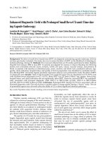
Neutrophil tissue infiltration
Representative tissue sections of lung, heart, liver, kidney,
and bowel are depicted in Figure 4. Histopathological
evaluation did not reveal tissue damage. However, count-
ing of CHE positive cells/HPF revealed increase of neu-
trophil numbers in the tissues. All SMC animals
exhibited neutrophil infiltration of the lungs versus con-
trol (SMC range: 101-500, n = 12; control range: 6-10, n =
5). Animals undergoing LIM treatment exhibited only a
weak infiltration (11-20, n = 9; 21-50, n = 3). The LIM-
mediated limitation of neutrophil infiltration was also
found in heart (left ventricle), liver, kidneys (glomeruli),
and bowel. However, the differences between SMC and
LIM groups were less evident than in the lung.
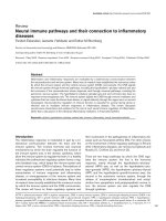
HO-1 expression, lipid peroxidation, and apoptosis
HO-1 gene and protein expression as a counter-regula-
tion mechanism of oxidative stress was found to be
induced in bowels, lungs, and livers in animals that
underwent hemorrhagic shock/resuscitation compared
to control animals that did not undergo hemorrhagic
shock (Figure 5). Both HO-1 gene (Figure 5A) and protein
(Figure 5B) expression was lower in the LIM group as
compared with SMC. In addition, MDA values that indi-
cate lipid peroxidation and thus tissue damage were sig-
nificantly lower in the bowels and slightly lower in the
lungs of animals in the LIM group compared with the
SMC group after shock (Figure 6A). Lipid peroxidation
was not found in the livers of animals of either group
when compared with control animals.
The putative contribution of apoptosis within bowels,
lungs, and livers was studied by TUNEL staining. The
numbers of TUNEL positive cells as the percentage from
DAPI positive cells were calculated. Results are depicted
as relative countings (Figure 6B) and qualitatively as
microphotographs (Figure 6C). Apoptosis was lower in
the lamina propria of the bowels (p < 0.05) and in the
lungs (not significant) of animals in the LIM group com-
pared with the SMC group. No Apoptosis was found by
TUNEL staining in the liver.
Figure 2 Time kinetics of leukocyte (A), neutrophil (B), monocytes (C), and lymphocytes (D) counts for the SMC group and LIM group; n =
12 per group. Mean ± SEM.
a
- Statistically significant (p < 0.05) between SMC and LIM group.
b
- Statistically significant (p < 0.05) difference compared
with end of shock value (70 minutes) in SMC group.
c
-Statistically significant (p < 0.05) difference compared with end of shock value (70 minutes) in
LIM group.
AB
DC
Lögters et al. Journal of Inflammation 2010, 7:18
/>Page 7 of 13
Discussion
In our porcine hemorrhagic shock/resuscitation model
we observed impaired hemodynamics, neutrophil tissue
infiltration, lipid peroxidation in the bowel, lung, and
liver during an observation period of 72 hours Extracor-
poreal immune therapy targeting neutrophil Fas amelio-
rated shock-related pathophysiology. The ability of the
mouse-anti-human agonistic anti-Fas IgM used in this
study to induce porcine neutrophil apoptosis and to
impair the effector functions was shown in earlier studies
[22,25]. In previous experiments and in experiments that
were done to establish this model, mini circuits without
antibody coating were run to exclude effects mediated by
the circuit itself. In these tests hemodynamics and leuko-
Figure 3 Time kinetics of mean arterial pressure (MAP) in the shock and resuscitation phase for the LIM group and the SMC group; n = 12
per group; Mean ± SEM. Mean arterial pressure is expressed as mmHg.
0
10
20
30
40
50
60
70
80
90
0
30
60
90
120
150
180 210 240
270
Time [min]
MAP [mm Hg]
SMC
LIM
shock phase resuscitation phase with SMC/LIM
Table 1: Time kinetics of hemodynamic parameters
0 h 48 h 72 h
SMC LIM SMC LIM SMC LIM
MAP [mmHg] 75.7 ± 2.57 75.2 ± 3.11 44.9 ± 2.64
a
40.3 ± 4.86
a
43.8 ± 2.63
a
52.9 ± 2.54
ab
HR [beats/
min]
86.7 ± 3.41 96.2 ± 4.37 91.9 ± 6.59 105.9 ± 6.63 95.6 ± 9.77 90.0 ± 5.00
CO [l/min] 3.0 ± 0.13 3.1 ± 0.12 2.3 ± 0.23 2.3 ± 0.30 2.2 ± 0.08
a
3.1 ± 0.24
b
CVP [mmHg] 3.3 ± 0.70 3.8 ± 0.55 1.1 ± 0.69 5.8 ± 2.19
b
3.4 ± 1.70 4.8 ± 1.24
svO
2
[%] 86.9 ± 0.95 82.9 ± 2.69 56.0 ± 2.47
a
57.7 ± 6.44
a
49.1 ± 3.70
a
63.1 ± 5.77
PCWP [mmHg] 7.3 ± 1.23 8.2 ± 0.57 3.8 ± 0.88 5.2 ± 0.89 5.9 ± 1.43 5.9 ± 1.12
MPAP [mmHg] 14.8 ± 1.22 17.8 ± 1.49 7.3 ± 1.01
a
10.9 ± 1.10
ab
12.2 ± 2.10
a
13.7 ± 1.05
a
MAP: mean arterial pressure, HR: heart rate; CO: cardiac output; CVP: central venous pressure; svO
2
: central venous oxygen saturation; PCWP:
pulmonary capillary wedge pressure; MPAP: mean pulmonary arterial pressure; n = 12 per group at time point 0; at 48 h and 72 h, n = 6; Mean
± SEM. a-statistically significant (p < 0.05) over-time; b-statistically significant (p < 0.05) between SMC and LIM group.
Lögters et al. Journal of Inflammation 2010, 7:18
/>Page 8 of 13
Table 2: Time kinetics of metabolic and organ specific parameters
0 h End shock 24 h 48 h 72 h
SMC LIM SMC LIM SMC LIM SMC LIM SMC LIM
Lactate 3.3 ± 0.26 3.4 ± 0.38 3.4 ± 0.28 4.0 ± 0.43
a
n.d. n.d. 2.1 ± 0.39 2.4 ± 0.78 2.3 ± 0.22
a
1.6 ± 0.31
a
BE 3.1 ± 0.58 5.0 ± 0.57
b
1.4 ± 0.91 1.4 ± 0.71
a
n.d. n.d. 4.3 ± 0.58 4.8 ± 1.59 5.2 ± 0.92 6.1 ± 1.05
Creatinine [1.1-1.8] 1.0 ± 0.03 0.9 ± 0.05
b
1.0 ± 0.05 0.9 ± 0.06 1.2 ± 0.07
a
1.2 ± 0.16 0.9 ± 0.09 1.1 ± 0.18 1.1 ± 0.06 0.8 ± 0.04
b
AST [23-54] 56 ± 6.8 40 ± 2.8 37 ± 4
a
31 ± 2.91 912 ± 193
a
1853 ± 572
a
378 ± 120
a
854 ± 515
a
62 ± 6.8 64 ± 9.7
ALT [50-90] 60 ± 5.68 51.1 ± 3.0 31 ± 3.3
a
26 ± 1.28
a
203 ± 25.3
a
258 ± 33.8
a
178 ± 19.2
a
213 ± 30.0
a
108 ± 5.84
a
123 ± 16.5
a
CK [251-810] 1643 ± 220 1183 ± 87 982 ± 134
a
716 ± 56
a
58420 ± 9767
a
77653 ± 14960
a
15851 ± 4185
a
29439 ± 15529
a
2338 ± 233 1431 ± 305
b
CK-MB 180 ± 20 151 ± 6 95 ± 13
a
97 ± 10
a
767 ± 84
a
969 ± 144
a
294 ± 33 467 ± 152
a
156 ± 12
a
134 ± 31
Troponin T [< 0.05] 0.03 ± 0.01 0.02 ± 0.003 0.04 ± 0.01 0.04 ± 0.01
a
0.08 ± 0.03 0.15 ± 0.05
a
0.02 ± 0.004 0.04 ± 0.021 0.02 ± 0.006 0.01 ± 0.00
Reference Ranges in []. Lactate [mmol/l]; BE [mmol/l]: base excess; creatinine [mg/dl]; AST [U/l]: aspartate aminotransaminase; ALT [U/l]: alanine aminotransaminase; CK [U/l]: creatine
phosphokinase; CK-MB [U/l]: „MB"-type isoenzyme of creatine phosphokinase; Troponin T [ng/ml]; n = 12 per group, 72 h n = 6; Mean ± SEM; a-statistically significant (p < 0.05) over-time; b-
statistically significant (p < 0.05) between SMC and LIM group. n.d. = not determined.
Lögters et al. Journal of Inflammation 2010, 7:18
/>Page 9 of 13
cyte counts were similar to the SMC group. However, in
the current study we may not totally exclude LIM effects
that are not dependent on Fas activation on neutrophils.
Our working hypothesis was that posthemorrhagic tar-
geting of circulating neutrophil Fas may rapidly impair
neutrophil effector functions and thus may prevent their
prolonged hyperactivation and neutrophil-mediated tis-
sue damage. We previously found that binding of neutro-
phils to membrane-bound but not soluble FasL
inactivated neutrophils within minutes even before signs
of apoptosis were detectable [29], leading us to the
assumption that immobilized agonistic anti-Fas may be
used to therapeutically limit hyperactivation of neutro-
phils. In addition, functionalized biocompatible surfaces
with agonistic anti-Fas in extracorporeal immune therapy
may be more suitable than systemic application of anti-
Fas because the latter approach has been shown to have
severe side effects such as liver toxicity and pulmonary
fibrosis [32,33].
Therefore, in order to effectively inactivate neutrophils
in an early phase of posthemorrhagic immune deregula-
tion, an extracorporeal circuit with a neutrophil inhibi-
tion module (LIM) on the functional basis of immobilized
agonistic anti-Fas IgM was used in a porcine hemorrhagic
shock/resuscitation model. The proof of concept of such
an approach had been previously shown in patients
undergoing cardiac surgery [24,25].
In this study, the efficacy of LIM has been shown by the
relative reduction of neutrophil counts during the treat-
ment phase. Histopathological analyses of post hemor-
rhagic organs clearly revealed lower numbers of
neutrophils within the pulmonary tissues and slightly less
numbers in heart, liver, kidney and bowel in animals of
the LIM group versus SMC. In addition, we found evi-
dence of improved pulmonary, cardiac, and kidney func-
tion in the LIM group as indicated by partially higher
svO
2
, and better cardiac output, respectively. Moreover,
CK values were lower in the LIM group, however, only
after 72 hours. Due to high SEM values at 24 and 48
hours, the interpretation of these data has to be done
carefully. Overall, the obtained evidence that posthemor-
rhagic hemodynamics and metabolism may be better in
the LIM group versus SMC should be confirmed by
future studies. In addition, the unexpected reduction of
monocyte counts by LIM treatment requires further
studies.
Although controversial reports exist regarding activa-
tion or inhibition of different cell types by Fas stimulation
[34] we never observed increased activity upon challeng-
ing neutrophils ex vivo with immobilized agonistic Fas.
One possible mechanistic explanation of our findings
from this in vivo study may be that LIM treatment
impairs the motility of circulating neutrophils which may
partly result in the failure of neutrophils to transmigrate
into tissues. Consequently, the well known neutrophil-
Figure 4 Chloracetatesterase staining of paraffin sections from heart, lung, liver, kidney, and bowel. Representative tissue samples for untreat-
ed healthy control pigs, pigs undergoing hemorrhage/resuscitation (SMC), and pigs undergoing hemorrhage/resuscitation with treatment (LIM). Ex-
cept for control animals, organs were harvested 48 h after shock.
Lögters et al. Journal of Inflammation 2010, 7:18
/>Page 10 of 13
mediated disruption of the integrity of endothelial/epi-
thelial layers, impairment of microcirculation, induction
of oxidative stress with subsequent lipid peroxidation
[35,36] might be limited by LIM. Indeed, neutrophil
chemotactic activity has been shown previously to be
reduced after LIM treatment [23]. It has been shown pre-
viously that blood cells made apoptotic by extracellular
exposure to psoralen and UV light exerted anti-inflam-
matory effects in a graft-versus-host disease model [37].
It would be of interest to find out whether similar anti-
inflammatory mechanisms may also exist upon Fas-
mediated neutrophil apoptosis. Further evidence that
apoptotic cells have anti-inflammatory and immunosup-
pressive effects when given systemically in a model of
murine LPS-induced endotoxic shock has been reported
[38].
Herein, shock/resuscitation-induced hemoxygenase-1
(HO-1) expression, probably as a consequence of pos-
themorrhagic oxidative stress [39,40], was clearly limited
in the LIM group in lung, liver, and bowel, organs that
frequently are impaired after trauma [41]. HO-1 is known
to be induced by oxidative stress and has been shown by
others to protect from hemorrhagic shock-induced tissue
injury [39]. The finding that gene and protein expression
of HO-1 was found to be lower in the LIM group may be a
result of limited neutrophil infiltration and neutrophil-
mediated oxidative stress.
Shock-induced lipid peroxidation was only observed in
the bowels. However, there seems to be no direct correla-
tion between the amount of lipid peroxidation and infil-
trated neutrophils within the bowel since only low
neutrophil numbers could be detected in the bowel after
shock. In contrast, high numbers of apoptotic cells were
found in the lamina propria of the bowel in the SMC but
not in the LIM group suggesting that inhibition of
peripheral inhibition of circulating neutrophils during
posthemorrhagic inflammation may result in protection
of the bowel. Similarly, shock-induced apoptosis in the
lung tissue was also largely prevented by LIM. The under-
lying mechanisms remain to be defined. One possible
explanation might be that LIM protects from the previ-
ously described no-reflow phenomenon associated with
Figure 5 Heme oxygenase-1 (HO-1) gene expression (A), and HO-1 protein expression (B) in control (white bars), SMC (grey bars), and LIM
(black bars) animals.
Control
SMC
LIM
GAPDH
Bowel
Lung
Liver
Bowel
Lung
Liver
HO-1
+LIM
-LIM
control
Actin
Bowel
Lung
Liver
Bowel
Lung
Liver
HO-1
HO-1
Bowel
Lung
Liver
0.0
0.5
1.0
1.5
2.0
control
-LIM
+LIM
relative protein expression
[arbitrary units]
HO-1
Bowel
Lung
Liver
0
2
4
6
control
-LIM
+LIM
relative gene expression
[arbitrary units]
Lögters et al. Journal of Inflammation 2010, 7:18
/>Page 11 of 13
Figure 6 Lipid peroxidation (A) and apoptosis (B) in bowel, lung, and liver as determined by means of malondialdehyde (MDA) assay and
Tdt-mediated dUTP Nick-End Labeling (TUNEL), respectively. Data is shown for control (white bars), SMC (grey bars), and LIM (black bars) animals.
*Statistically significant (p < 0.05) difference. Positive controls indicate staining with 4'-6-Diamidin-2'-phenylindol-dihydrochlorid (DAPI; left) and
TUNEL (right) after incubation of tissue with DNase.
Lögters et al. Journal of Inflammation 2010, 7:18
/>Page 12 of 13
neutrophils that are sequestered in the capillaries of the
tissues, thus damaging the tissue in the absence of overt
neutrophil tissue infiltration [41].
Conclusions
From our data we conclude that targeting of neutrophil
Fas during the early posthemorrhagic or posttraumatic
time period may ameliorate inflammation-mediated
sequelae and thus may be of therapeutic benefit for
trauma patients. Due to the small sample size the conclu-
sions have to be made carefully. As usual for explorative
studies that have the main objective in the identification
of the best primary end point for subsequent confirma-
tive studies, multiple testing of different parameters and
time points had to be done, resulting in a reduction of the
robustness of the tests performed. Nevertheless, the
results obtained provide an interesting basis encouraging
further evaluation.
However, the timing of neutrophil inhibition has to be
critically considered since inhibition of neutrophil activa-
tion might impair anti bacterial phagocytic effects of neu-
trophils which are essential to prevent sepsis [42,43]. On
the other hand, the early prevention of neutrophil-medi-
ated disruption e.g. of the intestinal or pulmonary epithe-
lium might in turn prevent bacterial dissemination and
sepsis. Further studies investigating potential clinical
benefits of neutrophil Fas-directed immune therapy in
patients after hemorrhagic shock or severe trauma are
encouraged.
Competing interests
JA and MS receive salary from and hold shares of LEUKOCARE. None of the
other authors have anything to declare.
Authors' contributions
TL conducted the experiments and draft the manuscript. AP-G, MS, IW, AO, SS,
JB, JB-M, AS participated in the experiments including surgical preparation and
data collection. WM participated in the histological analysis. AP-G, JA, JW par-
ticipated in the study design and revised the manuscript critically for impor-
tant intellectual content. TJ was in charge of he statistical evaluation. MSch
conceived of the study, and participated in its design and coordination and
draft the manuscript. All authors read and approved the final manuscript.
Acknowledgements
This study relied on financial resources of the University of Düsseldorf. It was
partly supported by the German "Bundesministerium für Wirtschaft" (ProInno)
and the Deutsche Forschungsgemeinschaft (SCHO-612/3-1). The authors
thank Samira Seghrouchni and Jutta Schneider for the excellent technical
assistance.
Author Details
1
Department of Trauma and Hand Surgery, University Hospital, Düsseldorf,
Germany,
2
Department of Thoracic and Cardiovascular Surgery, University
Hospital, Frankfurt am Main, Germany,
3
Department of Vascular Surgery,
Faculdade Medicina Marilia (FAMEMA), Marilia, Brasil and
4
Pathology Group
Starnberg, Starnberg, Germany
References
1. Moore FA, McKinley BA, Moore EE: The next generation in shock
resuscitation. Lancet 2004, 363:1988-1996.
2. Baue AE, Durham R, Faist E: Systemic inflammatory response syndrome
(SIRS), multiple organ dysfunction syndrome (MODS), multiple organ
failure (MOF): are we winning the battle? Shock 1998, 10:79-89.
3. Zhang Q, Raoof M, Chen Y, Sumi Y, Sursal T, Junger W, Brohi K, Itagaki K,
Hauser CJ: Circulating mitochondrial DAMPs cause inflammatory
responses to injury. Nature 2010, 464:104-107.
4. Bone RC: Toward a theory regarding the pathogenesis of the systemic
inflammatory response syndrome: what we do and do not know about
cytokine regulation. Crit Care Med 1996, 24:163-172.
5. Brown KA, Brain SD, Pearson JD, Edgeworth JD, Kewis SM, Treacher DF:
Neutrophils in development of multiple organ failure in sepsis. Lancet
2006, 368:157-169.
6. Fan J, Li Y, Levy RM, Fan JJ, Hackam DJ, Vodovotz Y, Yang H, Tracey KJ,
Billiar TR, Wilson MA: Hemorrhagic shock induces NAD(P)H oxidase
activation in neutrophils: role of HMGB1-TLR4 signaling. J Immunol
2007, 178:6573-6580.
7. Hoesel LM, Neff TA, Neff SB, Younger JG, Olle EW, Gao H, Pianko MJ,
Bernacki KD, Sarma JV, Ward PA: Harmful and protective roles of
neutrophils in sepsis. Shock 2005, 24:40-47.
8. Lenz A, Franklin GA, Cheadle WG: Systemic inflammation after trauma.
Injury 2007, 38:1336-1345.
9. Scholz M, Cinatl J, Schädel-Höpfner M, Windolf J: Neutrophils and the
blood-brain barrier dysfunction after trauma. Med Res Rev 2007,
27:401-416.
10. Shimizu T, Tani T, Endo Y, Hanasawa K, Tsuchiya M, Kodama M: Elevation
of plasma peptidoglycan and peripheral blood neutrophil activation
during hemorrhagic shock: plasma peptidoglycan reflects bacterial
translocation and may affect neutrophil activation. Crit Care Med
2002,
30:77-82.
11. Weiss SJ: Tissue destruction by neutrophils. N Engl J Med 1989,
320:365-276.
12. Wesche DE, Lomas-Neira JL, Perl M, Chung CS, Ayala A: Leukocyte
apoptosis and its significance in sepsis and shock. J Leukoc Biol 2005,
78:325-337.
13. Roesner JP, Petzelbauer P, Koch A, Tran N, Iber T, Vagts DA, Scheeren TW,
Vollmar B, Nöldge-Schomburg GE, Zacharowski K: Bbeta15-42 (FX06)
reduces pulmonary, myocardial, liver, and small intestine damage in a
pig model of hemorrhagic shock and reperfusion. Crit Care Med 2009,
37:598-605.
14. Mori T, Yamamoto H, Tabata T, Shimizu T, Endo Y, Hanasawa K, Fujimiya M,
Tani T: A free radical scavenger, edaravone (MCI-186), diminishes
intestinal neutrophil lipid peroxidation and bacterial translocation in a
rat hemorrhagic shock model. Crit Care Med 2005, 33:1064-1069.
15. Thorburn K: Bacterial translocation and intestinal neutrophil lipid
peroxidation in a hemorrhagic shock model Rat race or rat trap? Crit
Care Med 2005, 33:1167-1169.
16. Toda Y, Takahashi T, Maeshima K, Shimizu H, Inoue K, Morimatsu H, Omori
E, Takeuchi M, Akagi R, Morita K: A neutrophil elastase inhibitor,
sivelestat, ameliorates lung injury after hemorrhagic shock in rats. Int J
Mol Med 2007, 19:237-243.
17. Zakaria el R, Campbell JE, Peyton JC, Garrison RN: Postresuscitation tissue
neutrophil infiltration is time-dependent and organ-specific. J Surg Res
2007, 143:119-125.
18. Simon H-U: Neutrophil apoptosis pathways and their modifications in
inflammation. Immunological Reviews 2003, 193:101-110.
19. Casatella MA: Neutrophil-derived proteins: selling cytokines by the
pound. Adv Immunol 1999, 73:369-509.
20. Dibbert B, Weber M, Nikolaizik WH, Vogt P, Schöni MH, Blaser K, Simon HU:
Cytokine-mediated Bax deficiency and consequent delayed neutrophil
apoptosis: a general mechanism to accumulate effector cells in
inflammation. Proc Natl Acad Sci USA 1999, 96:13330-13335.
21. Saba S, Soong G, Greenberg S, Prince A: Bacterial stimulation of
epithelial G-CSF and GM-CSF expression promotes PMN survival in CF
airways. Am J Respir Cell Mol Biol 2002, 27:561-567.
22. Abdel-Rahman U, Margraf S, Aybek T, Loegters T, Moreno JB, Francischetti
I, Kranert T, Gruenwald F, Windolf J, Moritz A, Scholz M: Inhibition of
neutrophil activity improves cardiac function after cardiopulmonary
bypass. J Inflamm 2007, 4:21-29.
Received: 6 August 2009 Accepted: 20 April 2010
Published: 20 April 2010
This article is available from: 2010 Lögters et al; licensee BioMed Central Ltd. This is an Open Access article distributed under the terms of the Creative Commons Attribution License ( ), which permits unrestricted use, distribution, and reproduction in any medium, provided the original work is properly cited.Journal of Inflammation 2010, 7:18
Lögters et al. Journal of Inflammation 2010, 7:18
/>Page 13 of 13
23. Scholz M, Cinatl J: Fas/FasL interaction: A novel immune therapy
approach with immobilized biologicals. Med Res Rev 2005, 25:331-342.
24. Scholz M, Cinatl J, Barros RT, Lisboa AC, Genevcius CF, Margraf S,
Francischetti I, Oremek G, Windolf J, Simon A, Moritz A, Bitu-Moreno J:
First efficacy and safety results with the antibody containing leukocyte
inhibition module in cardiac surgery patients with neutrophil
hyperactivity. ASAIO J 2005, 51:144-147.
25. Scholz M, Simon A, Berg M, Schuller AM, Hacibayramoglu M, Margraf S,
Theisen A, Windolf J, Wimmer-Greinecker G, Moritz A: In vivo inhibition of
neutrophil activity by a FAS (CD95) stimulation module: Arterial in-line
application in a porcine cardiac surgery model. J Thorac Cardiovasc Surg
2004, 127:1735-1742.
26. Salmen S, Teran G, Borges L, Goncalvez L, Albarran B, Urdaneta H, Montes
H, Berrueta L: Increased Fas-mediated apoptosis in pölymorphonuclear
cells from HIV-infected patients. Clin Exp Immunol 2004, 137:166-172.
27. Ayub K, Laffafian I, Dewitt S, Hallett MB: Ca influx shutdown in
neutrophils induced by Fas (CD95) cross-linking. Immunology 2004,
112:454-460.
28. Peter ME, Budd RC, Desbarats J, Hedrick SM, Hueber AO, Newell MK, Owen
LB, Pope RM, Tschopp J, Wajant H, Wallach D, Wiltrout RH, Zörnig M, Lynch
DH: The CD95 receptor: apoptosis revisited. Cell 2007, 129:447-450.
29. Cinatl J Jr, Blaheta R, Bittoova M, Scholz M, Margraf S, Vogel JU, Cinatl J,
Doerr HW: Decreased neutrophil adhesion to human cytomegalovirus-
infected retinal pigment epithelial cells is mediated by virus-induced
up-regulation of Fas ligand independent of neutrophil apoptosis. J
Immunol 2000, 165:4405-4413.
30. Paunel-Görgülü A, Zörnig M, Lögters T, Altrichter J, Rabenhorst U, Cinatl J,
Windolf J, Scholz M: Mcl-1-mediated impairment of the intrinsic
apoptosis pathway in circulating neutrophils from critically ill patients
can be overcome by Fas stimulation. J Immunol 2009, 183:6198-206.
31. Cox G, Crossley J, Xing Z: Macrophage engulfment of apoptotic
neutrophils contributes to the resolution of acute pulmonary
inflammation in vivo. Am J Respir Cell Mol Biol 1995, 12:232-237.
32. Chang B, Nishikawa M, Sato E, Inoue M: Mice lacking inducible nitric
oxide synthase show strong resistance to anti-Fas antibody-induced
fulminant hepatitis. Arch Biochem Biophys 2003, 411:63-72.
33. Hagimoto N, Kuwano K, Miyazaki H, Kunitake R, Fujita M, Kawasaki M,
Kaneko Y, Hara N: Induction of apoptosis and pulmonary fibrosis in
mice in response to ligation of Fas antigen. Am J Respir Cell Mol Biol
1997, 17:272-278.
34. Strasser A, Jost PJ, Nagata S: The many roles of FAS receptor signaling in
the immune system. Immunity 2009, 30:180-192.
35. Schuller AM, Windolf J, Blaheta R, Cinatl J, Kreuter J, Wimmer-Greinecker G,
Moritz A, Scholz M: Degradation of microvascular brain endothelial cell
beta-catenin after co-culture with activated neutrophils from patients
undergoing cardiac surgery with prolonged cardiopulmonary bypass.
Biochem Biophys Res Commun 2005, 329:616-623.
36. Gatza E, Rogers CE, Clouthier SG, Lowler KP, Tawara I, Liu C, Reddy P,
Ferrara JL: Extracorporeal photopheresis reverses experimental graft-
versus-host disease through regulatory T cells. Blood 2008,
112:1515-1521.
37. Ren Y, Xie Y, Jiang G, Fan J, Yeung J, Li W, Tam PK, Savill J: Apoptotic cells
protect mice against lipopolysaccharide-mediated shock. J Immunol
2008, 180:4978-4985.
38. Engler R, Covell JW: Granulocytes cause reperfusion ventricular
dysfunction after 15 min ischaemia in the dog. Circ Res 1987, 61:20-28.
39. Rensing H, Jaeschke H, Bauer I, Pätau C, Datene V, Pannen BH, Bauer M:
Differential activation pattern of redox-sensitive transcription factors
and stress-inducible dilator systems heme oxygenase-1 and inducible
nitric oxide synthase in hemorrhagic and endotoxic shock. Crit Care
Med 2001, 29:1962-1971.
40. Douzinas EE, Kollias S, Tiniakos D, Evangelou E, Papalois A, Rapidis AD,
Tsoukalas GD, Patsouris E, Roussos C: Hypoxemic reperfusion after 120
mins of intestinal ischemia attenuates the histopathologic and
inflammatory response. Crit Care Med 2004, 32:2279-2283.
41. Engler R, Schmid-Schonbein GW, Pavelec R: Leukocyte capillary
plugging in myocardial ischemia and reperfusion in the dog. Am J
Pathol 1983, 111:98-111.
42. Maier B, Lefering R, Lehnert M, Laurer HL, Steudel WI, Neugebauer EA,
Marzi I: Early versus late onset of multiple organ failure is associated
with differing patterns of plasma cytokine biomarker expression and
outcome after severe trauma. Shock 2007, 28:668-674.
43. Sauer M, Altrichter J, Kreutzer HJ, Lögters T, Scholz M, Nöldge-Schomburg
G, Schmidt R, Mitzner SR: Extracorporeal cell therapy with granulocytes
in a pig model of Gram-positive sepsis. Crit Care Med 2009, 37:606-13.
doi: 10.1186/1476-9255-7-18
Cite this article as: Lögters et al., Extracorporeal immune therapy with
immobilized agonistic anti-Fas antibodies leads to transient reduction of cir-
culating neutrophil numbers and limits tissue damage after hemorrhagic
shock/resuscitation in a porcine model Journal of Inflammation 2010, 7:18