Báo cáo y học: "Proposed protective mechanism of the pancreas in the ra" pot
Bạn đang xem bản rút gọn của tài liệu. Xem và tải ngay bản đầy đủ của tài liệu tại đây (7.93 MB, 10 trang )
Axelsson et al. Journal of Inflammation 2010, 7:24
/>Open Access
RESEARCH
© 2010 Axelsson et al; licensee BioMed Central Ltd. This is an Open Access article distributed under the terms of the Creative Commons
Attribution License ( which permits unrestricted use, distribution, and reproduction in
any medium, provided the original work is properly cited.
Research
Proposed protective mechanism of the pancreas in
the rat
Jakob BF Axelsson*
1
, Hamid Akbarshahi
1
, Katarzyna Said
1
, Anders Malmström
2
and Roland Andersson
1
Abstract
Background: Heparan sulphate is known to have various functions in the animal body, including surveillance of tissue
integrity. Administered intraperitoneally, it induces a systemic inflammatory response syndrome and when given
locally in the pancreas it initiates a protective inflammatory response. The aim of the present study was to investigate
the underlying mechanisms behind cell recruitment following intra-ductal infusion of heparan sulphate.
Methods: Rats were subjected to intraductal-infusion of heparan sulphate, lipopolysaccharide and phosphate
buffered saline into the pancreas. Pancreatic tissue was harvested 1, 3, 6, 9 or 48 hours after infusion and stained
immunohistochemically for myeloperoxidase, ED-1, CINC-1 and MCP-1, as well as using eosin hematoxylin staining.
Furthermore, MPO activity and MCP-1 and CINC-1 concentrations of tissue homogenates were measured. All
differences were analyzed statistically using the Mann-Whitney U-test.
Results: During HS infusion, a rapid influx of macrophages/monocytes, as visualized as ED-1 positive cells, was seen
reaching a maximum at 6 hours. After 48 hours, the same levels of ED-1 positive cells were noted in the pancreatic
tissue, but with different location and morphology. Increased neutrophil numbers of heparan sulphate treated animals
compared to control could be detected only 9 hours after infusion. The number of neutrophils was lower than the
number of ED-1 positive cells. On the contrary, LPS infusion caused increased neutrophil numbers to a larger extent
than heparan sulphate. Furthermore, this accumulation of neutrophils preceded the infiltration of ED-1 positive cells.
Chemokine expression correlates very well to the cell infiltrate. MCP-1 was evident in the ductal cells of both groups
early on. MCP-1 preceded monocyte infiltration in both groups, while the CINC-1 increase was only noticeable in the
LPS group.
Conclusions: Our data suggest that heparan and LPS both induce host defense reactions, though by using different
mechanisms of cell-recruitment. This implies that the etiology of pancreatic inflammation may influence how the
subsequent events will develop.
Background
Despite that acute pancreatitis is a common clinical prob-
lem, with a yearly incidence of about 300/10
6
inhabitants
[1], the initial events are poorly understood. The lack of
knowledge is in part due to that sampling and investiga-
tion of e.g. human tissue during the early stage of acute
pancreatitis has not been possible.
The exocrine pancreas is subjected to various noxious
agents, which all may produce tissue injury leading to the
development of acute pancreatitis. Thus, the pancreas
produces digestive enzymes such as protesases and
lipases, which expose the ductal epithelium to digestive
enzymes, which may by partial activation, attack the duc-
tal membrane. Furthermore, in biliary duct obstruction it
is also argued that the exocrine part of the pancreas can
be exposed to bile. Although more controversial, it has
been proposed in the past that bacteria can migrate into
the pancreatic ductal system (for discussion of this topic
see [2]). A rapidly responding and well-tuned defense
against all of these noxious stimuli need to be present in
order to protect the vulnerable pancreatic gland.
A poorly regulated defense against ruptured cells and
microbes of the pancreas can lead to inflammation of the
gland. In order to obtain rapidly acting defense systems,
sensors of the epithelial surface are of central importance.
Heparan sulphate proteoglycans (HSPGs) substituted
* Correspondence:
1
Department of Clinical Sciences Lund, Lund University, BMC, D12, SE-221 84
Lund, Sweden
Full list of author information is available at the end of the article
Axelsson et al. Journal of Inflammation 2010, 7:24
/>Page 2 of 10
with polysaccharides sulphated to different degrees are
found anchored in the plasma membrane of epithelial
cells in the pancreas. These PGs have been suggested to
represent signaling molecules of membrane integrity [3]
by eliciting an inflammatory response in their soluble
form, making them candidates of these protective signal-
ing events. Administration of purified HS has been
shown to cause both local pancreatic defense reactions
[4], as well as systemic reactions [5]. The antithrombotic
properties of heparin have been utilized clinically for a
long time, but the more recently discovered, pro-inflam-
matory properties of HS have found clinical applicability
by lowering the labor times in women [6]. Heparin is a
highly sulphated GAG shown to possess anti-inflamma-
tory properties, whereas HS, a less sulphated GAG has
been shown both in vivo and in vitro to be pro-inflamma-
tory [5]. The mechanisms of which HS is capable of
inducing inflammatory responses are yet to be eluci-
dated. During bile reflux into the pancreas following gall-
stone obstruction, HSPGs may be cleaved and solubilized
from its membrane location. Pancreatic enzymes may
also make HS available for binding to receptors and other
biological actions otherwise not available when bound to
the epithelial wall [4].
As proposed, soluble HS can act as an endogenous
inducer of an inflammatory response of the pancreatic
epithelial cells. This phenomenon of HS-induced inflam-
mation was actually identified following intra-ductal
infusion of HS in the pancreas [4]. However, the underly-
ing mechanisms of the inflammation and what effector-
cells that actually are involved are still unknown [4]. To
study the underlying mechanisms of initiation and propa-
gation of HS as a trigger of inflammation in the pancreas,
the response was further studied. Previous studies both in
vitro and in vivo have shown the inflammatory response
of HS to be Toll-like receptor-4 (TLR4)-dependant [5].
The lack rats genetically modified in the TLR4 pathway
made us investigate known downstream mediators and
cellular events of TLR4 activity. As a positive control,
lipopolysaccharide (LPS), a known inducer of inflamma-
tion and an agonist of the TLR4, was included. The pro-
inflammatory effects of LPS have previously been studied
on preparations of both pancreatic acinar cells [7], as well
as on pancreatic stellate cells (PSCs) [8].
Based on previous results [4], as well as current knowl-
edge the pro-inflammatory properties of HS [5], we stud-
ied the recruitment of two inflammatory cell types,
monocytes and neutrophils. The aim of this study was to
elucidate this cell-recruitment in more detail.
Materials and methods
Animals and experimental design
Sprague-Dawley rats (SD, Scanbur BK AB, Sollentuna,
Sweden), weighing approximately 180 g, were used in this
study. All animals were kept under standard conditions
(12 hours dark/light cycle, 22°C) for 5 days prior to the
experiment. The rats had free access to water and rodent
chow (R34, Lactamin AB, Kimstad, Sweden). The animals
were kept in standard laboratory cages, with 3 animals in
each cage. The study was approved in all parts by the
local Ethics Animal Research Committee (Malmö/Lund
animal research ethics committee).
96 animals were randomized into three groups and
phosphate-buffered saline (PBS, 50 mM), heparan sul-
phate (HS3, 500 μg/ml) and LPS (2.5 μg/ml, lipopolysac-
charides from Escherichia coli 0111:B4, Sigma, S:t Louis,
MO, USA), respectively, were infused into the bilio-pan-
creatic duct. Another group of healthy, not operated, ani-
mals was added as a control. Each group was harvested at
1, 3, 6 or 9 hours after infusion (8 rats per time point). To
investigate the localization of cell infiltrates at a later time
point after HS-infusion, an additional group of rats were
analyzed 48 hours after HS-administration.
Polysaccharide preparation procedures
HS was prepared from bovine lung according to previ-
ously described methods [9,10]. Briefly, heparin by-prod-
ucts from beef lungs (Glaxo, Middlesex, UK) were
subjected to papain digestion. The crude material was
treated with copper sulphate at high pH to remove der-
matan sulphate and fractionated in ethanol to remove
chondroitin sulphate. The HS was further fractionated by
solubilization of the cetylpyridinium complexes at 0.6,
0.8, 1.0, 1.2 and 2.1 M sodium chloride to obtain the dif-
ferent HS preparations (HS2-HS6). A fraction of low sul-
fatation (HS3, 1.00 sulphate/unit compared to 2.40 of
heparin) was used for the experiments. This fraction has
much lower anti-coagulant properties than heparin, of 8
British Pharmacopoeial (BP) units/mg as evaluated by
measuring the increase in clotting time per mg sulphated
glycosaminoglycan compared to 157 BP units/mg of hep-
arin [6,10].
Animal model
The animal model and surgical procedures used has been
described in detail previously [4]. The animals were anes-
thetized using isoflurane (Isoba vet., Scherling-Plough,
Stockholm, Sweden), a midline laparotomy was per-
formed, the proximal end of the biliary duct clamped and
the biliary-pancreatic duct was cannulated. After infu-
sion of 200 μl PBS, HS or LPS into the bilio-pancreatic
duct during the course of 5 minutes, the catheter and
clamp were removed and the abdomen was closed in two
layers. Biopsies of the duodenal lobe of the pancreas were
harvested 1, 3, 6, 9 or 48 hours after infusion and snap-
frozen in liquid nitrogen or fixed in 4% phosphate-buff-
ered formalin (PFA).
Axelsson et al. Journal of Inflammation 2010, 7:24
/>Page 3 of 10
Analytical procedures
Myeloperoxidase (MPO) was measured essentially
according to Koike et al. [11], as briefly outlined below,
with some modifications. Tissue samples were homoge-
nized and washed in gradually increasing concentrations
of PBS. The supernatant was mixed with 3,3',5,5'-tetram-
ethylbenzidine in the presence of hydrogen peroxide
(H
2
O
2
) and the reaction was allowed to run for 3 minutes
on a 96-well plate. The reaction was stopped using sulfu-
ric acid (2 M, H
2
SO
4
) by adding equal amounts of H
2
SO
4
to the reaction mixture, after which the colour shift was
analyzed in a spectrophotometer at 450 nm (and 540 nm
as control wavelength). Horseradish peroxidase (HRP)
was used as standard and the results were expressed as
μU/ml.
Histological and immunohistochemical (IHC) tech-
niques were followed according to prevalent procedures.
Fixed tissue biopsies were dehydrated, paraffin-embed-
ded and 5 μm sections were routinely stained using hae-
matoxylin and eosin (HE). For IHC, two protocols were
used, either regular immunostaining, using primary anti-
bodies (Ab) directed against either cytokine-induced
neutrophil chemoattractant-1 (CINC-1) or monocyte
chemotactic protein-1 (MCP-1), or double-staining using
ED-1 Ab (a clone recognizing an epitope on rat mono-
cytes and macrophages) and Ab against MPO. Details
concerning the antibodies are summarized in table 1. The
single stained slides were then incubated with appropri-
ate secondary Ab (1:400, ABC Vectastain, Vector Labora-
tories, Burlingame, CA, USA) and visualized using 3,3'-
diaminobenzidine (DAB, DAB peroxidase substrate Kit,
Vectastain; Vector Laboratories). The double stained
slides were incubated with corresponding secondary anti-
bodies and then visualized using ABC followed by DAB
and streptavidin (DakoCytomation) followed by New
Fucsin. For blocking endogenous peroxidase, phos-
phatase and biotin, H
2
O
2
/methanol, Levamisol (DakoCy-
tomation) and avidin/biotin blocking (Vector
Laboratories), respectively, were used. To check specific-
ity of the staining, the primary Ab was either pre-incu-
bated with the epitope (when available) or excluded. The
slides were photographed using Nikon Eclipse E800
microscope, Olympus DP70 camera and appropriate soft-
ware. The total number of ED-1 and MPO positive cells
on the entire sections was calculated and the total area
was measured using ImageJ 1.38 (National Institute of
Health, USA). Cell counts were expressed as total cells
per mm
2
tissue. Cells staining positive for ED-1 or both
ED-1 and MPO were regarded as macrophages/mono-
cytes [12] and cells staining positive only for MPO were
regarded as neutrophils.
Enzyme-linked immunosorbent assay (ELISA) was used
to determine concentrations of MCP-1 and CINC-1 of
pancreas homogenates. Homogenates were prepared by
homogenizing pancreatic tissue in HEPES buffer (20 mM,
pH 7.4) supplemented with EDTA (1.5 mM) and protease
inhibitors (Complete, Roche Diagnostics GmbH, Man-
nheim, Germany). Commercially available ELISA kits
were used according to the manufacturer's instructions
(GE Healthcare, Buckinghamshire, UK and R&D Sys-
tems, Minneapolis, USA, respectively).
Statistics
The statistical analysis of the data was performed using
the Mann-Whitney U-test. A p-value < 0.05 was consid-
ered statistically significant and no corrections for multi-
Table 1: Details regarding antibodies used
Primary antibody
(dilution, manufacturer)
Secondary antibody Visualization
CINC-1
(1:10, R&D Systems)
Biotinylated anti-Gt
ABC Vectastain
DAB
Desmin
(1:400, Sigma)
Biotinylated anti-Ms
ABC Vectastain
DAB
ED-1
(1:400, Serotec)
Biotinylated anti-Ms
ABC Vectastain
DAB
MCP-1
(1:100, Abcam)
Biotinylated anti-Rb
ABC Vectastain
DAB
MPO
(1:900, DakoCytomation)
Biotinylated anti-Rb
ABC Vectastain
New Fuchsin
α-SMA
(1:200, Sigma)
*New Fuchsin
cytokine-induced neutrophil chemoattractant-1 (CINC-1), monocyte chemotactic protein-1 (MCP-1), myeloperoxidase (MPO), α-smooth
muscle actin (α-SMA), * alkaline phosphatase-conjugated primary antibody, no secondary antibody used.
Axelsson et al. Journal of Inflammation 2010, 7:24
/>Page 4 of 10
ple comparisons were made. All statistical analyses were
done using SPSS 16.0 (SPSS Inc., Chicago, Ill., USA). Out-
liers were defined as >1.5 times the inter-quartile range
and excluded from the figures, but included in all calcula-
tions.
All comparisons in the treatment groups were made to
the PBS group at the corresponding time point.
Results
It has previously been shown that HS causes inflamma-
tion when infused into the pancreas accompanied by a
rapid recruitment of inflammatory cells [4]. Despite
knowledge about this phenomenon, it is still not known
which cells that are triggered and how the signal trans-
duction pathway is activated. Upon closer inspection of
which cell types are present in the infiltrate, we found
monocytes and neutrophils to be the dominant cell spe-
cies. To elucidate the mechanism initiating these events
we have studied the synthesis of chemoattractants for
monocytes and neutrophils (MCP-1 and CINC-1, respec-
tively) and the following infiltration pattern of these two,
for the innate immune response, very important cell
types.
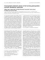
Early inflammatory cell infiltrate
Infiltration of ED-1 positive monocytes/macrophages
(brown staining) is an early event in HS-induced
response occurring between 1-6 hours after HS-infusion
(Figure 1). First after 9 hours after stimulation can neu-
trophils be seen (red staining). LPS stimulation gives a
much different cell infiltration pattern of early presence
of both monocytes/macrophages and neutrophils
Monocyte infiltration
Intra-ductal infusion of HS results in small but still signif-
icant effects on monocyte counts already at 1 and 3 hours
(p = 0.041 and p = 0.026, respectively); (Figure 2A). A 4-
fold increase start to appear at 6 hours (p = 0.002), rising
from a median count of 4.1 monocytes/mm
2
in controls
to 13.4 monocytes/mm
2
. At 9 hours after HS-infusion the
difference is even more prominent, rising to 17.0 mono-
cytes/mm
2
as compared to 1.2 monocytes/mm
2
in control
(p = 0.002).
LPS-infusion showed a different pattern of cell infiltra-
tion. LPS, at the presently used concentration, also gives
rise to increased monocyte numbers (Figure 2A), but in a
more linear fashion over time and is preceded by signifi-
cant elevation in neutrophils. The monocyte count
increases from 4.1 monocytes/mm
2
in controls to 12.1
monocytes/mm
2
(p < 0.001) at 6 hours and is elevated
even further to 30.5 monocytes/mm
2
(p = 0.02) at 9
hours.
When HS and LPS are compared they do not signifi-
cantly differ at any time point.
Neutrophil infiltration
After HS-stimulation a different pattern of neutrophil
infiltration compared to monocytes was seen (Figure 1,
red stained cells). No increase in neutrophil numbers
could be detected 1-6 hours after HS-infusion, in contrast
to the LPS group where the neutrophil infiltration was an
early event (Figure 2B). The increase of neutrophils was
not significantly increased until 9 hours after HS-infusion
(p = 0.041).
Three hours after LPS-stimulation the numbers of neu-
trophils had risen from 0.2 neutrophils/mm
2
in controls
to 2.0 neutrophils/mm
2
(p = 0.009), at 6 hours the num-
bers had increased to 6.5 neutrophils/mm
2
(p = 0.05) and
at 9 hours the count was at the same level, 5.9 neutro-
phils/mm
2
(p = 0.002); Figure 2B).
Comparison of neutrophil counts between HS and LPS
stimulation revealed differences between the groups in all
time points except 1 h after infusion.
Late stage inflammatory cells
The elevated numbers of monocytes seen 9 hours after
HS-stimulation persist for the coming 48 hours and the
median count at this time point is 22.8 monocytes/mm
2
(Figure 3A). After 48 hours the number of neutrophils
had returned to levels found in healthy animals (median
numbers in both groups 0.0 neutrophils/mm
2
; Figure 3B).
The localization of the ED-1 positive infiltrate of HS
treated animals differs dramatically between the early
time points (up to 9 hours; Figure 3C) and the 48 hour
group (Figure 3D). In the HS exposed animals the infil-
trate at the time points up to 9 hours are mainly restricted
to the interstitial space, while at 48 hours the ED-1 posi-
tive cells are predominantly found among acinar cells.
The morphology of the ED-1 positive cells are also differ-
ent than seen at earlier time points in that they have a
fully differentiated macrophage appearance at 48 hours,
while they are round and monocyte-like at 1-9 hours.
Neutrophil activation
The above findings of neutrophil infiltration after HS-
stimulation were confirmed by enzymatical measurement
of MPO activity in tissue homogenates (Figure 4). The
increase of MPO activity was only significantly (p =
0.002) elevated in the pancreatic tissue 9 hours following
HS infusion. LPS, on the other hand, seem to result in
more rapid effects and show elevated levels already after
6 hours (p = 0.003), an effect that sustained at 9 hours (p
= 0.009). Interestingly, the MPO activity was twice as
high in the HS exposed animals 9 hours after infusion
compared to LPS exposed animals in spite of the fact that
the number of neutrophils is clearly lower (p = 0.041) in
animals given HS-infusion. Forty-eight hours after HS-
infusion, the median activity of MPO had decreased from
30 μU/ml (at 9 hours) to 1.5 μU/ml (data not shown).
Axelsson et al. Journal of Inflammation 2010, 7:24
/>Page 5 of 10
No presence of active fibroblasts
Fibroblasts or pancreatic stellate cells have been sug-
gested to be involved in the inflammatory process of
acute pancreatitis [13]. To test the hypothesis of pancre-
atic stellate cells possibly being involved in the initial
events, staining of desmin and α-smooth muscle actin (α-
SMA), both used PSC markers [14], was performed. Up
to the measured 48 hours after HS-infusion, no co-local-
ization of chemoattractants and desmin positive cells
could be seen (data not shown). Furthermore, no staining
Figure 1 Histology of the pancreas 1-9 hours after infusion of phosphate buffered saline (PBS), heparan sulphate (HS) and lipopolysaccha-
ride (LPS). ED-1 positive cells brown and MPO positive cells red (arrows).
Axelsson et al. Journal of Inflammation 2010, 7:24
/>Page 6 of 10
of α-SMA could be seen outside vessels, which were spe-
cifically stained, indicating that pancreatic stellate cells
were not activated during the measured time span. This
out rules them as active participants in the early inflam-
matory response.
Chemokines
MCP-1
Expression of MCP-1 (Figure 5) was evident in the ductal
epithelial cells already 1 hour after infusion of HS and
LPS but not at later time points. Constitutive MCP-1
expression could be seen in vascular endothelial cells, as
well as in islet cells during the entire time period studied.
No systematic difference in acinar cells could be detected.
At later time points, pronounced MCP-1 staining was
detected in the invading inflammatory cells.
Quantitative measurements using ELISA showed no
significant differences comparing the HS group to PBS
but significant differences between LPS and PBS were
present 1 hour after exposure (data not shown).
CINC-1
Using immunohistochemistry, an increase of CINC-1
could only be detected in the inflammatory cell infiltrate
after HS-infusion. No expression could be seen in ductal
cells or other resident pancreatic cells during the first 9
hours after stimulation.
Consistent with the IHC observations, no elevated tis-
sue concentrations as measured using ELISA could be
demonstrated in the HS group (Figure 6). LPS-infusion,
on the other hand, induced a pronounced increase of
CINC-1 after 1 and 3 hours after infusion compared to
control (p = 0.015 and p = 0.041, respectively), but
returned to baseline concentrations 6 hours after stimula-
tion.
LPS concentrations
Possible LPS contamination is an important consider-
ation in the current studies. Therefore we taken measures
to minimize LPS contamination and struggled to have an
LPS free environment as possible. Measurements of LPS
show a low concentration of LPS present in the HS prepa-
rations (<100 pg/mg), resulting in a final concentration
<50 pg/ml in the pancreatic duct. This is a concentration
lower than would be expected to influence the inflamma-
tory response. The different responses elicited by HS and
LPS also suggest that LPS-contamination is not a major
contributor to the HS-induced inflammatory response.
Combined, this insures us that the inflammatory
response is a true response induced by HS and not LPS.
Discussion
HS-induced inflammatory response of the pancreas
seems to be a process mainly mediated by monocytes/
macrophages during the first 6 hours after stimulation,
while the LPS-initiated response seems to involve both
monocytes/macrophages and neutrophils. This observa-
tion makes sense, as the HS-induced response causes a
rapid influx of monocytes, a cell type whose main func-
tion is to phagocyte and clear the inflamed area of dam-
aged cells, and to prevent them from triggering a
powerful innate immune response. LPS, however, causes
recruitment of neutrophils, which efficiently eliminate
bacteria by oxidative burst. This finding may be of impor-
tance for the treatment of acute pancreatitis patients with
different etiologies as the two initiators investigated show
a distinct difference in cellular response. The HS-induced
inflammatory response can be hypothesized to corre-
spond to the aseptic acute pancreatitis-initiation, during
e.g. premature zymogen activation following biliary duct
stasis, and the LPS-induced inflammatory response cor-
responds, although controversial, to the scenario of retro-
grade migration of enteric bacteria into the pancreas or
septic complications of the manifest acute pancreatitis.
Figure 2 Infiltrate of the pancreas. (A) Number of ED-1 positive
(monocytes) cells/mm2. (B) Number of myeloperoxidase (MPO) posi-
tive cells (neutrophils)/mm2. PBS = phosphate buffered saline, HS =
heparan sulphate, LPS = lipopolysaccharide. Statistical significance de-
noted as * = p < 0.05, ** = p < 0.01.
!
!
Axelsson et al. Journal of Inflammation 2010, 7:24
/>Page 7 of 10
Clinical studies investigating potential relationships
between etiology and severity are conflicting, but clinical
studies are numerous confounded by their heterogeneous
material.
The later phase (48 hours) of the HS-induced pancre-
atic inflammation was also investigated and showed two
distinct differences as compared to the early phase (1-9
hours). After 48 hours the ED-1 positive cells had
migrated from the interstitial space of the pancreas to a
more peri-acinar location. When monocytes extravasate
and migrate into tissues they differentiate and become
multifunctional tissue macrophages. The macrophage
migration is governed by numerous mediators including
granulocyte-macrophage colony-stimulating factor (GM-
CSF) [15] and interleukine-6 (IL-6) [16]. These morpho-
logical changes could be confirmed in the present HS-
model and dramatic changes could be observed at 48
hours.
Immunohistological staining for MCP-1 and CINC-1,
chemoattractants for murine monocytes and neutrophils,
respectively, showed early up-regulation of MCP-1, but
not CINC-1 in ductal cells. Invading inflammatory cells
stained positive for both cytokines. These findings sug-
gest that HS stimulation of the ductal epithelium induce
MCP-1 secretion, which in turn recruits monocytes to
the pancreatic tissue. The invading cells produce MCP-1,
a known chemoattractant, but also an activator of mono-
cytes [17], resulting in an even more pronounced mono-
cyte recruitment. These cells also produce CINC-1,
which is chemotactic for neutrophils, causing a later sec-
ondary influx of neutrophils. This explains the biphasic
influx of the two cell types during HS-induced response.
Figure 3 Cell infiltrate of pancreas tissue 9 hours and 48 hours after infusion of heparan sulphate (HS). A. ED-1 positive cell counts 9 hours and
48 hours after infusion of HS. No difference of cell counts can be seen. B. Myeloperoxidase (MPO) positive cell counts 9 hours and 48 hours after infu-
sion of HS. C and D. Morphology of pancreas 9 and 48 hours infusion of HS. Statistical significance denoted as * = p < 0.05, ** = p < 0.01.
A. B.
C. D.
Axelsson et al. Journal of Inflammation 2010, 7:24
/>Page 8 of 10
The same expression pattern of MCP-1, but not CINC-
1, of rat acinar cells has been shown after caerulein stim-
ulation [18]. The fact that the acinar cells seem unaf-
fected in the current study and that they are specifically
affected in the caerulein model may be due to the differ-
ent cells that the two models target. The chemokine
changes could be detected mainly through immunohis-
tochemistry but not quantitatively using ELISA of tissue
homogenates. This fact is interpreted as that the local tis-
sue concentration is large enough to cause chemotaxis,
while the total concentrations is not enough for detection
of any differences in the total tissue analyzed using
ELISA.
In contrast to HS-stimulation, LPS induces early
CINC-1 expression. Already 1 hour after LPS infusion, a
small increase of CINC-1 is seen. This suggests that epi-
thelial or adjacent cells recruit neutrophils. It is therefore
reasonable to believe that HS and LPS induce two differ-
ent responses via different transduction pathways when
infused into the pancreatic duct. Several possible mecha-
nisms are present and of these, two are particularly
appealing. Either different cell types are responsible of
the recruitment of the different cell populations or two
different signaling pathways are activated within the same
cell. Following the first line of reasoning, it is reasonable
to hypothesize that monocytes are recruited by epithelial
cells, which in turn recruit neutrophils. The current study
demonstrates an early transcription of MCP-1 in the epi-
thelial cells, capable of recruiting monocytes and a later
expression of CINC-1 of the invading monocytes, which
in turn can attract neutrophils. The opposite may be true
in the LPS-induced early infiltration of neutrophils, a cell
type recently shown to possess the ability to recruit
monocytes [19]. The other possibility is that different
pathways are possible within the same cell. TLR4 is most
likely involved in the signaling cascade that is evoked by
Figure 4 Myeloperoxidase (MPO) activity of pancreatic tissue 1,
3, 6 and 9 hours after initiation of heparan sulphate-induced pan-
creatitis. PBS = phosphate buffered saline, HS = heparan sulphate, LPS
= lipopolysaccharide. Statistical significance denoted as * = p < 0.05, **
= p < 0.01.
Figure 5 MCP-1 expression 1 hour after infusion of PBS, HS and
LPS. HS-infusion (middle) causes increased MCP-1 expression of the
ductal epithelial cells (arrow) as compared to PBS control (top). Note
that the different epithelial cells respond to different extent. LPS also
induce a MCP-1 response of the epithelial cells (bottom). Other visible
structures of the histological images include veins (V), islets of Langer-
hans (I) and acinar cells (A).
Axelsson et al. Journal of Inflammation 2010, 7:24
/>Page 9 of 10
the two ligands, HS and LPS. In a clinical study investi-
gating the impact of two TLR4 mutations, TLR4
Asp299Gly and TLR4 Thr399Ile, a tendency of higher
frequency of the mutations were found in the group of
severe acute pancreatitis compared both to the group of
mild acute pancreatitis and the control group [20]. The
lack of statistical significance the authors explain by the
low frequency of the mutations in the population and
they suggest both mutations to be a risk factor for the
development severe acute pancreatitis. This clinical find-
ing is important and may suggest that TLR4 has a protec-
tive effect against uncontrolled inflammation of the
pancreas. In the rat, TLR4 has been detected in the ductal
epithelial cells, the first cells exposed to the ligands when
using this model, as well as in vascular endothelium and
islet beta cells [21-23]. TLR4 has also been described in
rat pancreatic stellate cells [24].
In order to elucidate involvement of two other resident
cell types, pancreatic stellate cells and resident mac-
rophages, during the early events we stained for markers
of both cell types as well as for chemokines. Pancreatic
stellate cells are distinguished from normal fibroblasts by
the presence of desmin, glial fibrillary acidic protein and
intracellular fat droplets. Upon activation, expression of
α-smooth muscle actin is seen. Desmin positive cells
morphologically similar to previously published descrip-
tions of pancreatic stellate cells were found [14,25] but no
co-localization of either chemokine stained for could be
demonstrated. Expression of α-smooth muscle actin was
restricted to the vessels. This suggests that pancreatic
stellate cells are neither activated nor active participants
during the initiation of the HS-induced response. They
are, however, likely to play an important role in the repair
phase after the response studied in these experiments.
Very few resident ED-1 positive cells could be detected
in the PBS control groups and in the early treatment
groups. This suggests that resident macrophages are
either absent or at least very rare in the healthy pancreas
and therefore unlikely to play a role in the initiating
events. The numbers increase dramatically at 6 hours
after HS administration and monocytes/macrophages are
clearly involved from this point and onward.
As discussed, the present model is most likely relevant
to the clinical situation, taken into account that two
ligands, HS and LPS, are possibly present during bile duct
obstruction. Both HS, shed from the ductal epithelium, as
well as LPS, set free from enteric bacteria entering the bil-
iary-pancreatic duct during occlusion, are relevant in the
event of bile duct occlusion. Ligation-induced acute pan-
creatitis in the rat shows a similar pattern of infiltration of
macrophages and neutrophils, where higher numbers of
macrophages precede neutrophils [26]. The shift in time
for the onset of the inflammation may be explained by the
delay of increasing HS levels in the duct and the fact that
the pattern is not identical to our HS data may be due to
other factors, such as elevated intraductal pressure,
which was not present in our model.
At present, studies to elucidate the mechanisms behind
the initiating events after HS administration is under-
taken by using mice lacking TLR4 or its adapter proteins.
Conclusions
Conclusions to be drawn from this study is that during
HS stimulation the pancreas responds by recruiting
monocytes and, at a later time point, neutrophils are the
important invading cells and that neutrophils plays a less
dominant role in the initiation of the inflammatory pro-
cess.
List of abbreviations
(α-SMA): α-smooth muscle actin; (Ab): antibody; (CINC-
1): cytokine-induced neutrophil chemoattractant-1;
(ELISA): enzyme-linked immunosorbent assay; (HE):
haematoxylin and eosin; (HS): heparan sulphate; (HSPG):
heparan sulphate proteoglycan; (HRP): horseradish per-
oxidase; (IHC): immunohistochemistry; (LPS): lipopoly-
saccharide; (MCP-1): monocyte chemotactic protein-1;
(MPO): myeloperoxidase; (PSCs): pancreatic stellate cells;
(PFA): phosphate-buffered formalin; (PBS): phosphate-
buffered saline; (PG): proteoglycan; (TLR4): Toll-like
receptor-4.
Competing interests
The authors declare that they have no competing interests.
Authors' contributions
JA, HA, KS were involved in the design of the experiment and carried out the
experimental work. AM and RA were involved in the design the study as well as
funding it and writing the manuscript.
Acknowledgements
These experiments were in part financed by a grant of Jakob Axelsson from
Fredrik and Ingrid Thuring's Foundation, Stockholm, Sweden.
Figure 6 CINC-1 concentrations in pancreatic tissue 1-9 hours af-
ter infusion of HS, PBS or LPS. PBS = phosphate buffered saline, HS =
heparan sulphate, LPS = lipopolysaccharide. Statistical significance de-
noted as * = p < 0.05, ** = p < 0.01.
Axelsson et al. Journal of Inflammation 2010, 7:24
/>Page 10 of 10
Author Details
1
Department of Clinical Sciences Lund, Lund University, BMC, D12, SE-221 84
Lund, Sweden and
2
Department of Experimental Medical Science, Lund
University, BMC, D12, SE-221 84 Lund, Sweden
References
1. Yadav D, Lowenfels AB: Trends in the epidemiology of the first attack of
acute pancreatitis: a systematic review. Pancreas 2006, 33:323-330.
2. Axelsson J: Initiation of experimental acute pancreatitis and
modulation of inflammatory response. In PhD thesis Lund University,
Department of Clinical Sciences; 2008.
3. Johnson GB, Brunn GJ, Kodaira Y, Platt JL: Receptor-mediated monitoring
of tissue well-being via detection of soluble heparan sulphate by Toll-
like receptor 4. J Immunol 2002, 168:5233-5239.
4. Axelsson J, Norrman G, Malmström A, Weström B, Andersson R: Initiation
of acute pancreatitis by heparan sulphate in the rat. Scand J
Gastroenterol 2008, 43:480-489.
5. Johnson GB, Brunn GJ, Platt JL: Cutting edge: an endogenous pathway
to systemic inflammatory response syndrome (SIRS)-like reactions
through Toll-like receptor 4. J Immunol 2004, 172:20-24.
6. Ekman-Ordeberg G, Malmström A: Use of sulphated
glycosaminoglycans for establishing effective labor in women.
International Patent SEO200005-7 2003.
7. Vaccaro MI, Calvo EL, Suburo AM, Sordelli DO, Lanosa G, Iovanna JL:
Lipopolysaccharide directly affects pancreatic acinar cells: implications
on acute pancreatitis pathophysiology. Dig Dis Sci 2000, 45:915-926.
8. Vonlaufen A, Xu Z, Daniel B, Kumar RK, Pirola R, Wilson J, Apte MV:
Bacterial Endotoxin: A Trigger Factor for Alcoholic Pancreatitis?
Evidence From a Novel, Physiologically Relevant Animal Model.
Gastroenterology 2007, 133:1293-1303.
9. Fransson LA, Sjoberg I, Havsmark B: Structural studies on heparan
sulphates. Characterization of oligosaccharides; obtained by periodate
oxidation and alkaline elimination. Eur J Biochem 1980, 106:59-69.
10. Westergren-Thorsson G, Onnervik PO, Fransson LA, Malmstrom A:
Proliferation of cultured fibroblasts is inhibited by L-iduronate-
containing glycosaminoglycans. J Cell Physiol 1991, 147:523-530.
11. Koike K, Moore FA, Moore EE, Poggetti RS, Tuder RM, Banerjee A:
Endotoxin after gut ischemia/reperfusion causes irreversible lung
injury. J Surg Res 1992, 52:656-662.
12. Dijkstra CD, Dopp EA, Joling P, Kraal G: The heterogeneity of
mononuclear phagocytes in lymphoid organs: distinct macrophage
subpopulations in the rat recognized by monoclonal antibodies ED1,
ED2 and ED3. Immunology 1985, 54:589-599.
13. Kishi S, Takeyama Y, Ueda T, Yasuda T, Shinzeki M, Kuroda Y, Yokozaki H:
Pancreatic duct obstruction itself induces expression of alpha smooth
muscle actin in pancreatic stellate cells. J Surg Res 2003, 114:6-14.
14. Bachem MG, Schneider E, Gross H, Weidenbach H, Schmid RM, Menke A,
Siech M, Beger H, Grunert A, Adler G: Identification, culture, and
characterization of pancreatic stellate cells in rats and humans.
Gastroenterology 1998, 115:421-432.
15. Akagawa KS: Functional heterogeneity of colony-stimulating factor-
induced human monocyte-derived macrophages. Int J Hematol 2002,
76:27-34.
16. Chomarat P, Banchereau J, Davoust J, Palucka AK: IL-6 switches the
differentiation of monocytes from dendritic cells to macrophages. Nat
Immunol 2000, 1:510-514.
17. Rollins BJ, Walz A, Baggiolini M: Recombinant human MCP-1/JE induces
chemotaxis, calcium flux, and the respiratory burst in human
monocytes. Blood 1991, 78:1112-1116.
18. Bhatia M, Brady M, Kang YK, Costello E, Newton DJ, Christmas SE,
Neoptolemos JP, Slavin J: MCP-1 but not CINC synthesis is increased in
rat pancreatic acini in response to cerulein hyperstimulation. Am J
Physiol Gastrointest Liver Physiol 2002, 282:G77-85.
19. Soehnlein O, Zernecke A, Eriksson EE, Rothfuchs AG, Pham CT, Herwald H,
Bidzhekov K, Rottenberg ME, Weber C, Lindbom L: Neutrophil secretion
products pave the way for inflammatory monocytes. Blood 2008,
112:1461-1471.
20. Hofner P, Balog A, Gyulai Z, Farkas G, Rakonczay Z, Takacs T, Mandi Y:
Polymorphism in the IL-8 gene, but not in the TLR4 gene, increases the
severity of acute pancreatitis. Pancreatology 2006, 6:542-548.
21. Li Y, Zhou ZG, Xia QJ, Zhang J, Li HG, Cao GQ, Wang R, Lu YL, Hu TZ: Toll-
like receptor 4 detected in exocrine pancreas and the change of
expression in cerulein-induced pancreatitis. Pancreas 2005, 30:375-381.
22. Vives-Pi M, Somoza N, Fernandez-Alvarez J, Vargas F, Caro P, Alba A, Gomis
R, Labeta MO, Pujol-Borrell R: Evidence of expression of endotoxin
receptors CD14, toll-like receptors TLR4 and TLR2 and associated
molecule MD-2 and of sensitivity to endotoxin (LPS) in islet beta cells.
Clin Exp Immunol 2003, 133:208-218.
23. Li Y, Zhou ZG, Zhang J, Chen YD, Li HG, Gao HK, Wang R, Hu TZ:
Microcirculatory detection of Toll-like receptor 4 in rat pancreas and
intestine. Clin Hemorheol Microcirc 2006, 34:213-219.
24. Masamune A, Kikuta K, Watanabe T, Satoh K, Satoh A, Shimosegawa T:
Pancreatic stellate cells express Toll-like receptors. J Gastroenterol 2008,
43:352-362.
25. Apte MV, Haber PS, Applegate TL, Norton ID, McCaughan GW, Korsten
MA, Pirola RC, Wilson JS: Periacinar stellate shaped cells in rat pancreas:
identification, isolation, and culture. Gut 1998, 43:128-133.
26. Meyerholz DK, Samuel I: Morphologic characterization of early ligation-
induced acute pancreatitis in rats. Am J Surg 2007, 194:652-658.
doi: 10.1186/1476-9255-7-24
Cite this article as: Axelsson et al., Proposed protective mechanism of the
pancreas in the rat Journal of Inflammation 2010, 7:24
Received: 8 April 2009 Accepted: 18 May 2010
Published: 18 May 2010
This article is available from: 2010 Axelsson et al; licensee BioMed Central Ltd. This is an Open Access article distributed under the terms of the Creative Commons Attribution License ( ), which permits unrestricted use, distribution, and reproduction in any medium, provided the original work is properly cited.Journal of Inflammation 2010, 7:24