Báo cáo y học: "The role of Qa-2, the functional homolog of HLA-G, in a Behcet''''s disease-like mouse model induced by the herpes virus simplex" pptx
Bạn đang xem bản rút gọn của tài liệu. Xem và tải ngay bản đầy đủ của tài liệu tại đây (1.06 MB, 12 trang )
Lee et al. Journal of Inflammation 2010, 7:31
/>
Open Access
RESEARCH
The role of Qa-2, the functional homolog of HLA-G,
in a Behcet's disease-like mouse model induced by
the herpes virus simplex
Research
Meeyoung Lee†1, Bunsoon Choi†1, Hyuk Jae Kwon1, Ju A Shim1, Kyung Sook Park2, Eun-So Lee3 and
Seonghyang Sohn*1,4
Abstract
Background: It has been suggested that the HLA-G molecule is a genetic risk factor for Behcet's disease (BD). In this
study, we evaluated the level of Qa-2, a murine nonclassical class I MHC molecule and possible functional homolog of
HLA-G, to determine if it was associated with various symptoms of BD-like mice. In addition, we investigated siRNA
(small interfering RNA) treatment to determine if it inhibited Qa-2 expression, thereby changing the symptoms of mice.
Methods: RNA interference (RNAi) and vector transfection were employed to manipulate gene expression in vivo in
mice. siRNA (small interfering RNA) or Qa-2 expression vector was applied to inhibit or up-regulate Qa-2 expression,
respectively.
Results: The Qa-2 levels in granulocytes were lower in BD-like mice than in normal controls. The silencing of Qa-2 by
intravenous injection of siRNA (500 nmol/mouse, 4 times at 3-day intervals) specifically reduced the Qa-2 levels and
worsened the BD-like symptoms.
Conclusions: Silencing Qa-2 by injecting siRNA into mice resulted in deterioration of symptoms in BD-like mice.
Background
Since HLA-G (human leukocyte antigen-G) was first
detected by Geraghty et al. [1], it has been reported that
HLA-G protein is expressed at the feto-maternal interface during pregnancy [2] and on a subset of thymic epithelial cells [3], and that it is also involved in maintenance
of tolerance of the maternal immune system toward the
semi-allogeneic fetus. HLA-G is also expressed in other
tissues such as intestinal mucosa [4] and PBMC [5].
Numerous studies have evaluated the relevance of HLAG under pathologic conditions such as transplantation,
autoimmunity, cancer, and hematologic malignancies [6].
HLA-G interacts with different natural killer (NK) cell
receptors and is able to inhibit NK and T-cell cytotoxicity,
as well as T-cell proliferation [7]. Interestingly, HLA-G
has been described as a unique ligand of the killer cell
inhibitory receptor, KIR2DL4, which is expressed on the
* Correspondence:
1
Laboratory of Cell Biology, Ajou University Institute for Medical Sciences,
Suwon, Korea
† Contributed equally
Full list of author information is available at the end of the article
surface of all NK cells [8]. Furthermore, HLA-G inhibits
the transendothelial migration of NK cells [9], shifts the
cytokine balance toward Th2 dominance [10], and suppresses the proliferation of allogeneic CD4+ T lymphocytes [11,12]. Taken together, HLA-G exerts specific
inhibitory effects against immune cells. In addition,
recent studies indicate unexpected expression of HLA-G
proteins in chronic cutaneous inflammatory diseases,
such as psoriasis [13] and atopic dermatitis [14].
Behcet's disease (BD) is a chronic multi-systemic disorder that involves the gastrointestinal, mucocutaneous,
ocular, vascular, central nervous, and articular systems.
BD has a chronic course that includes periodic exacerbations and progressive deterioration [15]. Although the
etiology of BD is unclear, viral infection has long been
postulated as one of its main factors. The viral hypothesis
has been verified by detection of the virus in saliva [16],
intestinal ulcers [17], and genital ulcers [18] of patients
with BD since it was first proposed by Hulỷsi Behỗet [19].
Furthermore, inoculation of the earlobe of ICR mice with
herpes simplex virus (HSV) enables development of a
© 2010 Lee et al; licensee BioMed Central Ltd. This is an Open Access article distributed under the terms of the Creative Commons Attribution License ( which permits unrestricted use, distribution, and reproduction in any
medium, provided the original work is properly cited.
Lee et al. Journal of Inflammation 2010, 7:31
/>
BD-like animal model [20]. Manifestations in mice following HSV inoculation involve multiple symptoms such
as oral ulcers, genital ulcers, skin ulcers, eye symptoms,
gastrointestinal ulcers, arthritis, and neural involvement,
as well as skin crusting. The frequency of these symptoms
is similar to that of patients with BD [21]. In addition to
viral causes of BD, several studies have identified lymphocyte dysfunction as a possible cause [22,23]. Thus, attention has been focused on the T helper (Th) 1 and Th2
cytokines, with Th1 cells perhaps playing a more important role in the immunopathogenesis of BD [24]. When
the Th2 adjuvant, aluminium hydroxide (alum), was
mixed with ovalbumin (OVA) and injected into mice suffering from BD, their cutaneous symptoms were
improved [25].
Park et al. [26] reported that the frequency of haplotypes containing a HLA-G 3741_3754 14 base pair insertion and 1597*delC was increased in BD patients.
Moreover, individuals who were homozygous with the
3741_3754*ins14/*ins14 genotype were found to have a
risk of BD that was 2.7-times greater than that of the controls. The HLA-G 3741*+14bp induces a significantly
lower expression level than the complete HLA-G mRNA
isoforms. In addition, the HLA-G 3741_3754 14-base
pair insertion allele was found to occur significantly more
frequently in BD patients with ocular, arthritis, and CNS
symptoms than in controls, and this insertion was found
to be related to the lower serum level of HLA-G [26]. The
authors who presented these findings suggested that
these HLA-G allelic variants are genetic risk factors for
BD. In addition, the HLA-G*010101 alleles have been
shown to have a significantly lower frequency in BD
patients than in control subjects [27].
As a result, it is important to determine if HLA-G contributes to the pathogenesis of BD. To accomplish this,
Qa-2 expression, the functional homolog of HLA-G in
mice, was identified and modulated by small interfering
RNA (siRNA) and the Qa-2 expression vector. The results
of this study confirmed that decreased Qa-2 levels are
related to changes in the disease pattern and deterioration of BD-like symptoms.
Methods
Animals, induction of BD-like symptoms, and scoring of BD
activity
Five-week-old ICR male mice were used in this study. To
induce a BD-like disease in mice, their earlobes were
scratched with a needle and then inoculated with 1.0 ×
106 plaque forming units/ml of HSV type 1 (F strain).
Virus inoculation was performed twice with a 10-day
interval, after which the mice were observed for 30
weeks. Mice were housed in conventional temperatureand light-controlled rooms (20-22°C, 12 h light cycle
starting at 8:00 a.m.) and had free access to food and
Page 2 of 12
water. During the experiment, the animals were observed
closely. Mice were handled in accordance with the protocols approved by our institutional animal care committee.
Manifestations in mice after HSV inoculation involved
multiple symptoms including oral ulcers, genital ulcers,
skin ulcers, eye symptoms, intestinal ulcers, arthritis, and
neural involvement, as well as skin crusting. Oral, genital,
and other skin ulcers (including bulla and crust), and eye
symptoms were all classified as major symptoms, while
other symptoms were classified as minor symptoms [20].
Overall, 15% of the HSV-injected mice developed BD-like
symptoms. The disappearance of symptoms and decrease
in lesion size constituted an improvement, similar to in
human patients.
The animals were observed once a week after HSV
inoculation, at which time the severity of BD was determined according to the BD activity index, as outlined in
the Behcet's Disease Current Activity Form 2006 http://
www.behcet.ws/pdf/BehcetsDiseaseActivityForm.pdf.
The occurrence of the following symptoms in the mouse
model were selected for analysis: mouth ulceration, genital ulceration, erythema, skin pustules, skin ulceration,
joints-arthritis, diarrhea, red eye (right, left), reduced
vision (right, left), loss of balance, discoloration, and
swelling of the face. The score of each symptom was one,
and the total score before and after treatment was used to
determine the severity of BD. Mice exhibiting significantly reduced symptoms were photographed to document improvement after treatment.
Synthesis and in vitro test of siRNA
Qa-2 siRNA oligonucleotides with the following sense
and anti-sense sequences were designed and synthesized
by Dharmacon (Chicago, IL, USA). The Qa-2 protein was
encoded by four genes in the Q region, Q6, Q7, Q8 and
Q9. These genes have a typical class I MHC gene structure involving exon 1 (leader peptide), exon 2 (α1
domain), exon 3 (α2 domain), exon 4 (α3 domain), exon 5
(transmembrane domain), and exons 6, 7 and 8 (cytoplasmic domains). As shown in Table 1, we selected four
sequences located in each domain to synthesize siRNA.
To confirm the function of interference, the synthesized
siRNA was tested in vitro in peripheral blood mononuclear cells (PBMC). To accomplish this, PBMCs were isolated from 5-6 week-old ICR mice and cultured at 1 × 10⁵
cells/ml in DMEM medium with 1% antibiotics and 10%
FBS. siRNA (200 nM) was incubated with 3 μL of oligofectamin (Gibco-Invitrogen, Rockville, MD) in 200 μL of
DMEM medium. After 24 h of treatment with siRNA, the
PBMCs were harvested and subjected to RT-PCR.
In vivo siRNA injection
For application to mice, 500 nM of siRNA in 200 μL of 5%
glucose, including transfection reagent jetPEI (Polyplus,
Lee et al. Journal of Inflammation 2010, 7:31
/>
Table 1: Qa-2 siRNA oligonucleotide sequences
Qa-2 domain
siRNA oligonucleotides sequences
Leader peptide
domain
5'-CAACACUCGCAAUAUU-3'(sense)
3'-GUUGUGAGCGACGUUAUAA-5'(antisense)
α3 domain
5'-AGGUCUUAUGGUGCUGUCAUU-3'(sense)
3'-UUUCCAGAAUACCACGACAGU5'(antisense)
Transmembrane
domain
5'-UGUGAUGAAUAGGAGGUGAUU-3'(sense)
3'-UUACACUACUUAUCCUCCACU5'(antisense)
Cytoplasmic
membrane domain
5'-UAGAGCUCUGAUAGAUCUCUU-3'(sense)
3'-UUAUCUCGAGACUAUCUAGAG5'(antisense)
France, Illkirchcedex), was intravenously injected into
mice one to four times with a three day interval between
injections. Two-days after the last injection, mice were
photographed and the PBMCs were analyzed using a fluorescence-activated cell sorter (FACS). The control group
was injected with 200 μL of 5% glucose. Qa-2 leader peptide domain siRNA did not down-regulate the Qa-2
mRNA level in in vitro PBMC cultures when compared to
other domains; therefore, the leader peptide domain
siRNA was injected as a control. For in vivo administration to mice, 1.5 μL of transfection reagent was mixed
with 5% glucose and siRNA. The Qa-2 siRNA was mixed
with α3 domain, transmembrane domain and cytoplasmic domain in equal amounts, after which it was administered to mice.
Flow cytometry
To analyze the Qa-2 expression, cells were harvested and
fixed with 4% formaldehyde in 1% fetal bovine serum
containing PBS for 20 min at room temperature, after
which they were incubated with FITC-conjugated antiQa-2 antibody (eBioscience, San Diego, CA, USA).
Stained cells were analyzed in FACS Vantage using the
Cell Quest software (Becton Dickinson, Franklin Lakes,
NJ, USA) by collecting at least 10,000 gated lymphocytes
[7].
Reverse transcription PCR (RT-PCR)
Total RNA was isolated using TRIzol (Life Technologies,
Helgerman, CT) according to the manufacturer's recommendations. Two μg of total RNA were used as a template
for cDNA synthesis, which was conducted using a SuperScript III First-Strand Synthesis System for RT-PCR kit
(Invitrogen, Carlsbad, CA). The cDNA was amplified by
PCR using the following primers: Qa-2, Sense: 5' AGGTCTTAT GGTGCTGTCAC-3', Anti sense: 5'- TGT
Page 3 of 12
GTAATTCTGCTCCTTCC -3'; β-actin, Sense: 5'-TG
GAATCCTGTGGCATCCATGAAAC -3', Antisense: 5'TAAAACGCAGCTCAGTAACAGTCCG-3';
IFNγ,
Sense:
5'-AGCGGCTGACTGAACTCAGATTGTAG
CTTGTACCTTTACTTCACTG-3', Antisense: 5'-GTC
ACAGTTTTCA GCTGTATAGGG-3'. Amplified PCR
products were visualized on 1.2% agarose gels.
Real Time PCR
For real-time SYBR Green RT-PCR, a 20-μl reaction containing 10 μl of 2× Quantitect SYBR Green Master Mix
(Qiagen, Valencia, CA, USA) was employed. The master
mix was composed of hot start Taq polymerase, a 0.4 μL
mix of 2 reverse transcriptases, 0.5 μL (10 ng/μL) of template and 0.8 μL of primers. An ABI 7900 HT thermal
cycler (Lab Centraal B.V., Haarlem, The Netherlands) was
used for all real-time RT-PCR assays. Reverse transcription was conducted at 50°C for 30 min, followed by denaturation at 95°C for 15 min. DNA was amplified by
subjecting the samples to 40 cycles of 95°C (30 s), 55°C
(30 s), and 72°C (30 s). Real-time RT-PCR data were collected for 15 sec at 75°C to avoid non-specific fluorescence due to the formation of primer dimers at low
template concentrations. For generation of standard
quantitation curves, the cycle threshold values were plotted proportionally against the logarithm of the input copy
numbers. Negative controls were included in each run.
Qa-2 vector construction
Qa-2 cDNA was amplified from total RNA extracted
from ICR mice lymph nodes by reverse transcriptase polymerase chain reaction (RT-PCR) using the following
primers: sense 5'-CGGGATCCCGATGGCTCTAACAA
TGCTGC-3', antisense 5'-CGGAATTCCGCTTCGTGTGAAAGTATGGAG-3'. The sense primer included the
BamH1 restriction site and the antisense primer included
the EcoR1 restriction site. The cDNA was subsequently
digested with BamHI and EcoRI and then inserted into
eukaryotic expression vector pcDNA3.1 (Invitrogen,
Carlsbad, CA, USA). Verification of the recombinant
construct was performed by DNA sequencing. The
empty vector pcDNA3.1 was used as a control. All plasmids were purified by two rounds of passage through
Endo-Free columns (Qiagen, Chatsworth, CA, USA), as
described elsewhere [28].
Qa-2 vector transfection to HeLa cells
HeLa cells were maintained in Dulbecco's modified Eagle
medium (DMEM) supplemented with 2 mM glutamine,
100 units/ml penicillin, 100 μg/ml streptomycin, and 5%
(v/v) dextran-charcoal-treated fetal bovine serum at 37°C
in 5% CO2. Cells were plated at 106 cells/10 cm dish the
day before transfection, after which they were transfected
using a lipofectimine kit (Invitrogen, Paisley, UK) accord-
Lee et al. Journal of Inflammation 2010, 7:31
/>
Page 4 of 12
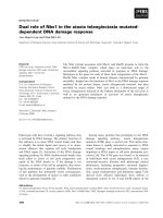
Figure 1 Qa-2 expression in PBMC of BD. A. RT-PCR demonstrated that mRNA expression was lower in PBMC of BD skin than in BD normal mice. B.
The frequency of Qa-2 in PBMC of normal healthy controls, BD asymptomatic (BD normal) mice, BD mucocutaneous symptomatic mice (BD skin), and
BD mucocutaneous and ocular symptomatic mice (BD eye) as determined by FACS analysis. In lymphocytes, the Qa-2 levels in BD eye mice were significantly lower than in normal healthy mice (p = 0.036). These levels were also lower than in BD skin mice, although this difference was not significant.
In granulocytes, the Qa-2 levels in BD eye mice were significantly lower than in normal healthy mice (p = 0.016). The Qa-2 levels in BD eye mice were
lower than in normal and BD skin mice, although this difference was not statistically significant. Qa-2 levels in BD skin were significantly lower than in
normal controls (p = 0.024). C. The portion of Qa-2 positive cells in lymphocytes or granulocytes. The frequency of Qa-2 positive cells in the granulocytes of BD skin and BD eye mice was lower than in normal controls and BD normal mice (BDN). The frequencies of Qa-2 positive cells in BD eye mice
were significantly lower than those in normal controls (p = 0.001).
ing to the manufacturer's instructions. The vector
pcDNA3.1 was transfected into HeLa cells as a control.
Administration of Qa-2 vector to mice
Normal and BD mice were intraperitoneally injected once
with 50 ng of pcDNA 3.1 or pcDNA 3.1 Qa-2 vector per
mouse, and their splenocytes or macrophages were isolated three days later and analyzed by flow cytometry.
Vector mixed with transfection reagent jetPEI was
injected into mice and the frequency of Qa-2 protein
expression was analyzed by FACS.
Statistical analysis
All data are presented as the mean ± SE. Statistical differences between groups were determined using a Student's
t test and the Bonferroni correction. Statistical analysis
was conducted using MedCalc® version 9.3.0.0.
Results
Qa-2 mRNA and Qa-2 positive PBMCs were lower in BD
symptomatic mice than in normal healthy mice
RT-PCR revealed that Qa-2 mRNA expression in peripheral blood mononuclear cells (PBMC) of mucocutaneous
Lee et al. Journal of Inflammation 2010, 7:31
/>
Page 5 of 12
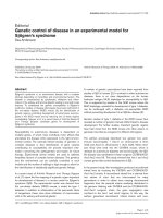
Figure 2 Qa-2 siRNA reduced Qa-2 mRNA and the frequency of Qa-2 positive cells in PBMC of normal mice. PBMC isolated from mice were
transfected with Qa-2 siRNAs with different Qa-2 domains for 24 hrs, and the expression of Qa-2 was then determined by reverse transcriptase-PCR
and FACS analysis. Lanes 4, 5 and 6 (α3 domain, transmembrane domain, and cytoplasmic domain, respectively) showed that siRNA effectively reduced the Qa-2 mRNA levels. Lane 3 (leader peptide) did not decrease the Qa-2 level. Lane 7 (a mixture of leader peptide, α3 domain, transmembrane
domain, and cytoplasmic domain) also did not decrease the Qa-2 level. Lane 1, control (not treated); Lane 2, 5% glucose treated; Lane 3, leader peptide
200 nmole; Lane 4, α3 domain 200 nmole; Lane 5, transmembrane domain 200 nmole; Lane 6, cytoplasmic domain 200 nmole; Lane 7, mixed 200
nmole (leader peptide + α3 domain+ transmembrane domain + cytoplasmic domain).
symptomatic BD mice was down-regulated when compared to asymptomatic BD mice, despite HSV inoculation (BD normal, BDN) (Figure 1A). Next, Qa-2 levels in
PBMCs obtained from normal healthy mice, BD asymptomatic mice (BDN), BD skin symptomatic mice (BD
skin), and BD eye symptomatic mice (BD eye) were analyzed by flow cytometry. The symptoms of BD skin consisted of typical mucocutaneous symptoms in mice
without ocular symptoms, while those of BD eye mice
consisted of ocular symptoms with mucocutaneous
symptoms. After FACS staining, lymphocytes and granulocytes were separated by gating. In lymphocytes, Qa-2
positive cells accounted for 94.78 ± 3.56% in normal
healthy mice, 92.56 ± 6.13% in BD normal mice, 91.73 ±
5.96% in BD skin, and 84.49 ± 11.95% in BD eye mice. BD
eye mice were found to have a statistically lower number
of Qa-2 positive cells than normal healthy mice (p =
0.036). In granulocytes, Qa-2 positive cells were 87.01 ±
7.97% in normal healthy mice, 82.29 ± 17.47% in BD normal mice, 67.9 ± 21.42% in BD skin mice, and 56.00 ±
30.49% in BD eye mice. BD skin and BD eye mice showed
significantly lower levels of Qa-2 positive cells than normal healthy mice (p = 0.024, p = 0.016 each) (Figure 1B).
The portion of Qa-2 positive cells in the granulocytes of
BD skin and BD eye mice was lower than that of normal
control and BD normal (BDN) mice. The portion of Qa-2
positive cells in the granulocytes of BD eye mice was significantly lower than that of normal controls (p = 0.001)
(Figure 1C). As shown in Figure 1, the decreased level of
Qa-2 was related to the BD symptoms.
RNA interference of Qa-2 transcription in vitro; Qa-2 siRNA
reduced Qa-2 mRNA levels in PBMCs of normal mice
PBMCs isolated from normal mice were transfected for
24 h with Qa-2 siRNA with different domains, after
which the expression of Qa-2 was determined by reverse
transcriptase-PCR. siRNA for the α3 domain, transmembrane domain, and cytoplasmic domain inhibited the Qa2 level; however, the leader peptide domain did not.
Mixed siRNA consisting of equal amounts each of these
four domains did not downregulate the Qa-2 mRNA
level. Flow cytometric analysis also showed a decreased
frequency of Qa-2 expression in the Qa-2 siRNA domaintreated groups, except for the leader peptide domain (Figure 2).
Downregulation of Qa-2 by intravenous injection of siRNA
into BD mice
Next, an siRNA mixture composed of the siRNA of the
α3 domain, transmembrane domain and the cytoplasmic
domain was injected into BD mice. Five to six individual
BD mice in each group were intravenously injected once
Lee et al. Journal of Inflammation 2010, 7:31
/>
Page 6 of 12
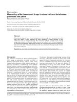
Figure 3 Down-regulation of Qa-2 after intravenous injection of siRNA into BD mice. A. BD mice were injected once with control siRNA or Qa2 siRNA (500 nmol/mouse), which was composed of the α3 domain, transmembrane domain, and cytoplasmic domain. PBMC collected from the orbital sinus before and 1 day after injection were analyzed by flow cytometry. Qa-2 siRNA effectively reduced the Qa-2 levels in the PBMC of BD mice.
B. Two days after injection, the frequency of Qa-2 positive lymphocytes and granulocytes was analyzed in Qa-2 siRNA injected BD mice. In lymphocytes
and granulocytes, Qa-2 siRNA significantly reduced the number of Qa-2 positive cells when compared to glucose-injected control mice. C. The frequency of Qa-2 positive cells in mice that were injected with Qa-2 siRNA four times. Specifically, 500 nmol control siRNA or Qa-2 siRNA in 200 μl of 5%
glucose solution was intraperitoneally injected four times with a three day interval between injections, and the PBMC were analyzed by FACS two
days after the last injection. In lymphocytes and granulocytes, Qa-2 siRNA significantly reduced the Qa-2 positive cells when compared to glucose
injected control mice.
with 5% glucose or 500 nmol of Qa-2 siRNA or control
siRNA, and their PBMCs were analyzed one day and two
days later by flow cytometry. One day after Qa-2 siRNA
injection, the number of Qa-2 positive granulocytes was
32.18 ± 14.64%, which was significantly lower (p = 0.049)
than that of mice treated with 5% glucose (54.21 ± 1.89%)
Table 2: Changes in symptoms after Qa-2 siRNA injection
into BD mice
siRNA
Deteriorated number/
total number
Qa-2 siRNA
3/6
Leader peptide siRNA
1/6
Glucose
0/7
The symptoms of BD mice deteriorated following treatment with
Qa-2 siRNA. Qa-2 siRNA was intravenously injected into BD mice
four times with a three day interval between injections.
Deterioration occurred in three of six BD mice.
or leader peptide (61.32 ± 12.27%) (Figure 3A). In lymphocytes, the Qa-2 positive cell counts did not differ significantly among groups. Two days later, the frequency of
Qa-2 positive cells was 84.12 ± 10.34% in Qa-2 siRNA
injected mice, while it was 94.23 ± 3.86% of glucose
injected control mice in lymphocytes (p = 0.029). In granulocytes, the frequency of Qa-2 positive cells was 42.18 ±
28.40% in Qa-2 siRNA injected mice, while it was 75.65 ±
23.59% in glucose injected control mice (p = 0.008).
These findings demonstrated that Qa-2 siRNA effectively
reduced the frequencies of Qa-2 positive cells in lymphocytes and granulocytes in BD mice (Figure 3B). To determine if repeated administration can reduce the Qa-2 level
more efficiently, the frequency of Qa-2 positive cells in
BD mice that were injected with Qa-2 siRNA four times
was analyzed. To accomplish this, 500 nmol control
siRNA or Qa-2 siRNA in 200 μl of 5% glucose solution
was intraperitoneally injected four times with a three day
interval in between injections. Two days after the last
injection, the PBMCs were analyzed by FACS. In lymphocytes and granulocytes, Qa-2 siRNA led to a significant
reduction in Qa-2 positive cells when compared to glu-
Lee et al. Journal of Inflammation 2010, 7:31
/>
Page 7 of 12
Figure 4 Qa-2 siRNA deteriorated BD symptoms. For each mouse, 500 nmol each of control siRNA or Qa-2 siRNA in 200 ml of 5% glucose solution
was intraperitoneally injected four times with three day intervals, and the symptoms were photographed (A) and the severity score was analyzed (B)
two days after the last injection. The severity was lower in Qa-2 siRNA injected BD mice when compared to control siRNA injected BD mice, although
this change was not significant. The disease score was estimated according to the Patients Index Score, Behcet's disease current activity form 2006,
ICBD. The symptoms of BD mice deteriorated after treatment with Qa-2 siRNA. Deterioration occurred in three of six BD mice (A). When treated with
control siRNA, the deterioration occurred in one of six mice, while no change was observed in any of the mice injected with 5% glucose.
cose injected control mice (p = 0.05, p = 0.02 each) (Figure 3C). However, the reduction in Qa-2 level observed in
response to one and four injections did not differ significantly.
The change in symptoms after Qa-2 siRNA injection into BD
mice
To determine if down-regulation of Qa-2 could influence
the symptoms of BD, changes in symptoms (Table 2) and
the disease severity score were examined after administration of siRNA to BD mice. Specifically, Qa-2 siRNA
was intravenously injected into BD mice four times with a
three day interval between treatments. After the injection
of siRNA, deterioration occurred in three of six BD mice
(Figure 4A). However, in mice treated with control
siRNA, the deterioration only occurred in one of the six
mice. In addition, there was no change in symptoms
observed in any of the seven BD mice injected with 5%
glucose. The change in symptoms was scored according
to the severity score of BD, which is outlined in the BD
Current Activity Form. As shown in Figure 4B, the score
of the Qa-2 siRNA-injected group increased from 5.66 ±
1.21 to 7.16 ± 2.04, although this change was not statistically significant (p = 0.07). In contrast, the score in the
control siRNA injected group increased to 4.0 ± 3.08
from 3.8 ± 2.68, while that of the glucose injected group
changed from 4.0 ± 1.41 to 3.4 ± 0.89.
Qa-2 siRNA increased IFNγ mRNA levels in spleens of BD
mice
Recent in vitro studies have suggested that some duplex
siRNA sequences have non-specific effects and can
induce an IFN response, particularly at high concentrations [29,30]. However, further studies are needed to
determine if these series of reactions can occur in vivo
and if this can occur in response to our siRNA sequences
[31]. Xie et al. reported that non-viral siRNA delivery to
diseased tissue does not elicit an immune response [32].
To determine the IFNγ mRNA expression, the spleen tissues of BD mice that were injected with siRNA four times
were subjected to reverse transcriptase PCR (RT-PCR)
(Figure 5A) and real time PCR (Figure 5B). The IFNγ
mRNA expressions were increased in the Qa-2 siRNAinjected mice when compared to the control siRNA or
glucose injected group. Increased IFNγ was not due to
siRNA, but rather to suppressed Qa-2 expression because
control siRNA did not increase the level of IFNγ. These
findings are in accordance with the finding that HLA-Gexpressing cells showed significantly reduced levels of
IFNγ [33].
Lee et al. Journal of Inflammation 2010, 7:31
/>
Page 8 of 12
2 gene of pGEM-Qa-2 into pcDNA3.1 vector was confirmed by digestion with EcoRI and BamHI (Figure 6A),
after which the inserted sequence was confirmed by
sequencing using T7 promoter (Figure 6B). The vector
was intraperitoneally injected once into mice, and peritoneal macrophages and splenocytes were isolated four
days later. As shown in Figure 7, the frequency of Qa-2
expressing cells in splenocytes increased to 94.53 ± 0.64%
in the Qa-2 vector injected mice, while it was 89.83 ±
2.66% in control vector injected mice (p = 0.45). Additionally, their frequency in macrophages increased to
82.25 ± 5.62% in the Qa-2 vector injected mice, while it
was 67.53 ± 4.66% in control vector injected mice (p =
0.003). The IFNγ levels in macrophages of Qa-2 vectorinjected mice also decreased to 16.60 ± 6.11%, while they
were 66.24 ± 7.28% in control mice (p < 0.001) (Figure 8).
Qa-2 expression vector appeared to work in macrophages, and these effects were accompanied by a
decrease in IFNγ.
The frequency of NK cells in BD and BDN mice
Figure 5 The expression of IFN-γ mRNA in the spleens of glucose,
control siRNA, and Qa-2 siRNA-injected BD mice. The expression of
IFN-γ mRNA as shown by RT-PCR (A) and real time PCR (B). IFNγ mRNA
expression was increased in the Qa-2 siRNA-injected mice.
Qa-2 expression vector decreased the frequency of IFNγ
stained macrophages in BD mice
To confirm if Qa-2 could influence IFNγ expression, Qa2 vector was constructed in PC3.1 vector and then
administered to normal and BD mice. Cloning of the Qa-
To confirm the relationship between HLA-G and the NK
cells, the frequency of NK cells was observed in BD and
BDN mice using flow cytometry. As shown in Figure 9,
the frequency of NK cells in splenocytes was 13.8 ± 2.2%
in BD mice (n = 9) when compared to BDN mice (5.4 ±
0.3%) (n = 5, p < 0.001) and normal mice (8.9 ± 1.1%) (n =
7, p < 0.001). The frequency of NK cells in BD mice was
higher than BDN. These findings indicate that down-regulation of HLA-G may influence the higher frequency of
NK cells in BD mice.
Figure 6 Construction of a Qa-2 expression vector. A. pcDNA3.1-Qa-2 was constructed by insertion of the full length mouse Qa-2 gene into the
EcoR1 and BamH1 restriction site (expected size: 1.36 kb + 5.43 kb). The inserted Qa-2 gene was confirmed by digestion with EcoRI and BamHI. B. Vector inserted Qa-2 was sequenced using T7 promoter.
Lee et al. Journal of Inflammation 2010, 7:31
/>
Page 9 of 12
Figure 7 Expression of pc3.1DNA Qa-2 vector in vivo in normal mice. The frequency of Qa-2 protein in splenocytes and macrophages isolated
from normal mice injected with 50 μg pc3.1DNA Qa-2 vector as determined by FACS analysis. The macrophages isolated from mice injected with
pc3.1DNA Qa-2 vector showed a higher frequency of Qa-2 positive cells when compared to the non-injected control (p = 0.01) and pc3.1 DNA vectorinjected mice (p = 0.003). The splenocytes also showed a higher frequency of Qa-2 positive cells when compared to the non-injected control and
pc3.1 DNA vector injected mice, although this difference was not significant.
Discussion
In this study, Qa-2 expression in HSV-induced BD mice
was investigated and compared to that of normal mice
and BD asymptomatic mice. The number of Qa-2 positive
granulocytes in PBMC was lower in BD mice than in BD
asymptomatic or normal healthy mice. Among BD mice,
the Qa-2 frequency of PBMC in BD eye mice was lower
than in BD skin mice, and the differences were larger in
granulocytes than lymphocytes. mRNA expression also
showed a pattern similar to the FACS frequency. Furthermore, we found that the in vivo injection of Qa-2 siRNA
reduced the Qa-2 mRNA and protein levels in PBMC of
BD mice and deteriorated BD symptoms. Taken together,
these findings indicate that down-regulation of Qa-2
could be an important factor in worsening of BD symptoms.
It has been reported that genetic variants with a 14-bp
deletion polymorphism in the HLA-G region are associated with Kawasaki disease [34], juvenile idiopathic
arthritis [35], ulcerative colitis, and Crohn's disease [36].
In patients with Behcet's disease, the frequency of haplotypes containing the HLA-G 3741_3754 14 base pair
insertion and 1597*delC was found to increase, and this
insertion was associated with a lower serum level of
HLA-G [26]. In the present study, we found that Qa-2
mRNA and Qa-2 positive PBMCs were significantly
lower in BD symptomatic mice than in normal healthy
mice.
RNA interference has emerged as a powerful tool to
inhibit protein expression [37], and we previously
reported that TNF alpha siRNA and IL-6 siRNA inhibited
the serum protein level of TNF alpha and IL-6 in vivo in
the BD mouse model [38,39]. In the present study, Qa-2
siRNA was found to reduce Qa-2 mRNA levels and protein expression in vitro in PBMCs isolated from normal
mice, and intravenous injection of siRNA into BD mice
down-regulated the frequency of Qa-2 expression in lymphocytes and granulocytes of BD mice. Treatment of BD
mice with Qa-2 siRNA resulted in deterioration of symptoms such as skin ulcer and arthritis, and decreased Qa-2
Lee et al. Journal of Inflammation 2010, 7:31
/>
Page 10 of 12
Figure 8 Expression of Qa-2 and IFNγ in the macrophages of Qa-2 vector injected BD mice as shown by FACS analysis. BD mice were intraperitoneally injected once with 50 μg of pcDNA 3.1 vector or pcDNA 3.1 Qa-2 vector and their macrophages were analyzed four days later by flow cytometry. Qa-2 positive cells were increased in the Qa-2 vector injected mice, whereas IFNγ positive cells decreased.
levels were found to be related to changes in the disease
progression. Control siRNA injection to BD mice did not
change the BD symptoms and disease severity score. The
inhibitory function of HLA-G might be important in regulation of the immune responses [40].
HLA-G also influences the Th cytokine balance toward
Th2 by promoting the secretion of IL-3, IL-4 and IL-10
while down-regulating the production of IFNγ and TNFα
[41-43]. In the present study, Qa-2 siRNA increased the
IFNγ mRNA levels in the spleens of BD mice, whereas
control siRNA did not increase the IFNγ mRNA levels.
The increase in IFNγ mRNA levels after injection of Qa-2
siRNA to BD mice was not due to a non-specific immune
response, but rather to down-regulation of Qa-2. In addition, the present results showed that the injection of Qa-2
Figure 9 The frequency of NK cells in BD and BDN mice. The frequency of NK cells was analyzed in BD and BDN mice using flow cytometry.
expression vector decreased IFNγ-stained macrophages
in BD mice.
It has been suggested that genetic, immunologic and
inflammatory factors play a significant role in susceptibility to BD [44]. NK cells play a role in induction and/or
regulation of various types of immune responses, including several autoimmune diseases, through cytotoxicity
and cytokine production [45]. Several studies have shown
natural killer (NK)-mediated cytotoxicity, and cytokine
secretion is believed to play roles in the immunopathogenesis of Behcet's disease [46,47]. Functionally, HLA-G
directly inhibits the cytolytic function of peripheral blood
NK cells [48]. The frequency of NK cells was found to be
higher in BD mice than BDN mice. Increased numbers of
NK cells have been reported in patients with BD [49]. The
down-regulation of Qa-2 by siRNA might increase the
number of NK cells, and the increase of NK cells might
play an important role in the pathogenesis of BD.
Conclusions
Qa-2 levels were lower in the PBMC of BD mice than in
the PBMC of normal mice. In addition, Qa-2 levels were
lower in BD mice with eye involvement than in BD mice
with mucocutaneous involvement and BD asymptomatic
mice. Qa-2 siRNA effectively reduced Qa-2 mRNA
expression in PBMC culture and the frequency of Qa-2
positive PBMC in BD mice, indicating that Qa-2 siRNA
effectively reduced Qa-2 expression both in vitro and in
vivo. Qa-2 siRNA was capable of modulating BD-like
symptoms, leading to deterioration of BD mice. The
results of this study confirmed that decreased Qa-2 levels
are related to changes in the disease pattern and deterioration of BD-like symptoms.
Lee et al. Journal of Inflammation 2010, 7:31
/>
Competing interests
The authors declare that they have no competing interests.
Authors' contributions
ML conducted the molecular and flow cytometric portions of the study, participated in the in vivo experiment, and drafted the manuscript. BC participated in
the vector construction and in vitro experiment. HJK participated in making the
BD mouse model. JAS conducted the experiments on the NK cells. KSP and ESL
participated in the design of the study and discussion of data analysis. SS conceived of the study, participated in its design and coordination, and helped
draft the manuscript. All authors read and approved the final manuscript.
Acknowledgements
This study was supported by grant No. R01-2008-000-20474-0 from the Basic
Research Program of the Korea Science & Engineering Foundation and KRF2008-531-E00024 from the Korea Research Foundation.
Author Details
of Cell Biology, Ajou University Institute for Medical Sciences,
Suwon, Korea, 2Department of Biology, Sungshin University, Seoul, Korea,
3Department of Dermatology, Ajou University School of Medicine, Suwon,
Korea and 4Brain Korea 21 Project for Medical Science, Ajou University, Suwon,
Korea
1Laboratory
Received: 8 January 2010 Accepted: 24 June 2010
Published: 24 June 2010
© 2010 Lee is available article distributed under the terms of the Creative Commons
This is an Open Access from: Attribution License ( which permits unrestricted use, distribution, and reproduction in any medium, provided the original work is properly cited.
Journal of Inflammation 2010, 7:31
article et al; licensee BioMed Central Ltd.
References
1. Geraghty DE, Koller BH, Orr HT: A human major histocompatibility
complex class I gene that encodes a protein with a shortened
cytoplasmic segment. Proc Natl Acad Sci USA 1987, 84:9145-9149.
2. Ellis SA, Sargent IL, Redman CW, McMichael AJ: Evidence for a novel HLA
antigen found on human extravillous trophoblast and a
choriocarcinoma cell line. Immunology 1986, 59:595-601.
3. Crisa L, McMaster MT, Ishii JK, Fisher SJ, Salomon DR: Identification of a
thymic epithelial cell subset sharing expression of the class Ib HLA-G
molecule with fetal trophoblasts. J Exp Med 1997, 186:289-298.
4. Torres MI, Lopez-Casado MA, Luque J, Rios A: New advances in celiac
disease: serum and intestinal expression of HLA-G. Int Immunol 2006,
18:713-718.
5. Rizzo R, Hviid TV, Stignani M, Balboni A, Grappa MT, Melchiorri L, Baricordi
OR: The HLA-genotype is associated with IL-10 levels in activated
PBMCs. Immunogenetics 2005, 57:172-181.
6. Carosella ED, Favier B, Rouas-Freiss N, Moreau P, Lemaoult J: Beyond the
increasing complexity of the immunomodulatory HLA-G molecule.
Blood 2008, 111:4862-70.
7. Riteau B, Rouas-Freiss N, Menier C, Paul P, Dausset J, Carosella ED: HLA-G2,
-G3 and -G4 isoforms expressed as nonmature cell-surface
glycoproteins inhibit NK and antigen-specific CTL cytolysis. J Immunol
2001, 166:5018-5026.
8. Rajagopalan S, Long EO: A human histocompatibility leukocyte antigen
(HLA)-G-specific receptor expressed on all natural killer cells. J Exp Med
1999, 189:1093-1099.
9. Dorling A, Monk NJ, Lechler RI: HLA-G inhibits the transendothelial
migration of human NK cells. Eur J Immunol 2000, 30:586-593.
10. Kanai T, Fujii T, Unno N, Yamashita T, Hyodo H, Miki A, Hamai Y, Kozuma S,
Taketani Y: Human leukocyte antigen-G-expressing cells differently
modulate the release of cytokines from mononuclear cells present in
the decidua versus peripheral blood. Am J Reprod Immunol 2001,
45:94-99.
11. Riteau B, Menier C, Khalil-Daher I, Sedlik C, Dausset J, Rouas-Freiss N,
Carosella ED: HLA-G inhibits the allogeneic proliferative response. J
Reprod Immunol 1999, 43:203-211.
12. Bainbridge DR, Ellis SA, Sargent IL: HLA-G suppresses proliferation of
CD4+ T lymphocytes. J Reprod Immunol 2000, 48:17-26.
13. Aractingi S, Briand N, Le Danff C, Viguier M, Bachelez H, Michel L,
Dubertret L, Carosella ED: HLA-G and NK receptor are expressed in
psoriatic skin: a possible pathway for regulating infiltrating T cells? Am
J Pathol 2001, 159:71-77.
Page 11 of 12
14. Khosrotehrani K, Le Danff C, Reynaud-Mendel B, Dubertret L, Carosella ED,
Aractingi S: HLA-G expression in atopic dermatitis. J Invest Dermatol
2001, 117:750-752.
15. Shimizu T, Ehrlich GE, Inaba G, Hayashi K: Behcet disease (Behcet
syndrome). Semin Arthritis Rheum 1979, 8:223-260.
16. Lee S, Bang D, Cho YH, Lee ES, Sohn S: Polymerase chain reaction reveals
herpes simplex virus DNA in saliva of patients with Behỗet's disease.
Arch Dermatol Res 1996, 288:179-183.
17. Lee ES, Lee S, Bang D, et al.: Herpes simplex virus detection by
polymerase chain reaction in intestinal ulcer of patients with Behcet's
disease. In Proceedings of 7th International Conference on Behcet's Disease
Edited by: Hamza M. Tunis: Pub Adhoua; 1997:71-73.
18. Bang D, Cho YH, Choi HJ, et al.: Detection of herpes simplex virus DNA
by polymerase chain reaction in genital ulcer of patients with Behcet's
disease. In Proceedings of 7th International Conference on Behcet's Disease
Edited by: Hamza M. Tunis: Pub Adhoua; 1997:74-76.
19. Behcet H: Ueber rezidivierende, apthöse, durch ein virus verusachte
geschwüre am mund, am auge und an den genitalen. Dermatol
Wochenschr 1937, 36:1152-1157.
20. Sohn S, Lee ES, Bang D, Lee S: Behcet's disease-like symptoms induced
by the herpes simplex virus in ICR mice. Eur J Dermatol 1998, 8:21-23.
21. Sohn S, Bang D, Lee ES, Kwon HJ, Lee SI, Lee S: Experimental studies on
the antiviral agent famciclovir in Behcet's disease symptoms in ICR
mice. Br J Dermatol 2001, 145:799-804.
22. Sohn S, Bang D, Lee SI, Kim YA, Lee ES, Ha JY, Kim JH, Choi SY, Lee S:
Combined treatment with colchicine and Herba Taraxaci (Tarazacum
mongolicum Hand.-Mazz.) attenuates Behcet's disease-like symptoms
in mice and influences the expressions of cytokines. Int
Immunopharmacol 2003, 3:713-721.
23. Lee ES, Kim YA, Kwon HJ, Bang D, Lee S, Sohn S: Thalidomide upregulates macrophage inflammatory protein-1 in herpes simplex virusinduced Behcet's disease-like animal model. Arch Derm Res 2004,
296:175-181.
24. Frassanito MA, Dammacco R, Cafforio P, Dammacco F: Th1 polarization of
the immune response in Behcet's disease: a putative pathogenetic role
of interleukin-12. Arthritis Rheum 1999, 42:1967-1974.
25. Sohn S, Lee ES, Kwon HJ, Lee SI, Bang D, Lee S: Expression of Th2
cytokines decreases the development of and improves Behỗet's
disease-like symptoms induced by herpes simplex virus in mice. J
Infect Dis 2001, 15:1180-1186.
26. Park KS, Nam JH, Lee ES, Choi JS, Bang D, Lee S: Increased risk of human
leukocyte antigen-G gene variants in Behỗet's disease. Clin Exp
Rheumatol 2006, 24:S126-S127. Erratum in: Clin Exp Rheumatol. 2007,
25:507-508
27. Park KS, Park JS, Nam JH, Bang D, Sohn S, Lee ES: HLA-E*0101 and HLAG*010101 reduce the risk of Behcet's disease. Tissue Antigens 2007,
69:139-144.
28. Ulker N, Lewis KD, Hood LE, Stroynowski I: Activated T cells transcribe an
alternatively spliced mRNA encoding a soluble form of Qa-2 antigen.
EMBO J 1990, 9:3839-4387.
29. Jackson AL, Bartz SR, Schelter J, Kobayashi SV, Burchard J, Mao M, Li B,
Cavet G, Linsley PS: Expression profiling reveals off-target gene
regulation by RNAi. Nat Biotechnol 2003, 21:635-637.
30. Sledz CA, Holko M, de Veer MJ, Silverman RH, Williams BR: RNA
interference and double-stranded-RNA-activated pathways. Biochem
Soc Trans 2004, 32:952-956.
31. Hamar P, Song E, Kokeny G, Chen A, Ouyang N, Lieberman J: Small
interfering RNA targeting Fas protects mice against renal ischemiareperfusion injury. Proc Natl Acad Sci USA 2004, 101:14883-14888.
32. Xie FY, Woodle MC, Lu PY: Harnessing in vivo siRNA delivery for drug
discovery and therapeutic development. Drug Discov Today 2006,
11:67-73.
33. Rieger L, Hofmeister V, Probe C, Dietl J, Weiss EH, Steck T, Kämmerer U:
Th1- and Th2-like cytokine production by first trimester decidual large
granular lymphocytes is influenced by HLA-G and HLA-E. Mol Hum
Reprod 2002, 8:255-261.
34. Kim JJ, Hong SJ, Hong YM, Kim S, Kang MJ, Kim KJ, Seo EJ, Yoo HW,
Cheong HS, Shin HD, Park IS, Lee JK: Genetic variants in the HLA-G region
are associated with Kawasaki disease. Hum Immunol 2008, 69:867-871.
Lee et al. Journal of Inflammation 2010, 7:31
/>
35. Veit TD, Vianna P, Scheibel I, Brenol CV, Brenol JC, Xavier RM, DelgadoCañedo A, Gutierrez JE, Brandalize AP, Schuler-Faccini L, Chies JA:
Association of the HLA-G 14-bp insertion/deletion polymorphism with
juvenile idiopathic arthritis and rheumatoid arthritis. Tissue Antigens
2008, 71:440-446.
36. Glas J, Török HP, Tonenchi L, Wetzke M, Beynon V, Teshome MY, Cotofana
S, Schiemann U, Griga T, Klein W, Epplen JT, Folwaczny C, Folwaczny M,
Mussack T, Weiss EH: The 14-bp deletion polymorphism in the HLA-G
gene displays significant differences between ulcerative colitis and
Crohn's disease and is associated with ileocecal resection in Crohn's
disease. Int Immunol 2007, 19:621-626.
37. Pawar RM, Raj GD, Kumar TM, Raja A, Balachandran C: Effect of siRNA
mediated suppression of signaling lymphocyte activation molecule on
replication of peste des petits ruminants virus in vitro. VirusResearch
2008, 136:118-123.
38. Choi B, Hwang Y, Kwon HJ, Lee ES, Park KS, Bang D, Lee S, Sohn S: Tumor
necrosis factor alpha small interfering RNA decreases herpes simplex
virus-induced inflammation in a mouse model. J Dermatol Sci 2008,
52:87-97.
39. Shim J, Byun HO, Lee YD, Lee ES, Sohn S: Interleukin-6 small interfering
RNA improved the herpes simplex virus-induced systemic
inflammation in vivo Behcet's disease-like mouse model. Gene Ther
2009, 16:415-425.
40. Trowsdale J, Betz AG: Mother's little helpers: mechanisms of maternalfetal tolerance. Nat Immunol 2006, 7:241-246.
41. Fujii T, Hamai Y, Kozuma S Miki A, Yamashita T, Hyodo H, Unno N, Taketani
Y: Effects of sairei-to and tokishakuyaku-san on cytokine release from
peripheral blood mononuclear cells upon recognition of HLA-G
protein in the treatment of recurrent abortion. Methods Find Exp Clin
Pharmacol 1999, 21:261-264.
42. Rieger L, Hofmeister V, Probe C Dietl J, Weiss EH, Steck T, Kämmerer U:
Th1- and Th2-like cytokine production by first trimester decidual large
granular lymphocytes is influenced by HLA-G and HLA-E. Mol Hum
Reprod 2002, 8:255-261.
43. Carosella ED, Moreau P, Aractingi S, Rouas-Freiss N: HLA-G: a shield
against inflammatory aggression. Trends Immunol 2001, 22:553-555.
44. Zierhut M, Mizuki N, Ohno S, Inoko H, Gül A, Onoộ K, Isogai E:
Immunology and functional genomics of Behỗet's disease. Cell Mol Life
Sci 2003, 60:1903-1922.
45. Carnaud C, Lee D, Donnars O, Park SH, Beavis A, Koezuka Y, Bendelac A:
Cross-talk between cells of the innate immune system: NKT cells
rapidly activate NK cells. J Immunol 1999, 163:4647-4650.
46. Ahn JK, Chung H, Lee DS, Yu YS, Yu HG: CD8brightCD56+ T cells are
cytotoxic effectors in patients with active Behcet's uveitis. J Immunol
2005, 175:6133-6142.
47. Takeno M, Shimoyama Y, Kashiwakura J, Nagafuchi H, Sakane T, Suzuki N:
Abnormal killer inhibitory receptor expression on natural killer cells in
patients with Behỗet's disease. Rheumatol Int 2004, 24:212-216.
48. Rouas-Freiss N, Marchal RE, Kirszenbaum M, Dausset J, Carosella ED: The
alpha1 domain of HLA-G1 and HLA-G2 inhibits cytotoxicity induced by
natural killer cells: Is HLA-G the public ligand for natural killer cell
inhibitory receptors? Proc Natl Acad Sci USA 1997, 94:5249.
49. Kaneko F, Takahashi Y, Muramatsu R, Adachi K, Miura Y, Nakane A,
Minagawa T: Natural killer cell numbers and function in peripheral
lymphoid cells in Behcet's disease. Br J Dermatol 1985, 113:313-318.
doi: 10.1186/1476-9255-7-31
Cite this article as: Lee et al., The role of Qa-2, the functional homolog of
HLA-G, in a Behcet's disease-like mouse model induced by the herpes virus
simplex Journal of Inflammation 2010, 7:31
Page 12 of 12