Báo cáo y học: "Antioxidant activity of tuberosin isolated from Pueraria tuberose Linn" pot
Bạn đang xem bản rút gọn của tài liệu. Xem và tải ngay bản đầy đủ của tài liệu tại đây (395.56 KB, 8 trang )
RESEARC H Open Access
Antioxidant activity of tuberosin isolated from
Pueraria tuberose Linn
Nidhi Pandey, Yamini B Tripathi
*
Abstract
Antioxidant activity of Pueraria tuberose DC, (PT) Leguminosae (Fabaceae) has already been reported by us and here
an active compound has been isolated and its action on expression of iNOS protein has been explored by using
LPS induced changes in attached rat peritoneal macrophage cell culture. The pure compound was isolated by col-
umn chromatography and its structure was characterized by spectral studies , which was identified as tuberosin
(5 hydroxy 3,4,7,3’,4’ pentamethoxy flavone). Its antioxidant capacity was determined and compared with alcoholic
extract as EC
50
value for scavenging potential towards pre-generated monocation ABTS* radical, superoxide radi-
cals, hydroxyl radicals, metal chelation property and on lipid peroxidation. Further, rat peritoneal macrophages
were isolated, cultured and the attached macrophages wer e exposed to lipopolysaccharide (LPS) with different
concentrations of tuberosin (pretreatment for 30 min). After 17 h the released NO content, in culture supernatant,
was indirectly estimated as accumulated nitrite by Griess reagent. To understand the mechanism of action, the
extent of expression of inducible nitric oxide synthase genes, the iNOS protein was assessed in macrophage lysate
by using its antibody on western blot analysis. Tuberosin significantly scavenged all the species of FRs, described
above and it also inhibited the LPS induced release of NO and amount of iNOS protein in macrophages. All the
changes were significant and concentration dependent. Thus it could be suggested that tuberosin, is one of the
active principles of Pueraria tuberose, which directly scavenges various species of Free radicals (FRs) and also inhi-
bits LPS induced inflammatory changes in macrophages.
Background
In recent years, phyto-medicine is in great demand as
food supplement for age related chronic diseases,
because of their multi-targeted action and lesser side
effects [1] In fact, these diseases are associated with gen-
eration of excessive free radical (FR) [ 2] and associated
inflammation [3] and these herbal products are rich in
polyphenols, specially flavones and tannin s. Therefore,
search for potent antioxidants with anti-inflammatory
potential has always been in demand. In various coun-
tries, these herbs are used as a component of their alter-
native system of medicine [4] and in Ayurveda, an
Indian system of medicine, medicinal plants are well
documented for their therapeutic claims, with records of
long clinical use, for prevention and management of sev-
eral metabolic disorders [5].
Pueraria tuberosa Linn (PT), Leguminosae (Fabaceae),
knownasBidaarikand[6]isanextensiveperennial
climber, with palmately arranged leaves, blue colored
flowers and half inches thickbark[7],growingthrough-
out tropical parts of India, mostly in moist regions, mon-
soon forests and coastal tracts. Its tuberous root, which is
brown in color and slightly curved, is in clinical use for
rejuvenation therapy. Its microscopic picture reveals the
presence of prismatic calcium crystals and tanniniferous
cells. It’s major chemical constituents include flavones
[C-glycoside (5,7,3’,5’-tetrahydroxy-4’-methoxyflavone-3’-
O-a-Lrhamnopyranosyl1®3-O-b-D-galactopy ranoside)],
Isoflavones (Puerarone), Coumstan (Tuberostan, Puer-
arostan) [8], Epoxychalcanol [Puetuberosano l], (3’ -
hydroxy-4’-phenoxy-a,b-epoxychalcan-a’ol)] [9], Ptero-
carpanoids [Hydroxytuberosin, Anhydroxytuberosin
(3-O-meth ylanhydrotuberosin)] [10], and Tuberosin [11].
The powder of PT root-tubers are in clinic al use as anti-
aging and also as tonic, aphrodisiac, demulcent, lactago-
gue, purgative, cholagogue and also in scorpion sting.
Besides, it is also useful in emaciation of children, debility
and poor digestion [6,7]. O ther investigators have
reported it for skin care, as anti-fertility [12]. One of its
* Correspondence:
Department of Medicinal Chemistry, Institute of Medical Science, Banaras
Hindu University, Varanasi-221005, India
Pandey and Tripathi Journal of Inflammation 2010, 7:47
/>© 2010 Pandey and Tripathi; licen see BioMed Central Ltd. This is an Open Access article distributed under the terms of the Creative
Commons Attr ibution License ( which permits unrestricted use, distribution, and
reproduction in any medium, provided the original work is properly cited.
phytochemical, purerin, has been associated with anti-
diabetic property [13].
The presence of free transition metals in the biological
system leads to excessive generation of free radicals [14].
However when the natural antioxidant enzymes are not
sufficient to scavenge these active FRs, then their unu-
sual longer persistence in the cell, causes peroxidation
of cellular lipids and proteins, which resul ts to damage
of cell-organelles. Further these oxidized macromole-
cules b ehave as foreign proteins and af fect the immune
system. They may activate t he inflammatory cascade,
resulting in initiation of various degenerative diseases
and autoimmune disorders [15]. Therefore, these antiox-
idants have variety of other biological responses, because
of their indirect influence on inflammatory a nd immu-
nity pathway. To name a few, these includes eugenol,
gallic acid and quercetin [16-19].
As we have already reported the antioxidant property
of PT tuber extract [20], so here its active principle h as
been isolated and the role of inflammation has been
explored. Since alcoholic fraction of PT tuber had
shown most potent FR scavenging potential , therefo re it
was subjected to column chromatography and the
isolated compounds were tested for their antioxidant
potential and one of its most active compounds was
characterized by spectral analysis. Its property was com-
pared with its mother extract in terms of their EC
50
.
Further, its anti-inflammatory property was explored by
monitoring its inhibitory effect on LPS (Lipopolysac-
charide) induced expression of inducible nitric oxide
synthase (iNOS) and release of nitric oxide (NO) in the
culture supernatant, by attach ed rat peritoneal macro-
phages culture.
Methods
Material
2,2’ -azinobis-3-ethyl benzothiazoline-6-sulfonic acid
(ABTS*), Deoxyribose, were purchased from Sigma
Aldrich C o. USA. Nitrobluetetrazolium (NBT), Ribofla-
vin, L-methionine, t hiobarbituric acid, Ethylenediamine
tetra acetic acid (EDTA) were purchased from Hi-Media
Ltd,ferricchlorideanhydrous(FeCl
3
)ascorbicacid,
trichloro acetic acid, po tassium persulfate Vitamin C
were purchased from Merck Ltd. All the other reagents
were of analytical grade.
Isolation and characterization of Tuberosin
The root-tubers of Pueraria tuberose were purchased
from local market and its authenticity was rechecked on
pharmacognostical parameters. Its voucher specimen was
persevered in the dept. (No. YBT/MC/12/1-2007). The
dried root-tuber -powder was successively extracted with
hexane and then with ethanol in a soxhlet extractor. The
solvent free alcoholic extract (yield-12-18% w/w) was
saved for column chromatography. 8 g of this extract was
separated over silica gel column (80 × 4 cm) and eluted
with organic solvent with increasing polarity. The ethyl
acetate fraction was subjected to re-chromatography on a
smaller silica gel column (30 × 1.5 cm) by using benzene:
ethyl acetate (7:3) as elution solvent. The isolated com-
pound was re-crystallized from benzene, which furnished
whit e crystals, m.p. 271-272°C. Its purity was confirmed
by thin layer chromatography on silica gel G plate, where
it showed single spot of Rf value 0.45 with solvent, Ben-
zene: Chloroform (6:4). The spectral data of the isolated
compound (UV, IR and NMR) were compared with the
data of other compounds, isolated from PT extract and
reported in the literature [11]. Based on similarity, this
biologically active isolated compound was identified as
5 hydroxy 3,4,7,3’,4’ pentamethoxy flavone (Tuberosin).
2. Assay of antioxidant property
a. ABTS* radical scavenging activity
ABTS* radical scavenging activity of tuberosin was
determined according to Re et al. [21], where ABTS*
radicals were pre-generated by mixing solutions of
ABTS* (14 mM) and potassium persulphate (4.9 mM).
After mixing different concentrations of the test com-
pound with the ABTS* solution, the reduction in degree
of absorbance was recorded at 734 nm.
b. Lipid peroxidation assay
Lipid Peroxidation assay was carried out by modified
method to measure thiobarbituric acid-reactive sub-
stances (TBARS) [22], where FeSO
4
was used to induce
lipid peroxidation in egg yolk homogenates [23]. The
pink colour, developed after heating the reaction
mixture in water bath for 1 h, was read at 532 nm.
c. Superoxide radical scavenging property
Superoxide radical scavenging property was assessed by
monitoring the capacity o f test compounds to scavenge
instantly generated superoxides, through riboflavin
mediated photosensitive reaction. The added NBT
solution reacted with superoxide radicals and rate of
formation of its coloure d product was monitored at
560 nm [24].
d. Hydroxyl radical scavenging property
Similarly, hydroxyl radical scavenging potential was
measured by Non Site-specific hydroxyl radical-
mediated 2-deoxy-D-r ibose degradation. Here, the reac-
tion was carried out in presence of FeCl
3
and EDTA.
Here, its complex reacted with H
2
O
2
in presence of
ascorbic acid to produce OH radicals, which degraded
the deoxyribose to a coloured end product, which was
monitor ed at 532 nm. Finally to assess the metal chelat-
ing property of the test material, the Site-specific hydro-
xyl radical-mediated 2-deoxy-D-ribose degradation was
monitored, where the above reaction was carried out in
absence of EDTA. The difference i n the readings of the
Pandey and Tripathi Journal of Inflammation 2010, 7:47
/>Page 2 of 8
above 2 reactions were considered as degree of metal
chelation [25].
3. Effect on NO production
Inbred male rats of Charls foster (CF) strain of matched
age and weight were purchased from the central animal
house of Institute of Medical Sciences and acclimatized
in our laboratory conditions for 7 days. On the experi-
mental day, the rats were anaesthetized by injecting
ketamine and 10 ml of sterile ice -cold phosphate buffer
saline, devoid of calcium and magnesium ions was
injected in to the peritoneal cavity to each rat, through
a syringe [26]. The abdomen was squeeze d for 5 min,
and then the peritoneal fluid was aspirated out. It was
centrifuged and the cell pellet was washed 2 times with
serum free RPMI-1640 media to harvest the macro-
phages . This cell preparation was finally suspend ed in a
known volume of complete RPMI-1640 media supple-
mented with 5% fetal calf serum (FCS). The isolated
macrophages were counted by trypan blue exclusion
method in haemocytometer and appropriately diluted to
have 1 × 10
4
cells in 200 μl,whichwastakenineach
cavity of 96 well culture plate. The plate w as incubated
for 2 hr at 37°C in 5% CO
2
atmosphere to attach the liv-
ing macrophages [27,28] and then culture supernatant
was replaced w ith fresh c omplete media. The attached
macrophages were used for various experiments as
described in respective tables. All tests were carr ied out
in triplicate. In one set only drug vehicle (0.1% DMSO)
was added, in another set, quercetin was added as posi-
tive control and i n test wells, different concentrations of
tuberosin w ere added. After pre-incubation for 30 min,
LPS (20 ng/ml) was added to each well, mixed and incu-
bated overnight for 17 h ours to induce nitric oxide
(NO) production. Next day, accumulated nitrite in the
culture supernatant was monitored by using Griess
reagent [29] (1% sulf anilamide/0.1% naphthalene dia-
mine dihydrochloride 2.5% H
3
PO
4
). Absorbance was
read at 550 nm in an ELISA plate reader (Multiscan). It
is an indirect method to measure the accumulated
nitrite in the culture supernatant, which reflects the
concentration of released nitric oxide. The EC
50
value of
isolated compound (concentration of sample required to
inhibit 50% response of LPS for NO production) for
each parameter were determined by statistical formula,
given below in the method section.
4. Effect on iNOS expression by Western blot Analysis
After removing the culture supernatant for nitrite esti-
mation, the attached macrophages were washed with
PBS and then lysed by adding 200 μ llysisbuffer(20
mM Tris-Buffer (pH = 7 .4), containing 0.25 sucrose,
EDTA (1 mM), PMSF (100 μgml
−1
), aprotinin (10 μg
ml
−1
), leupeptin (10 μgml
−1
). The protein of this cell
lysate was estimated by Bradford method [30] and its 20
μg protein was run in each lane on 8% sodium dodecyl
sulphate-polyacrylamide gel electrophoresis (SDS-PAGE)
[31]. The separated protein bands were transferred to
nitrocellulose membrane by electro-blotting, washed
with TBS (Tris-buffered saline) containing 0.05% (v/v)
Tween 20 and blocked with 5% (wt/vol) dried non-fat
milk in TBS f or 2 hrs. Finally, the bl ot was incubated
with rabbit polyclonal anti-iNO S antibody (SC650, Santa
Cruz Biotechnology, 1/1000 in TBS-Tween-20 buffer) at
4°C overnight and visualized by alkaline phosphatase-
conjugated anti-rabbit IgG as the secondary antibody.
DAB (diamminobenzidine) was used as substrate [32].
The intensity of bands was analyzed by image analyzer-
2254. The equal loading of sample in each lane was con-
firmed by monitoring the expression of ß-actin.
5. Statistics
All data were expressed as means ± SD. Pearson’s corre-
lation analysis (SPSS 7.5 for Windows, SPSS Inc.) was
used to test for t he significance of relationship between
the concentra tion and percentage inhibition at a p <0.05
significa nce level. The EC
50
of for different parameters
were calculated by using the following formula
YABX
50
=+
Where, A = Mean of × - B (predicted Y value=, 50%)
B =
∑−∑∑
∑−∑
XY X Y N
YYN
.( )( )/
(/)
22
X = independent variable (Concentration of Drug)
Y = dependent variable (% inhibition)
Results
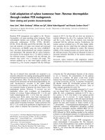
(1) Characterization of Tuberosin
The spectral data of the isolated compound for UV, IR,
1
H-NMR, and
13
C NMR (Table 1) were compared with
thedataavailableintheliteratureandbasedonthe
similarity, the isolated compound was identified as was
tuberosin (figure 1).
(2). ABTS* assay
Tuberosin scavenged the pre-generated ABTS* radicals
in concentration-dependent manner, with EC
50
values as
70 ng/ml, which was lower as compared to its mother
extract (alcoholic fraction of PT- 320 μmug/ml ). The
difference was in the range of 44.71 fold (Table 2).
(3). Superoxide scavenging assay
Tuberosin also scavenged the instantly generated super-
oxide radicals in a concentration-dependent manner
with EC
50
value at 156 μmug/ml (Table 2), which was
Pandey and Tripathi Journal of Inflammation 2010, 7:47
/>Page 3 of 8
1.5 times lo wer than it’ s alcoholic mother extract
(240 μmug/ml).
(4). Lipid Peroxidation Assay
There was significant and concentration-dependent inhi-
bition by tuberosin on FeSO
4
induced lipid peroxidation
(Table 3). Tuberosin had 7.95 fold lower EC
50
value
(98 μmug/ml) as compared to the alcoholic extract of
PT (780 μmug/ml).
(5). Non-site specific Hydroxyl radical scavenging assay
(With EDTA)
Tuberosin was found to be the more potent hydroxyl
radical scavenger with EC
50
values of (32 μmug/ml),
which was 9.6 time lower than it ’s alcoholic fraction
(EC
50
310 μmug/ml) (Table 4).
(6) Site specific Hydroxyl radical scavenging assay
(Without EDTA)
Further in the case of Site specific Hydroxyl radical
scavenging assay (without EDTA), EC
50
values of tuber-
osin was at 28 μmug/ml, which was lower than the
value obtained in case of non site specific reaction
(described above), suggesting its additional role as metal
chelation (Table 4).
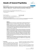
(7) Effect of tuberosin on LPS induced NO production and
iNOS-protein expression in macrophages
Tuberosin significantly inhibited LPS induced release of
nitric oxide (NO) by macrophages in concentration-
dependent manner (Table 5). It also inhibited the accu-
mulation of iNOS proteins in th e attached macrophages
(Figure 2).
Discussion
Various pure isolated phytochemicals or plant extracts
having natural cockt ail of va rious poly-phenolics, have
shown antioxidant and anti-infl ammatory property
[33,34]. They ar e also in use for the management of age
related chronic diseases such as diabetic complications
[35], atherosclerosis [36] and inflammation [37], as food
supplement or as add-on therapy with conventio nal
medicine.
The powder of PT r oot-tubers are already in clinical
use by Ayurvedic physicians of Indian system of medi-
cine [6], but neither its mechanism of action nor the
active principle for its antioxidant and anti-inflammatory
property has been explored so far. Interestingly, our data
has helped in characterizing the isolated compound as
tuberosin, which has already been reported [11], but no
biological activity related to LPS induced changes, has
been available in the literature.
Tuberosin has exhib ited direct FR trapping capacity in
a chemical reaction system, however, variability in its
potency towards various free radical species, could be
because of the difference in the electron potential of
these free radical species [38]. Further, the Fe i nduced
lipid peroxidation in presence of ascorbic acid, is an
example non-enzymatic process (Fe
++
/ascorbic acid),
therefore, the anti-lipid-peroxidative property of tub ero-
sin, described above, indicates its total antioxida nt capa-
city. As it has also shown metal chelation property
along with direct FR trapping property, therefore the net
response of inhibition towards lipid peroxidation could
be a combined effect of these 2 responses.
Table 1 Analytical data of isolated compound (Tuberosin; 5hydroxy 3,6,7,3’4’ pentamethoxy flavone)
Melting point 271-72°C
TLC pattern Solvent system: benzene:ethyl acetate (7:3)
RF value: 0.45
UV(MeOH) (log ε): 255(4.26),
274 (4.18) and
346 nm (4.21)
IR (KBr) cm
-1
3480, 1664 and 1559
1
H NMR (CDCl
3
) δ 12.62 (1 H, s, O - H), 7.75 (2 H, m, 2’ - H and 6’ - H), 7.01 (1 H, d, J = 9.0 Hz, 5’ - H), 6.51 (1 H, s, 8 - H), 3.98 (9 H, s, 3 ×
OCH
3
), 3.93 (3 H, s, OCH
3
) and 3.87 (3 H, s, OCH
3
).
13
C NMR δ 158.7 (C - 2), 132.4 (C - 3), 178.8 (C - 4), 155.7 (C-5), 138.8 (C-6), 152.7 (C-7), 90.3 (C- 8), 151.5 (C-9), 106.6 (C-10), 122.9 (C-1’),
111.7 (C-2’), 148.9 (C-3’), 152.2 (C-4’), 111.6 (C-5’), 122.1 (C-6’), 60.7 (6-OCH
3
), 56.1 (7-OCH
3
), 60.1 (3-OCH
3
), 56.2 (3’ - OCH
3
) and
55.9 (4’ - OCH
3
).
Figure 1 Structure of tuberosin.
Pandey and Tripathi Journal of Inflammation 2010, 7:47
/>Page 4 of 8
Table 2 Effect of tuberosin on pre-generated ABTS* radical and superoxide radical scavenging property
Concentration of
tuberosin (μM)
% decrease in absorbance at 734 nm (mean ± S.D.) for
ABTS* radical scavenging
% decrease in absorbance at 560 nm (mean ± S.D.) for
SO radical scavenging
50 8.42 ± 0.99* 5.78 ± 0.46**
125 24.09 ± 0.33* 18.98 ± 0.76**
200 45.16 ± 0.89* 27.89 ± 0.55**
250 60.00 ± 1.05* 54.78 ± 0.87**
375 77.25 ± 1.06* 67.11 ± 0.77**
500 93.08 ± 0.63 * 83.44 ± 0.63**
775 97.24 ± 0.89* 97.24 ± 1.22**
EC
50
198.67 μmuM 205.11 μmuM
EC
50
of Vit C- 220 μ muM, EC
50
for quercetin- 0.60 μmuM
n = 3, Level of significance: p* < 0.1 and p** < 0.001
Table 3 Inhibition of lipid peroxidation induced by FeSO
4
using egg yolk homogenates
Concentration of tuberosin (mM) Absorbance at 532 nm
(mean ± SD)
% decrease in absorbance
(mean ± SD)
Blank 0.380 –
control 0.355 –
12 0.320 ± .021* 7.08 ± 1.30
18 0.283 ± .020* 17.89 ± 1.00
25 0.248 ± .018** 27.98 ± 1.42
30 0.198 ± .015** 42.74 ± 0.96
40 0.131 ± .010** 62.05 ± 1.50
45 0.073 ± .002** 78.76 ± 1.16
50 0.049 ± .001** 85.83 ± 1.30
n=3EC
50
of tuberosin- 49.22 mM, EC
50
for quercetin- 0.60 μmuM
@
Level of significance: p* < 0.1 and p** < 0.0 01.
@
Reference 18
Table 4 Effect of tuberosin in the deoxyribose assay in the presence of EDTA (non-site specific) to assess the Hydroxyl
radical scavenging activity and absence of EDTA (site specific) to assess metal chelation property
Concentration of tuberosin (mM) Absorbance at 532 nm (mean ± S.D) % decrease in absorbance
(mean ± SD)
(Non site specific) (Site specific) (Non site specific) (Site specific)
Normal 0.310 ± .018 0.515 ± .028 ––
Blank 0.288 ± .017 0.485 ± .022 ––
0.25 0.280 ± 0.014* 0.400 ± .024* 2.60 ± 0.94 17.54 ± 1.25
0.50 0.256 ± .015* 0.356 ± .021** 11.22 ± 0.97 26.54 ± 1.15
0.75 0.235 ± .013** 0.311 ±.020** 18.35 ± 0.88 35.82 ± 0.91
1.00 0.199 ± .012** 0.271 ± .018** 30.73 ± 1.08 44.08 ± 0.96
1.25 0.156 ± .011** 0.190 ± .011** 45.94 ± 0.94 60.80 ± 0.94
1.80 0.125 ± .010** 0.119 ± .008** 56.72 ± 1.14 75.51 ± 0.99
2.50 0.092 ± .007** 0.01 ± .001** 67.94 ± 0.67 98.04 ± 0.62
n=3;EC
50
of tuberosin: Non site specific assay = 1.14 mM and site specific assay = 0.918 mM; EC
50
(μmuM) for quercetin - Non site specific assay 0.80 and site
specific assay- 0.50; Level of significance: p* < 0.1 and p** < 0.001.
Pandey and Tripathi Journal of Inflammation 2010, 7:47
/>Page 5 of 8
Tuberosine has shown lower EC
50
value on all tested
parameters than its mother alcoholic extract, which sug-
gests its higher potency, and therefore it could be co nsid-
ered as its active principle. However, it has been found to
be significantly less poten t than quercetin, which could
be because of structural difference in these two com-
pounds. It has been documented earlier that number and
position of hydroxyl groups in the flavones ring, regulates
its antioxidant potential and the presence of 3-OH makes
the compound more potent than that of 5-OH group
[39]. From the structural comparison of these 2 com-
pounds, it is clear that tuberosine has 5-OH group,
where as quercetin has 3-OH group. Thus, the higher
potency of quercetin over tuberosin could be explained.
Measurement of inhibitory property of a test compound
against LPS induced NO release is one of the standard
models to explore anti-inflammatory potential of any test
drug. LPS is known to induce iNOS through activation of
NF-kBandthisprocessinvolvesfreeradicals(FR)inits
early steps, just after interacting with its Toll-like receptor
(TLR) [40,41]. Therefore, free radical scavengers have been
reported earlier to inhibit t his process and our data has also
shown concentration-dependent inhibition of LPS induced
NO release. This trapping capacity of tuberosin, for variety
of free radical s pecies and also for metal chelation property
has been found in ou r in vitro testing on a chemical test
model. Thus, it c ould be suggested that tuberosin might be
acting on the initial steps of the signaling cascade of LPS
induced NO production, but it is still not clear, whether it
is directly inhibiting the activity of iNOS e nzyme or it is
suppressing the s ynthesis of this enzyme.
To target this question, we explored the effect of
tuberosin on i NOS protein in macrophages, when
exposed to LPS. Interestingly, our data show that tuber-
osin significantly inhibited the iNOS pro tein in western
blot analysis. The results suggested that tuberosin is
inhibiting the expression of iNOS genes, as amount of
iNOS proteins was significantly lower in tuberosin pre-
treated cells in concentration dependent manner.
Conclusion
From the above experimental results, it could be sug-
gested that tuberosin is one of the active principles of
Table 5 Effect of tuberosin on LPS induced NO production and iNOS expression by attached rat peritoneal
macrophages.
Concentration of tuberosin (ng/ml) NO production (μg/10
4
cells) Pixel value of iNOS bands in western blot
Normal Cells 10.11 ± 1.043 -
Only LPS (20 ng/ml) 39.89 ± 1.983 16023
LPS(20 ng/ml)+Tuberosin(ng/ml) -
100 38.09 ± 1.933 15878
200 36.09 ± 1.862** -
300 27.02 ± 1.698** 10678
400 21.16 ± 1.829* -
500 13.04 ± 1.904* -
600 10.09 ± 1.898* 5082
LPS + Quercetin(50 ng/ml) 9.98 ± 1.041 4223
EC
50
of tuberosin 399.68 ng/ml -
EC
50
of quercetin 190 ng/ml -
Values were significant (p* < 0.1, p** < 0.001) when compared with experimental control.
Figure 2 Effect of different concentrations of Tuberosin on LPS
induced iNOS expression in attached rat peritoneal
macrophages. The macrophages were pretreated with quercetin
and tuberosin as given below for 30 minutes and then LPS was
added (20 ng/ml) and incubated for 17 hrs. The normal cells were
exposed to 0.1% DMSO without any LPS. Lane-1: LPS(20 ng/ml),
Lane-2: Normal cells. Lane3:LPS+Quercetine(50 ng/ml), Lane4:LPS
+Tuberosine(100 ng/ml), Lane-5: LPS+Tuberosine(300 ng/ml), Lane-6:
LPS+Tuberosine(600 ng/ml). The bars depict densitometric analysis
of western blot (given in the inset). This picture represents one out
of total three experiments carried out separately.
Pandey and Tripathi Journal of Inflammation 2010, 7:47
/>Page 6 of 8
Pueraria tuberose for its claimed antioxidant property.
The tuberosine has direct scavenging potential for vari-
ety of free radicals w ith preference to ABTS* radicals
fol lowed by hydro xyl radicals and then superoxide radi-
cals. It has additional metal chelation property. Tube ro-
sin has potential to inhibit LPS induced NO production
in concentration-dependent manner, which is due to
inhibition in the expression of iNOS proteins.
Acknowledgements
Authors are thankful to Banaras Hindu University, for extending the
infrastructure and for fellowship of Ms Nidhi Pandey. We acknowledge the
help of Prof SK Upadhyay for statistical analysis, Prof SK Trigun for western
blot analysis. The financial help from an ongoing CSIR project is also
acknowledged for purchase of chemicals and glass wares.
Authors’ contributions
NP carried out the experimental works. YBT conceived of the study, and
participated in its design, discussion of results, over all coordination and
wrote the manuscript. All authors read and approved the final manuscript.
Competing interests
The authors declare that they have no competing interests.
Received: 30 June 2009 Accepted: 14 September 2010
Published: 14 September 2010
References
1. Tripathi YB, Tripathi P, Arjmandi BH: Nutraceuticals and cancer
management. Frontiers in Bioscience 2005, 10:1607-1618.
2. Winrow VR, Winyard PG, Morris CJ, Blake DR: Free radicals in inflammation:
second messengers and mediators of tissue destruction. British Medical
Bulletin 1993, 49:506-522.
3. Wilson JD, Robinson AJ, Kinghorn SA, Hicks DA: Implications of
inflammatory changes on cervical cytology. BMJ 1990, 10(Suppl
6725):638-640.
4. Gacche RN, Dholen A: Antioxidant and possible anti-inflammatory
potential of selected medicinal plants prescribed in the Indian
traditional system of medicine. Pharmaceutical biology 2006, 44:389-395.
5. Tripathi YB, Tripathi P, Korlagunta K, Chai SC, Smith BJ, Arjmandi BH: Role of
Sandhika: A Polyherbal Formulation on MC3T3-E1 Osteoblast-like Cells.
Inflammation 2008, 31(suppl 1):1-8.
6. Chopra RN, Nayar SL, Chopra IC: Glossary of Indian Medicinal Plants. CISR
New Delhi 1956, 256.
7. Pandey GS, Chunekar KC, Vidari K, (Eds): Bhav Prakash Nighantu.
ChaukambhaVidya Bhavan, Varanasi 1998, 1:388-89.
8. Ramakrishna KV, Khan RA, Kapil RS: A new isoflavone and Coumestan
from Pueraria tuberosa. Indian Journal of Chemistry, Section B: Organic
Chemistry including Medicinal Chemistry Central Drug Research Institute
Lucknow, India 1998, 27(3):285.
9. Pawan K, Khan RA, Agrawal , Kapil RS: Puetuberosanol an epoxychalcanol
from Pueraria tuberosa. Phytochemistry(Oxford) 1996, 42(1):243-244.
10. Prasad AVK, Kapil RS, Polpi SP: Structure of Pterocarponoids
anhydrotuberosin 3-O methylanhydrotuberosin and tuberostan from
Pueraria tuberosa. Indian journal of chemistry, section B, organic chemistry
including medicinal chemistry 1985, 24(3):236-239.
11. Joshi BN, Kamat VN, Govindachari TR: Structure of tuberosin, a new
pterocarpan from Pueraria tuberosa. India Indian Journal of Chemistry Ciba
Research Centre Bombay 1972, 10(11):1112-3.
12. Gupta RS, Sharma R, Choudhary R, Bhatnagar AK, Joshi YC: Antifertility
effect of Pueraria tuberosa root extract on male rats. Pharmaceutical
Biology 2004, 42(8):603-609.
13. Xiong FL, Sun XH, Gan L, Yang XL, Xu HB: Puerarin protects rat pancreatic
islets from damage by hydrogen peroxide. Eur J Pharmacol 2006,
529(1-3):1-7.
14. Ong WY, Halliwell B: Iron atheroscelosis, and neurodegeneration: a key
role for cholesterol in promoting iron- dependent oxidative damage?
Ann N Y Acad Sci 2004, 1012:51-64, (Review).
15. Halliwell B, Gutteridge JM, Blake D: Metal ions and oxygen radical
reactions in human inflammatory joint disease. Philos Trans R Soc Lond B
Biol Sci 1985, 311(1152):659-71.
16. Kroes BH, vanden Berg AJ, Quarles van Ufford HC, van Dijk, Labadie RP:
Anti-inflammatory activity of gallic acid. Planta Med 1992, 58(6):499-504.
17. Victoria García-Mediavilla Irene Crespo, Collado SPilar, Esteller Alejandro,
Sánchez-Campos Sonia, Tuñón JMaría, González-Gallego Javier: The anti-
inflammatory flavones quercetin and kaempferol cause inhibition of
inducible nitric oxide synthase, cyclooxygenase-2 and reactive C-protein,
and down-regulation of the nuclear factor kappaB pathway in Chang
Liver cells. European Journal of Pharmacology 2007, 557(2-3):221-229.
18. Verdrengh M, Jonsson IM, Holmdahl R, Tarkowski A: Genistein as an anti-
inflammatory agent. Inflamm Res 2003, 52(8):341-6.
19. Wang Y, Ho CT: Metabolism of Flavonoids Forum. Nutr 2009, , 61: 64-74.
20. Pandey Nidhi, Chaurasia JK, Tiwari OP, Tripathi Yamini B: Antioxidant
properties of different fractions of tubers from Pueraria tuberosa Linn.
Food Chemistry FOCHMS 2007, 105:219-222.
21. Re R, Pellegrini N, Proteggente A, Pannala A, Yang M, Rice-Evans C:
Antioxidant acitivity applying an improved ABTS radical cation
decolorizing assay. Free Radicals in Biology and Medicine 1999, , 26:
1231-1237.
22. Tiwari OP, Triapthi YB: Anti oxidant properties of different fractions of
Vitex negundo. Food Chemistry 2007, 100:1170-1176.
23. Ohkowa H, Ohisi N, Yagi K: Assay for lipid peroxides in animals tissue by
thiobarbituric acid reaction. Analytical Biochemistr 1979, 95:351-358.
24. Beauchamp C, Fridovich I: Superoxide dismutase: Improved assay and an
assay applicable to acrylamide gels. Analytical Biochemistry 1971,
44:276-287.
25. Halliwell B, Gutteridge MC, Aruoma OI: The deoxyribose method: a simple
test-tube assay for determination of rate constants for reactions of
hydroxyl radicals. Anal Biochem 1987, 165:215-219.
26. Satoh A, Shimosegawa T, Fujita M, Kimura K, Masamune A, Koizumi M,
Toyota T: Inhibition of nuclear factor- B activation improves the survival
of rats with taurocholate pancreatitis. GUT 1999, 44:253-258.
27. Machaiah JP, Vakil UK: Protein deficiency and age related alterations in
rat peritoneal macrophage lipids. Journal of Biosciences
1989, 14:4.
28. Pandey RS, Singh BK, Tripathi Yamini B: Effect of gum rasin of Boswellia
serrata L inhibits LPS induced NO production in rat Macrophages along
with hypolipidemic property. Indian J of Expt Biol 2005, 43:509-516.
29. Griess JP: Bemerkungen zu der Abhandlung der HH: Wesely und
Benedikt “Über einige Azoverbindungen”. Ber Deutsch Chem Ges 1879,
12:426-428.
30. Bradford MM: A rapid and sensitive for the quantitation of microgram
quantitites of protein utilizing the principle of protein-dye binding.
Analytical Biochemistry 1976, 72:248-254.
31. Laemmli : Cleavage of structural proteins during the assembly of the
head of bacteriophage T4. Nature 1970, 227(5259):680-685.
32. Towbin H, Staehelin T, Gordon J: Electrophoretic transfer of proteins from
polyacrylamide gels to nitrocellulose sheets: procedure and some
applications. Proc Natl Acad Sci USA 1979, 76(9):4350-4354.
33. Tripathi YB: BHUx: a patented poly herbal formulation to prevent
hyperlipidemia and atherosclerosis. Recent Pat Inflamm Allergy Drug Discov
2009, 3(1):49-57.
34. Gayathri B, Manjula N, Vinaykumar KS, Lakshmi BS, Balakrishnan A: Pure
compound from Boswellia serrata extract exhibits anti-inflammatory
property in human PBMCs and mouse macrophages through inhibition
of TNFa, IL-1b, NO and MAP kinases. International Immunopharmacology
2007, 7(4):473-482.
35. Carl-David A, Unne S, Ole T, Elisabet A: Effects of inhibition of glycation
and oxidative stress on the development of diabetic nephropathy in
rats. Journal of diabetes and its complications 2002, 6(16):395-400.
36. Tripathi YB, Reddy MM, Pandey RS, Subhashini J, Tiwari OP, Singh BK,
Reddanna P: Anti-inflammatory properties of BHUx, a polyherbal
formulation to prevent atherosclerosis. Inflammopharmacology 2004,
12(2):131-52.
37. Chaurasia S, Tripathi P, Tripathi YB: Antioxidant and anti-inflammatory
property of Sandhika: a compound herbal drug. Indian J Exp Biol 1995,
33(6):428-32.
Pandey and Tripathi Journal of Inflammation 2010, 7:47
/>Page 7 of 8
38. Pasha FA, Cho SJ, Beg Y, Tripathi YB: Quantum chemical QSAR study of
flavones and their radical-scavengingactivity. Medicinal Chemistry Research
2007, 16(7-9):408-417.
39. Akir E, Figen E, Nevin K: Theoretical investigation of quercetin and its
radicalisomers. Journal of Molecular Structure: THEOCHEM 2003, 631(1-
3):141-146.
40. Ikeda K, Kubo S, Hirohashi K, Kinoshita H, Kaneda K, Kawada N, Sato EF,
Inoue M: Mechanism that regulates nitric oxide production by
lipopolysaccharide-stimulated rat Kupffer cells. Physiol Chem Phys Med
NMR 1996, 28(4):239-53.
41. Je-Seong W, Yeong BI, Singh AK, Singh I: Dual role of cAMP in iNOS
expression in glial cells and macrophages is mediated by differential
regulation of p38-MAPK/ATF-2 activation and iNOS stability. Free Radical
Biology and Medicine 2004, 11(37):1834-1844.
doi:10.1186/1476-9255-7-47
Cite this article as: Pandey and Tripathi: Antioxidant activity of tuberosin
isolated from Pueraria tuberose Linn. Journal of Inflammation 2010 7:47.
Submit your next manuscript to BioMed Central
and take full advantage of:
• Convenient online submission
• Thorough peer review
• No space constraints or color figure charges
• Immediate publication on acceptance
• Inclusion in PubMed, CAS, Scopus and Google Scholar
• Research which is freely available for redistribution
Submit your manuscript at
www.biomedcentral.com/submit
Pandey and Tripathi Journal of Inflammation 2010, 7:47
/>Page 8 of 8