Báo cáo y học: " Effects of pro-inflammatory cytokines on expression of kynurenine pathway enzymes in human dermal fibroblasts" pdf
Bạn đang xem bản rút gọn của tài liệu. Xem và tải ngay bản đầy đủ của tài liệu tại đây (1.87 MB, 7 trang )
RESEARCH Open Access
Effects of pro-inflammatory cytokines on
expression of kynurenine pathway enzymes in
human dermal fibroblasts
Linnéa Asp
1
, Anne-Sofie Johansson
1
, Amandeep Mann
1
, Björn Owe-Larsson
2
, Ewa M Urbanska
3,4
, Tomasz Kocki
3
,
Magdalena Kegel
5
, Göran Engberg
5
, Gabriella BS Lundkvist
1
and Håkan Karlsson
1*
Abstract
Background: The kynurenine pathway (KP) is the main route of tryptophan degradation in the human body and
generates several neuroactive and immunomodulatory metabolites. Altered levels of KP-metabolites have been
observed in neuropsychiatric and neurodegenerative disorders as well as in patients with affective disorders. The
purpose of the present study was to investigate if skin derived human fibroblasts are useful for studies of
expression of enzymes in the KP.
Methods: Fibroblast cultures were established from cutaneous biopsies taken from the arm of consenting
volunteers. Such cultures were subsequently treated with interferon (IFN)-g 200 U/ml and/or tumor necrosis factor
(TNF)-a, 100 U/ml for 48 hours in serum-free medium. Levels of transcripts encoding different enzymes were
determined by real-time PCR and levels of kynurenic acid (KYNA) were determined by HPLC.
Results: At base-line all cultures harbored detectable levels of transcripts encoding KP enzymes, albeit with
considerable variation across individuals. Following cytokine treatment, considerable changes in many of the
transcripts investigated were observed. For example, increases in the abundance of transcripts encoding
indoleamine 2,3-dioxygenase, kynureninase or 3-hydroxyanthranilic acid oxygenase and decreases in the levels of
transcripts encoding tryptophan 2,3-dioxygenase, kynurenine aminotransferases or quinolinic acid
phosphoribosyltransferase were observed following IFN-g and TNF-a treatment. Finally, the fibroblast cultures
released detectable levels of KYNA in the cell culture medium at base-line conditions, which were increased after
IFN-g, but not TNF-a, treatments.
Conclusions: All of the investigated genes encoding KP enzymes were expressed in human fibroblasts. Expression
of many of these appeared to be regulated in response to cytokine treatment as previously reported for other cell
types. Fibroblast cultures, thus, appear to be useful for studies of disease-related abnormalities in the kynurenine
pathway of tryptophan degradation.
Keywords: human, fibroblast, kynurenine pathway, gene expression, cytokine
Introduction
The kynurenine pathway (KP) is the main route of tryp-
tophan degradation in the human body and generates
several neuroactive and immunomod ulatory metabol ites
[1,2]. KP activity has the potential to affect a range of
neurotransmitter systems in the brain including
glutamatergic, cholinergic and serotonergic transmissi on
[2-4]. Indeed, altered levels of KP-metabolites have been
observed in neuropsychiatri c and neurodegenerative dis-
orders [5-8] as well as in patients with affective disor-
ders [9-13]. While experimental studies support an
involvement of kynureni ne metabol ites in the pathogen-
esis of both psychiatric and neurodegen erative disorders
[14-20], the underlying cause of the dysregulation of
kynurenine metabolism in these disorders is not known.
* Correspondence:
1
Department of Neuroscience, Karolinska Institutet, Retzius väg 8, 171 77
Stockholm, Sweden
Full list of author information is available at the end of the article
Asp et al . Journal of Inflammation 2011, 8:25
/>© 2011 Asp et al; licensee BioMed Central Ltd. This is an Open Access article distributed under the terms of the Creative Commons
Attribution License ( which permits unrestricted use, distribution, and reproduction in
any medium, provided the original work is properly cited.
Several studies have sh own that infections activate the
KP, which thereby appear to serve both as a direct
defense mechanism and as a means of modulating the
immune response [1,21]. The enzyme indoleamine 2,3-
dioxygenase (IDO1) is the first and rate-limiting step of
this pathway and is highly induced by the pro-inflamma-
tory cytokine interferon (IFN)-g [22,23]. However, it is
not clear if pro-inflammatory cytokines affect expression
of genes encoding other enzymes o f the KP. While
human fibroblasts have previously been employed for
studying the role of IDO1 in controlling experimental
infections [24-26], expression or functionality of genes
encoding downstream enzymes in the KP have not been
investigated in such cells. Since alterations in the KP
may potentially reflect the pathophysiology of several
neuropsychiatric disorders, it is of major importance to
study the KP in primary cells obtained from humans. In
the present study, we have established human ex vivo
skin fibroblast cell cultures as a successful approach to
study the KP. We investigated if transcripts encoding
enzymes in the kynurenine pathway can be detected in
these cells and if their relativ e abundances are modu-
lated by IFN-g and/or tumor necrosis factor (TNF)-a.
Materials and methods
Tissue isolation and culture
To establish fibroblast cultures, a cutaneous biopsy was
taken from the arm of seven consenting volunteers
recruited at Karolinska University Hospital Huddinge.
Biopsies were minced and pl aced in 35 mm dishes
(Corning Incorporated, Corning NY, USA) under a ster-
ile glass cover slip and cultured in DMEM Glutamax, 10
mM HEPES, 1X MEM amino acids, 1X sodium pyruvate
supplemented with 100 U/ml penicillin, 100 μg/ml
streptomycin, 15% fetal calf serum (all from Invitrogen,
Paisley, UK), in a humidified 37°C, 5% CO
2
incubator.
The regional ethics c ommittee approved the study (04-
273/1, supplements 2006/637-32 and 2009-06-12).
Cytokine treatment
After 2 passages, cells were seeded into 6-well plates
(Corning Inc.). At confluence, cytokine treatment wa s
performed during 48 hours using human recombinant
TNF-a 100 U/ml or IFN-g 200 U/ml (PeproTech, Lon-
don, U.K.) in serum-free media, otherwise as above.
Experiments were ended by removal and freezing of the
supernatants and addition of lysis buffer to the cell
monolayer, see below.
RNA extraction and reverse transcription
Total RNA was extracted from the cells using the
RNeasy Mini kit (Qiagen, GmbH, Hilden, Germany).
The amount and purity of the RNA was assessed by
spectrophotometry using a Nanodrop ND-1000
(NanoDrop Technologies, Wilmington, DE, USA). Total
RNA (250 ng) was subsequently treated with 1 unit of
amplification grade DNase I (Invitrogen) for 15 min at
room temperature and inactivated by the addition of 2.5
mM EDTA followed by incubation at 65°C for 10 min
according to the manufacturer’ s instructions. The
DNase-treated RNA was subsequently reverse tra n-
scribed in 20 μl reactions containing the following
reagents from Invitrogen; 250 ng of Oligo(dT) primer, 1
× First Strand Buffer, 10 mM DTT and 500 μMofeach
dNTP and 100 U Superscript II. cDNA synthesis was
allowed to proceed for 1 h at 42°C before inactivation at
72°C for 10 min.
Real-time PCR and data analysis
One μl cDNA templates were added to triplicate 25 μl
reaction mixtures using Platinum SYBR Green qPCR
Supermix UDG (Invitrogen). An ABI Prism 7500 real-
time thermocycler was used (Applied Biosystems, Palo
Alto, CA, USA). Primers (Invitrogen) are provided in
Table 1. Threshold cycle (Ct) values from the exponen-
tial phase of the PCR amplification plot for each target
transcript were normalized to that encoding glyceralde-
hyd-3-phosphate dehydrogenase (GAPDH). From these
values, fold-difference s in the lev els of transcripts
Table 1 Transcripts analyzed by real-time PCR, gene
symbols and primer sequences
Target
transcript
Gene Polarity Sequence (5’®3’)
IDO1 INDO Sense GCATTTTTCAGTGTTCTTCGCATA
Anti-sense CATACACCAGACCGTCTGATAGCT
TDO TDO2 Sense GAACATCTTTTTATCATAACTCATCAAGCT
Anti-sense ACAACCTTAAGCATGTTCCTTTCAT
KMO KMO Sense TGTAATCCTCCAAGCTTCAATCTG
Anti-sense CTAGTAGATGCCCACTGAATATTTGTG
HAAO HAAO Sense GGACGTTCTGTTTGAGAAGTGGTT
Anti-sense AGCTGAAGAACTCCTGGATGATG
KAT1 CCBL1 Sense CCTGCTAAGGCTCAGGTATAACCT
Anti-sense GGACTCAAGCCTAAAGGCAACTC
KAT2 AADAT Sense CACATCTGGCAGCCAACAAG
Anti-sense CACTGGCAACATTAATAATGTTGCA
KAT3 CCBL2 Sense ACTATCAGCCATCCCCGTTTC
Anti-sense AATGAAGCAAAAACGCACAAACT
KAT4 GOT2 Sense TGTGGTGTGCAGCCTCTCAT
Anti-sense AAGCCTGAACCCAGCTAGCA
KYNU KYNU Sense ACAGGATCTGCCTCCAGTTGA
Anti-sense TGGCCCACTTATCTAGTTCTTCTTC
QPRT QPRT Sense ACACCGGCCATGGGTTAAC
Anti-sense GCCCCATTGGCCACTGA
GAPDH GAPDH Sense CACATGGCCTCCAAGGAGTAA
Anti-sense TGAGGGTCTCTCTCTTCCTCTTGT
Asp et al . Journal of Inflammation 2011, 8:25
/>Page 2 of 7
between individual untreated and treated cell cultures
were calculated according to the formula 2
-ΔΔCt
[27].
Analysis of kynurenic acid levels
Cell culture supernatants (1.0 ml) were collected and
kep t in -20°C until analysis. In order to precipitate resi-
dual protein, samples were centrifuged at 20800 g for 5
minutes and an equal volume of 0.4 M perchloric acid
was added to the supernatants. After a second centrifu-
gation 70% perchloric acid (300 μl) was added, and
thereafter the supernatants were centrifuged twice at
20800 g for 5 minutes.
Analysis of KYNA was performed using an isocratic
reversed-phase high-performance liquid chromatography
(HPLC) system, including a dual-piston, high-liquid
delivery pump (Bischoff Chromatography, Leonberg,
Germany), a ReproSil-Pur C18 column (150 × 4 mm,
Dr. Maisch GmbH, Ammerbuch, Germany) and a fluor-
escence detector (FP 2020, Jasco Ltd., Hachioji City,
Japan) with an excitation wavelength of 344 nm and an
emission wavelength of 398 nm (18 nm bandwidth). A
mobile phase of 50 mM sodium acetate (pH 6.2,
adjusted with acetic acid) and 7.0% acetonitrile was
pumped through the reversed-phase column at a flow
rate of 0.5 mL/min. Samples of 50 μL were manually
injected into a Rheodyne injector with a sample loop of
50 μl(Rheodyne,RhonertPark,CA,USA).Zincacetate
(0.5 M not pH adjusted) was delivered post column by a
peristaltic pump (P-500; Pharmacia, Uppsala, Sweden) at
a flow rate of 0.10 ml/hr. S ignals from the fluorescence
detector were transferred to a computer for analysis
with Datalys Azur software (Datalys, Grenoble, France).
The retention time of KYNA was about 7-8 minutes.
Initially, the sensitivity of the system was verified by
analysis of a standard mixture of KYNA with concentra-
tions from 1 to 30 nM, w hich resulted in a linear stan-
dard plot.
Statistics
Comparisons across treatments were done by repeated
measures ANOVA with Bonferroni’s Multiple Compari-
son Test using GraphPad (GraphPad Software, Inc., San
Diego, CA, USA).
Results
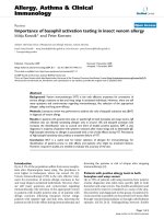
Detection of transcripts encoding KP enzymes
All the investigated kynurenine pathway transcripts
(IDO1, TDO, KAT1, KAT2, KAT3, KAT4, KMO,
KYNU, HAAO, QPRT) were detected in untreated
fibroblast cell cultures, Figure 1. The levels of expression
varied considerably across the different genes, with tran-
scripts encoding IDO1 detected at the lowest level and
those encoding KAT3 detected at the hig hest level (dif-
ference 8 × 10
3
fold). The variation across individual
cultures (n = 7 ), ranged from 2.5 (KAT3) to 145-fold
(KYNU).
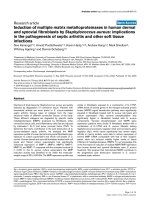
Modulation of transcript-levels by IFN-g and/or TNF-a
The potential effects of IFN-g, TNF-a, or a combin ation
of IFN-g and TNF-a on kynurenine pathway transcrip ts
were investigated in the fibroblast cell cultures, see Fig-
ure 2. The levels of transcripts encoding IDO1 were sig-
nifica ntly increased (> 10
5
-fold) in cultures treated with
IFN- g (p<0.001)aswellasIFN-g together with TNF-a
(p < 0.00 1) compared to untreated cultures although no
effect of TNF-a alone was observed (Figure 2A). Tran-
scripts encoding tryptophan 2,3-dioxygenase (TDO), on
the other hand, were significantly down-regulated in
cultures treated with a combination of IFN-g and TNF-
a (20-fold; p < 0.001) as compared to u ntrea ted cells or
cells treated with the individuals cytokines (Figure 2B).
Moreover, levels of t ranscri pts encoding the k ynurenine
aminotransferases (KATs) were either unaffected or
down-regulated by the cytokine treatments. Whereas
KAT2 was unaffected by cytokine treatment, KAT1 and
KAT3 transcript levels were reduced following treat-
ment with the com bination of IFN-g and TNF-a (2.6-
fold, p < 0.001 and 1.7-fold, p < 0.01 respectively, Figure
2C, D and 2E). Levels of transcripts encoding mitochon-
drial aspartate aminotransferase (mitAAT, i.e KAT4)
were significantly down regulated (1.5-fold) in cultures
treated with IFN-g (p < 0.05) and further decreased with
the combination of IFN-g and TNF-a (2.7-fold; p <
0.001, Figure 2F). Levels of transcripts encoding kynure-
nine 3-monooxygenase (KMO) observed in the
Figure 1 Relative levels of transcripts encoding enzymes in the
kynurenine pathway in human skin-derived fibroblasts from 7
individuals. Transcripts encoding the following enzymes were
investigated; Indoleamine 2,3-dioxygenase 1 (IDO1), Tryptophan 2,3-
dioxygenase (TDO), Kynurenine aminotransferases (KAT) 1-4,
Kynurenine 3-monooxygenase (KMO), Kynureninase (KYNU), 3-
Hydroxyanthranilic acid oxygenase (HAAO) and Quinolinic acid
phosphoribosyltransferase (QPRT).
Asp et al . Journal of Inflammation 2011, 8:25
/>Page 3 of 7
fibroblast cultures were not significantly affected by the
cytokine treatment (Figure 2G). Levels of transcripts
encoding kynureninase (KYNU) were up-regulated fol-
lowing IFN-g treatment (8-fold; p < 0.01) or with TNF-
a treatment (28-fold; p < 0.001). A further increase in
the levels of KYNU transcripts was observed with the
combination of IFN-g and TNF-a (650-fold; p < 0.001,
Figure 2H). Levels of transcripts encoding 3-
Figure 2 Relative levels of transcripts encoding enzymes in the kynurenine pathway (A-J) following treatment with IFN-g (200 U/ml),
TNF-a (100 U/ml) or the combination of these two cytokines (IFN-g+TNF-a) during 48 hrs in serum-free cell culture medium (n = 7).
Levels of all transcripts are normalized to levels observed in untreated control cells (base-line). *p < 0.05, **p < 0.01, ***p < 0.001.
Asp et al . Journal of Inflammation 2011, 8:25
/>Page 4 of 7
hydroxyanthranilate 3,4-dioxygenase (HAAO) were up
regulated only in cultures treated with the combination
of IFN-g and TNF-a (12-fold, p < 0.001, Figure 2I).
Levels of transcripts encoding quinolinate phosphoribo-
syltransferase (QPRT) were down-regulated by the com-
bination of IFN-g and TNF-a (5-fold, p < 0.001), but
unaffected by the individual cytokines (Figure 2J).
Effects on KYNA levels
To address potential functionality of the KP in these
human fibroblast cultures, we measured the accumula-
tion of KYNA, one of the end metabolites in the KP in
the supernatants. Levels of KYNA were detectable in
supernatants from untreated ex vivo fibroblast cultures
(3.4 ± 0.6 nmol/l). Significantly (p < 0.0001) higher
levels were detected in supernatants of cells treated with
IFN-g (27.2 ± 18 nmol/l) or with IFN-g and TNF-a
(39.8 ± 20.1 nmol/l) as compared to supernatants from
untreated cells. TNF-a alone did not cause a significant
increase in the accumulation of KYNA.
Discussion
We here report, for the first time, that human skin
fibroblast cultures express detectable levels of transcripts
encoding the different enzymes of the KP. Substantial
differences in the basal levels of expression across genes
and individuals were observed which are likely to be
explained by ge netic and epigenetic variation between
individual cultures. Following treatment with IFN-g,
these cultures exhibited relative increases of > 10
5
-fold
for transcripts encoding IDO1. We also found that
human skin fibroblast cultures can release KYNA, and
that this release was significantly increased following
IFN-g, but not TNF-a, treatment, indicating that at least
some of the transcriptional changes observed in
response to IFN-g are functional in these cells.
Thus, in agreement with previous reports [28,29],
human fibroblast cultu res appear to be able to increase
the rate of tryptophan degradation along the kynurenine
pathway in response to IFN-g treatment. Our present
findings support the notion that IDO1 is the major
determinant of this response in human fibroblasts, as is
alsothecaseinmanyothercelltypes,derivedboth
from the brain and from peripheral tissues [30]. For
example, Guillemin and co-workers reported increased
levels of KYNA and increased level s of transcripts
encoding IDO1 following IFN-g, but not following TNF-
a treatment of human fetal astrocytes [23]. More
recently, increased levels of KYNA and transcripts
encoding IDO1 were also observed in primary neurons
and neuroblastoma cells following IFN-g treatment [22].
While Heyes and colleagues [31] reported a small
increase in K MO activity in monocytes following IFN-g
treatment, we did not observe any significant effect on
transcripts encoding KMO following cytokine treatment.
Our observations are thus in agreement with the effects
of IFN-g observed in neuronal cells [22]. Whereas IFN-g
or TNF-a, alone or in combination, markedly increased
transcripts of KYNU and HAAO, we observed no effect
or even decreased levels of transcripts encoding KAT
enzymes b y these cytokines. Indeed there is no consen-
sus in earlier studies regardi ng the response of the KAT
enzymes to I FN-g treatment. Whereas increases in the
levels of KAT 1 and KAT 2 were observed in fetal astro-
cytes following IFN-g treatment [23], no effect on the
levels of transcripts encoding these enzymes was
observed in neuronal ce lls [22]. In neuroblastoma cells,
levels of transcripts encoding TDO were reduced by the
IFN-g treatment whereas no effects on the levels of tran-
scripts encoding KAT1, KAT2, KYNU, KMO, HAAO or
QPRT were observed [22]. Differences in transcription
of genes encoding enzymes involved in the KP in
response to IFN-g therefore most likely exist across cell
types. These differences probably also explain some of
the differences observed across cell types in their
enzyme activities and in their abilities to form kynure-
nine and quinolinic acid [31]. The physiological role of
the kynurenine pathway in skin-derived fibroblasts is
not known but may involve effects not primarily related
to acetylcholine or glutamate receptors such as effects
on cell proliferation [1], cytokine release [32] or micro-
bial growth [21,24-26,33] as described in other periph-
eral cell types.
The increases in KYNU and HAAO, and decrease in
levels of transcripts encoding QPRT, following IFN-g
and TNF-a treatment suggest that such treatment can
potentially alter the accumulation of other metabolites
generated by the KP, such as quinolinic acid. It should
also be noted that TNF-a treatment alone caused a pro-
nounced and sel ective increase (almost 30-fold) in levels
of transcripts encoding KYNU, suggesting a direct influ-
ence of TNF-a on expression of this gene. Thus, it
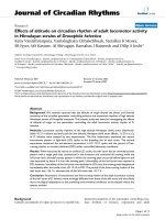
appears as i f certain cytokines can differentially affect
expression of genes in the KP, at least in fibroblasts (for
overview see Figure 3), and thereby potentially modulate
levels of individual metabolites.
Fibroblast cultures derived from patients and healthy
controls have previously been used to study a range of
CNS-diseases. For example, in fibroblasts from patients
with schizophrenia, alterations in pathways involved in
cell cycle regulation and RNA processing have been
identified [34]. Moreover, alterations in growth, mor-
phology, cell adhesion, apoptotic pathways, composition
of phospholipid fatty acids in the plasma membrane and
glutathione synthesis are reported [35-39]. Aberrant
amino acid transport has been identified in fibroblast
from patients with schizophrenia, bipolar disorder as
well as autism [40-42]. These reports suggest that
Asp et al . Journal of Inflammation 2011, 8:25
/>Page 5 of 7
peripheral tissues can be used to identify alterations at
the molecular level in patients with psychiatric disorders
and thus provide a useful method to investigate
mechanisms underlying such disorders. The advantage
of studying ex vivo cultures compared to postmortem
tissue or blood samples is that in such cultures con-
founding factors like medi cal treatments are minimized.
Furthermore, in contrast to using clinical samples, ex
vivo cell cultures can also be u sed to conduct well-con-
trolled studies of potential gene-environment interac-
tions. The present findings suggest that fibroblast
cultures can be used to study disease-related abnormal-
ities in the kynurenine pathway of t ryptophan
degradation.
Acknowledgements
The present study was supported by the Stanley Medical Research Institute,
the Swedish Research Council (HK, GSL), Fredrik och Ingrid Thurings Stiftelse
(LA, ASJ), Söderström-Königska and Wolffs stiftelser (GSL), and Swedish
Medical Society (GSL).
Author details
1
Department of Neuroscience, Karolinska Institutet, Retzius väg 8, 171 77
Stockholm, Sweden.
2
Department of Clinical Neuroscience, Karolinska
Institutet, Section of Psychiatry at Karolinska University Hospital Huddinge,
141 86 Stockholm, Sweden.
3
Department of Experimental and Clinical
Pharmacology, Medical University, Lublin, Jaczewskiego 8, 20-090 Lublin,
Poland.
4
Department of Toxicology, Institute of Agricultural Medicine, Lublin,
Jaczewskiego 2, 20-950 Lublin, Poland.
5
Department of Physiology and
Pharmacology, Karolinska Institutet, Nanna Svartz väg 2, 171 77 Stockholm,
Sweden.
Authors’ contributions
BOL performed biopsies. ASJ performed cell cultures. LA and AM carried out
the RNA analyses. MK carried out the KYNA analyses. LA performed all
statistical analyses. EMU, TK participated in the design of the study. HK, GE,
GSL and EMU conceived of the study, and participated in its design and
coordination. HK drafted the manuscript. All authors helped to revise the
first draft of the manuscript and all authors approved the final manuscript.
Competing interests
The authors declare that they have no competing interests.
Received: 6 May 2011 Accepted: 8 October 2011
Published: 8 October 2011
References
1. Moffett JR, Namboodiri MA: Tryptophan and the immune response.
Immunol Cell Biol 2003, 81(4):247-265.
2. Stone TW: Neuropharmacology of quinolinic and kynurenic acids.
Pharmacol Rev 1993, 45(3):309-379.
3. Hilmas C, Pereira EF, Alkondon M, Rassoulpour A, Schwarcz R,
Albuquerque EX: The brain metabolite kynurenic acid inhibits alpha7
nicotinic receptor activity and increases non-alpha7 nicotinic receptor
expression: physiopathological implications. J Neurosci 2001,
21(19):7463-7473.
4. Zmarowski A, Wu HQ, Brooks JM, Potter MC, Pellicciari R, Schwarcz R,
Bruno JP: Astrocyte-derived kynurenic acid modulates basal and evoked
cortical acetylcholine release. Eur J Neurosci 2009, 29(3):529-538.
5. Nilsson LK, Linderholm KR, Engberg G, Paulson L, Blennow K, Lindstrom LH,
Nordin C, Karanti A, Persson P, Erhardt S: Elevated levels of kynurenic acid
in the cerebrospinal fluid of male patients with schizophrenia. Schizophr
Res 2005, 80(2-3):315-322.
6. Schwarcz R, Rassoulpour A, Wu HQ, Medoff D, Tamminga CA, Roberts RC:
Increased cortical kynurenate content in schizophrenia. Biol Psychiatry
2001, 50(7):521-530.
7. Guidetti P, Luthi-Carter RE, Augood SJ, Schwarcz R: Neostriatal and cortical
quinolinate levels are increased in early grade Huntington’s disease.
Neurobiol Dis 2004, 17(3):455-461.
8. Erhardt S, Blennow K, Nordin C, Skogh E, Lindstrom LH, Engberg G:
Kynurenic acid levels are elevated in the cerebrospinal fluid of patients
with schizophrenia. Neurosci Lett 2001, 313(1-2):96-98.
9. Gabbay V, Klein RG, Katz Y, Mendoza S, Guttman LE, Alonso CM, Babb JS,
Hirsch GS, Liebes L: The possible role of the kynurenine pathway in
adolescent depression with melancholic features. J Child Psychol
Psychiatry 51(8):935-943.
10. Myint AM, Kim YK, Verkerk R, Scharpe S, Steinbusch H, Leonard B:
Kynurenine pathway in major depression: evidence of impaired
neuroprotection. J Affect Disord 2007, 98(1-2):143-151.
11. Myint AM, Kim YK, Verkerk R, Park SH, Scharpe S, Steinbusch HW,
Leonard BE: Tryptophan breakdown pathway in bipolar mania. J Affect
Disord 2007, 102(1-3):65-72.
12. Miller CL, Llenos IC, Dulay JR, Weis S: Upregulation of the initiating step of
the kynurenine pathway in postmortem anterior cingulate cortex from
individuals with schizophrenia and bipolar disorder. Brain Res 2006, 1073-
1074:25-37.
13. Olsson SK, Samuelsson M, Saetre P, Lindstrom L, Jonsson EG, Nordin C,
Engberg G, Erhardt S, Landen M: Elevated levels of kynurenic acid in the
cerebrospinal fluid of patients with bipolar disorder. J Psychiatry Neurosci
2010, 35(3):195-199.
14. Asp L, Holtze M, Powell SB, Karlsson H, Erhardt S: Neonatal infection with
neurotropic influenza A virus induces the kynurenine pathway in early
life and disrupts sensorimotor gating in adult Tap1-/- mice.
Int J
Neuropsychopharmacol 2009,
1-11.
15. Erhardt S, Schwieler L, Emanuelsson C, Geyer M: Endogenous kynurenic
acid disrupts prepulse inhibition. Biol Psychiatry 2004, 56(4):255-260.
Figure 3 Overview of the changes in levels of transcripts
encoding enzymes in the kynurenine pathway in human skin-
derived fibroblasts following treatment with IFN-g (white), TNF-
a (gray) or the combination of these two cytokines (black)
during 48 hrs. Squares indicate no significant change whereas
arrows indicate significant up- or down-regulation.
Asp et al . Journal of Inflammation 2011, 8:25
/>Page 6 of 7
16. Potter MC, Elmer GI, Bergeron R, Albuquerque EX, Guidetti P, Wu HQ,
Schwarcz R: Reduction of endogenous kynurenic acid formation
enhances extracellular glutamate, hippocampal plasticity, and cognitive
behavior. Neuropsychopharmacology 35(8):1734-1742.
17. Laugeray A, Launay JM, Callebert J, Surget A, Belzung C, Barone PR:
Peripheral and cerebral metabolic abnormalities of the tryptophan-
kynurenine pathway in a murine model of major depression. Behav Brain
Res 210(1):84-91.
18. Chess AC, Simoni MK, Alling TE, Bucci DJ: Elevations of endogenous
kynurenic acid produce spatial working memory deficits. Schizophr Bull
2007, 33(3):797-804.
19. Ting KK, Brew BJ, Guillemin GJ: Effect of quinolinic acid on human
astrocytes morphology and functions: implications in Alzheimer’s
disease. J Neuroinflammation 2009, 6:36.
20. Rahman A, Ting K, Cullen KM, Braidy N, Brew BJ, Guillemin GJ: The
excitotoxin quinolinic acid induces tau phosphorylation in human
neurons. PLoS One 2009, 4(7):e6344.
21. Pfefferkorn ER: Interferon gamma blocks the growth of Toxoplasma
gondii in human fibroblasts by inducing the host cells to degrade
tryptophan. Proc Natl Acad Sci USA 1984, 81(3):908-912.
22. Guillemin GJ, Cullen KM, Lim CK, Smythe GA, Garner B, Kapoor V,
Takikawa O, Brew BJ: Characterization of the kynurenine pathway in
human neurons. J Neurosci 2007, 27(47):12884-12892.
23. Guillemin GJ, Kerr SJ, Smythe GA, Smith DG, Kapoor V, Armati PJ, Croitoru J,
Brew BJ: Kynurenine pathway metabolism in human astrocytes: a
paradox for neuronal protection. J Neurochem 2001, 78(4):842-853.
24. Gupta SL, Carlin JM, Pyati P, Dai W, Pfefferkorn ER, Murphy MJ Jr:
Antiparasitic and antiproliferative effects of indoleamine 2,3-
dioxygenase enzyme expression in human fibroblasts. Infect Immun 1994,
62(6):2277-2284.
25. Dai W, Pan H, Kwok O, Dubey JP: Human indoleamine 2,3-dioxygenase
inhibits Toxoplasma gondii growth in fibroblast cells. J Interferon Res
1994, 14(6):313-317.
26. Spekker K, Czesla M, Ince V, Heseler K, Schmidt SK, Schares G, Daubener W:
Indoleamine 2,3-dioxygenase is involved in defense against Neospora
caninum in human and bovine cells. Infect Immun 2009, 77(10):4496-4501.
27. Livak KJ, Schmittgen TD: Analysis of relative gene expression data using
real-time quantitative PCR and the 2(-Delta Delta C(T)) Method. Methods
2001, 25(4):402-408.
28. Byrne GI, Lehmann LK, Kirschbaum JG, Borden EC, Lee CM, Brown RR:
Induction of tryptophan degradation in vitro and in vivo: a gamma-
interferon-stimulated activity. J Interferon Res 1986, 6(4):389-396.
29. Daubener W, MacKenzie CR: IFN-gamma activated indoleamine 2,3-
dioxygenase activity in human cells is an antiparasitic and an
antibacterial effector mechanism. Adv Exp Med Biol 1999, 467:517-524.
30. King NJ, Thomas SR: Molecules in focus: indoleamine 2,3-dioxygenase. Int
J Biochem Cell Biol 2007, 39(12):2167-2172.
31. Heyes MP, Chen CY, Major EO, Saito K: Different kynurenine pathway
enzymes limit quinolinic acid formation by various human cell types.
Biochem J 1997, 326(Pt 2):351-356.
32. Di Serio C, Cozzi A, Angeli I, Doria L, Micucci I, Pellerito S, Mirone P,
Masotti G, Moroni F, Tarantini F: Kynurenic acid inhibits the release of the
neurotrophic fibroblast growth factor (FGF)-1 and enhances proliferation
of glia cells, in vitro. Cell Mol Neurobiol 2005, 25(6):981-993.
33. Kuc D, Rahnama M, Tomaszewski T, Rzeski W, Wejksza K, Urbanik-
Sypniewska T, Parada-Turska J, Wielosz M, Turski WA: Kynurenic acid in
human saliva–does it influence oral microflora? Pharmacol Rep 2006,
58(3):393-398.
34. Wang L, Lockstone HE, Guest PC, Levin Y, Palotas A, Pietsch S, Schwarz E,
Rahmoune H, Harris LW, Ma D, Bahn S: Expression profiling of fibroblasts
identifies cell cycle abnormalities in schizophrenia. J Proteome Res 2010,
9(1):521-527.
35. Mahadik SP, Mukherjee S, Laev H, Reddy R, Schnur DB: Abnormal growth
of skin fibroblasts from schizophrenic patients. Psychiatry Res 1991,
37(3):309-320.
36. Mahadik SP, Mukherjee S, Wakade CG, Laev H, Reddy RR, Schnur DB:
Decreased adhesiveness and altered cellular distribution of fibronectin
in fibroblasts from schizophrenic patients. Psychiatry Res 1994, 53(1):87-97.
37. Catts VS, Catts SV, McGrath JJ, Feron F, McLean D, Coulson EJ, Lutze-
Mann LH: Apoptosis and schizophrenia: a pilot study based on dermal
fibroblast cell lines. Schizophr Res 2006, 84(1):20-28.
38. Gysin R, Kraftsik R, Sandell J, Bovet P, Chappuis C, Conus P, Deppen P,
Preisig M, Ruiz V, Steullet P, Tosic M, Werge T, Cuenod M, Do KQ: Impaired
glutathione synthesis in schizophrenia: convergent genetic and
functional evidence. Proc Natl Acad Sci USA 2007, 104(42):16621-16626.
39. Mahadik SP, Mukherjee S, Horrobin DF, Jenkins K, Correnti EE, Scheffer RE:
Plasma membrane phospholipid fatty acid composition of cultured skin
fibroblasts from schizophrenic patients: comparison with bipolar
patients and normal subjects. Psychiatry Res 1996, 63(2-3):133-142.
40. Persson ML, Johansson J, Vumma R, Raita J, Bjerkenstedt L, Wiesel FA,
Venizelos N: Aberrant amino acid transport in fibroblasts from patients
with bipolar disorder. Neurosci Lett 2009, 457(1):49-52.
41. Flyckt L, Venizelos N, Edman G, Bjerkenstedt L, Hagenfeldt L, Wiesel FA:
Aberrant tyrosine transport across the cell membrane in patients with
schizophrenia. Arch Gen Psychiatry 2001, 58(10):953-958.
42. Fernell E, Karagiannakis A, Edman G, Bjerkenstedt L, Wiesel FA, Venizelos N:
Aberrant amino acid transport in fibroblasts from children with autism.
Neurosci Lett 2007, 418(1):82-86.
doi:10.1186/1476-9255-8-25
Cite this article as: Asp et al.: Effects of pro-inflammatory cytokines on
expression of kynurenine pathway enzymes in human dermal
fibroblasts. Journal of Inflammation 2011 8:25.
Submit your next manuscript to BioMed Central
and take full advantage of:
• Convenient online submission
• Thorough peer review
• No space constraints or color figure charges
• Immediate publication on acceptance
• Inclusion in PubMed, CAS, Scopus and Google Scholar
• Research which is freely available for redistribution
Submit your manuscript at
www.biomedcentral.com/submit
Asp et al . Journal of Inflammation 2011, 8:25
/>Page 7 of 7