Báo cáo y học: "Parthenolide inhibits ERK and AP-1 which are dysregulated and contribute to excessive IL-8 expression and secretion in Cystic Fibrosis cells" ppsx
Bạn đang xem bản rút gọn của tài liệu. Xem và tải ngay bản đầy đủ của tài liệu tại đây (1.57 MB, 45 trang )
This Provisional PDF corresponds to the article as it appeared upon acceptance. Fully formatted
PDF and full text (HTML) versions will be made available soon.
Parthenolide inhibits ERK and AP-1 which are dysregulated and contribute to
excessive IL-8 expression and secretion in Cystic Fibrosis cells
Journal of Inflammation 2011, 8:26 doi:10.1186/1476-9255-8-26
Aicha Saadane ()
Jean Eastman ()
Melvin Berger ()
Tracey L Bonfield ()
ISSN 1476-9255
Article type Research
Submission date 13 October 2010
Acceptance date 12 October 2011
Publication date 12 October 2011
Article URL />This peer-reviewed article was published immediately upon acceptance. It can be downloaded,
printed and distributed freely for any purposes (see copyright notice below).
Articles in Journal of Inflammation are listed in PubMed and archived at PubMed Central.
For information about publishing your research in Journal of Inflammation or any BioMed Central
journal, go to
/>For information about other BioMed Central publications go to
/>Journal of Inflammation
© 2011 Saadane et al. ; licensee BioMed Central Ltd.
This is an open access article distributed under the terms of the Creative Commons Attribution License ( />which permits unrestricted use, distribution, and reproduction in any medium, provided the original work is properly cited.
1
Parthenolide inhibits ERK and AP-1 which are dysregulated and contribute to
excessive IL-8 expression and secretion in cystic fibrosis cells
Aicha Saadane†, Jean Eastman, Melvin Berger* and Tracey L. Bonfield*
Department of Pediatrics, Case Western Reserve University
11100 Euclid Avenue, BRB-822 Cleveland Ohio 44106, OH 44106
Phone: (216)368-4376. Fax: (216) 368-4223
†Corresponding author
*Co-Mentorship
Emails address:
AS:
JE:
MB:
TLB:
2
Abstract
Background: Excessive secretion of IL-8 characterizes cystic fibrosis (CF). This has
been attributed to excessive activation of epithelial cell I-κΒ Kinase and/or NFκΒ.
Maximum IL-8 production requires 3 cooperative mechanisms: 1) release of the promoter
from repression; 2) activation of transcription by NFκΒ and AP-1; 3) stabilization of
mRNA by p38-MAPK. Little is known about regulation of IL-8 by MAPKs or AP-1 in
CF.
Methods: We studied our hypothesis in vitro using 3-cellular models. Two of these
models are transformed cell lines with defective versus normal cystic fibrosis
transmembrane conductance regulator (CFTR) expression: an antisense/sense transfected
cell line and the patient derived IB3-1/S9. In the third series of studies, we studied
primary necropsy human tracheal epithelial cells treated with an inhibitor of CFTR
function. All cell lines were pretreated with parthenolide and then stimulated with TNFα
and/or IL-1β.
Results: In response to stimulation with TNFα and/or IL-1β, IL-8 production and mRNA
expression was greater in CF-type cells than in non-CF controls. This was associated with
enhanced phosphorylation of p38, ERK1/2 and JNK and increased activation of AP-1.
Since we previously showed that parthenolide inhibits excessive IL-8 production by CF
cells, we evaluated its effects on MAPK and AP-1 activation and showed that
parthenolide inhibited ERK and AP-1 activation. Using a luciferase promoter assay, our
studies showed that parthenolide decreased activation of the IL-8 promoter in CF cells
stimulated with TNFα/IL-1β.
3
Conclusions: In addition to NFκB MAPKs ERK, JNK and p38 and the transcription
factor AP-1 are also dysregulated in CF epithelial cells. Parthenolide inhibited both
NFκB and MAPK/AP-1 pathways contributing to the inhibition of IL-8 production.
4
Introduction
Cystic fibrosis (CF) is characterized by repeated and progressive airways infection,
inflammation, and obstruction. It is now clear that the immune and inflammatory
responses in CF lung are disproportionate to the threat posed by infection [1-6].
Bronchoalveolar lavage (BAL) fluid from patients with CF contains higher
concentrations of IL-8 and PMN than BAL from patients without CF but with similar
burdens of bacteria or LPS [7-9]. Considerable evidence indicates that activation of
NFκΒ is prolonged and excessive in epithelial cell lines, mice and humans with defective
CFTR expression or function [5, 6, 10-14]. NFκΒ clearly plays a key role in the
regulation of expression of pro-inflammatory cytokines, chemokines and mucins which
are important in CF. However, despite its major role; it is unlikely that NFκΒ alone
should be sufficient for maximal transcriptional activation or induction of all the genes
that are involved in this complex disease. Several studies suggest that expression of IL-8
is subject to multiple and coordinated regulation by mitogen activated protein kinases
(MAPKs) and AP-1, in addition to NFκΒ [12, 15-17]. Holtmann and colleagues have
proposed a model in which the extent of IL-8 production is the net result of 3 regulatory
mechanisms: 1) release of the promoter from repression; 2) transcriptional activation by
NFκΒ and AP-1; and 3) stabilization of mRNA by p38 MAPK [16]. IL-8 gene expression
is also regulated in part through remodeling of chromatin structure which is under the
control of various changes including histone acetylation and DNA methylation [18, 19-
20]. Recently, we showed that parthenolide, a naturally occurring sesquiterpene lactone
5
from the medicinal plant feverfew, inhibits IκΒ kinase (IKK), NFκB activation and IL-8
secretion by CF epithelial cells [14].
Very few studies focused on MAPKs activity [12, 15] but none of these showed the
prolonged activation of the MAPKs that could explain the prolonged and unresolved
inflammatory responses documented in CF patient. Moreover, to our knowledge no study
has shown excessive or prolonged activation of AP-1 in CF.
In the present study, we investigated the following two hypotheses: 1) To
determine if the signaling pathway going through the MAPKs: p38, extracellular-
regulated protein kinase (ERK), and Jun-N terminal protein kinase (JNK) to the
transcription factor activator-protein-1 (AP-1) signaling is dysregulated in CF epithelial
cells; 2) To determine whether this pathway can be manipulated by parthenolide. To
study MAPKs and AP-1 involvement in CF epithelial IL-8 production we used 3
different CF epithelial cell models and their corresponding controls. The first model is the
human bronchial epithelial cell line16 HBE, stably transfected with antisense
oligonucleotides, inhibiting the expression of CFTR (AS). The control for this cell line is
16HBE cells stably transfected with sense oligonucleotides (S). The second model uses
cells obtained from a patient with CF, this is designated IB3 and the control cell line
which has been corrected with full-length CFTR, this is designated S9 [22]. The third
model involved using human primary tracheal epithelial cells treated with CFTR inhibitor
172 (CFTR
inh
172) [21].
6
METHODS
Cell culture
We used two different sets of cell lines with CF defects and their corresponding controls
(Table 1). The first was 16 HBE human bronchial epithelial cell line stably transfected
with an anti-sense (AS) oligonucleotide which inhibits expression of CFTR; and a sister
cell line transfected with CFTR sense oligonucleotide, (S) as the control. These have
been described previously [21] and were kindly provided by Dr. Pamela Davis (Case
Western Reserve University, Cleveland). The second was IB3-1 cells from a patient with
CF, and the control cell line, S9 cells, which was rescued from CFTR deficiency by
stable transfection with full-length functional CFTR [22]. Cells were maintained in a 5%
CO
2
incubator at 37°C using MEM (Mediatech, Inc. Herndon, VA) for AS and S cell
lines and LHC-8 media (Biosource, Camarillo, CA) for IB3-1 and S9 cell lines. All
media contained penicillin/streptomycin and 10% fetal bovine serum.
We also used human tracheal epithelial cells (HTE) recovered from necropsy specimens,
as previously described [21]. Cells were grown in an air-liquid interface (ALI) on
collagen-coated, semi-permeable membrane as previously described. Cells were allowed
to differentiate for 3 to 4 weeks, then switched to liquid-liquid interface, (LLI) and
treated with either DMSO 1:1000 (vehicle control) or 20 µM CFTR
inh
172 [21]. Drugs
were added to both the apical and basolateral sides and refreshed every 24 hours. After 72
hours, cells were pretreated with 15µM parthenolide or vehicle alone for 1 hour before
treatment with TNFα at 100ng/ml for 3 h. The HTE cells were all from the same donor
specimens. Six filters were used for DMSO and six with CFTR
inh
172.
7
Table 1: Cell Lines
CF Wild Type
AS (16HBE stably transfected with an anti-
sense oligonucleotide)
S (16HBE stably transfected with CFTR
sense oligonucleotide)
IB3-1 (CF patient) S9 (CF patient-transfected with full length
CFTR)
HTE+CFTR
inh
172 (necropsy human
tracheal epithelial treated with CFTR
inhibitor 172)
HTE+DMSO (necropsy human tracheal
epithelial treated with DMSO)
Experimental conditions
Cell lines were plated at 2 x 10
6
cells/well on vitrogen-coated 6-well plates. Twenty-four
hours after plating, the cells were switched to serum-free medium for 18 h. IL-8
production was induced by treating AS and S cells with TNFα (100ng/ml, Sigma St.
Louis, MO) with or without IL-1β (100ng/ml, Sigma St. Louis, MO), and IB3-1 and S9
with TNFα at 30 ng/ml [2, 14]. Viability and possible cytotoxicity of the cytokines were
determined by trypan blue exclusion [14]. As in our previous studies, cell lines were
pretreated with 40 µM parthenolide for AS and S and 15 µM for IB3-1 and S9 (Sigma,
St. Louis, MO) [14] for 1 h before treatment with TNFα and/or IL-1β. Parthenolide was
dissolved in dimethylsulfoxide (DMSO), such that the final maximum concentration of
DMSO in the experimental media was 0.04 %. Controls contained the same concentration
of DMSO. At various times, media were harvested and assayed for IL-8 (R&D Systems,
Minneapolis, MN). Unstimulated controls were run in each experiment. After removal of
8
media, the epithelial cell monolayers were washed with ice cold PBS and harvested by
scraping, then pelleted at 600 x g for 5 min at 4°C. Separate cytosolic and nuclear
proteins extracts were prepared according to manufacturer’s instructions (BioVision
Research products, Mountain View, CA). Extracts were then used for analysis of NFκΒ
and AP-1. In another set of experiment cells were lysed using PhosphoSafe (Novagen,
Madison, WI) containing protease and phosphatase inhibitors. Protein concentrations
were determined using the Bradford method (Bio-Rad Laboratories) and data for IL-8
production were expressed as pg/mg cellular protein.
RNA isolation, reverse transcription and real-time PCR
RNA was extracted using Trizol or the RNeasy
®
Mini kit (Qiagen). For reverse
transcription, 5 µg total RNA was adjusted to 8µl. One µl of oligo (dt) (0.5 µg/µl) and 1
µl dNTP mix (10 mM each) were added. The mixtures were denatured at 65°C for 5 min
and chilled on ice. cDNA was produced by adding a mixture containing 200 mM Tris-
HCl, pH 8.4, 500 mM KCl), MgCl2, DTT, 1 µl RNase OUT, and 1 µl Superscript
TM
II
reverse transcriptase. The samples were incubated at 42°C for 50 min and at 70°C for 15
min then chilled on ice. RNase H (1 ul) was added, followed by incubation at 37°C for 20
min. The cDNA solution was subsequently diluted 20 times with water and stored at -
80°C. Primers were designed using Primer Express software (Applied Biosystems, Foster
City, CA). Transcript levels were normalized using the housekeeping gene GAPDH. We
have previously determined that GAPDH expression is constitutive in the cell lines and
was not s not altered by TNF-α or IL-1β treatment (data not shown). Samples were run in
triplicate on an ABI Prism 7700 sequence detector (Applied Biosystems, Foster City,
CA) according to the manufacturer's instructions for 40 cycles, and the average threshold
9
cycle (Ct) was determined. Changes in IL-8 mRNA levels were expressed as ∆Ct and
∆∆Ct and the expression level of IL-8 gene was represented as fold increase: 2
-∆∆CT
,
where ∆∆Ct = [∆Ct
sample stimulated
)] - [∆Ct
sample unstimulated
] and ∆Ct = [Ct
sample
]- [Ct
GAPDH
]
Electrophoretic Mobility Shift Assay (EMSA)
Aliquots of nuclear extracts from cell lines (5 µg) were suspended in binding
buffer [10 mM Tris-HCl, pH 7.5, 1 mM MgCl2, 50 mM NaCl, 0.5 mM EDTA, 0.5 mM
DTT, 4% glycerol, 0.5 µg poly (dI-dC)] at room temperature for 10 min then incubated
for an additional 20 min with
32
P-radiolabeled consensus oligonucleotides:
5’AGTTGAGGGGACTTTCCCAGGC-3’ for NFkB assays 5’-
CGCTTGATGAGTCAGCCGGAA-3’ for AP-1 (both from Promega, Madison, WI).
Protein binding of the oligonucleotides was analyzed using 6% non-denatured PAGE and
autoradiography [14]. Cold competitors were used to assure the specificity of binding of
each oligonucleotide. A Panomics Luminex based kit was also used to study NFκB and
AP-1 activation, according to manufacturer’s instructions (Panomics Fremont, CA).
Immunoblotting for MAP kinases: ERK, JNK and p38
Aliquots of cell extracts (15 µg protein) were separated by 10% SDS-PAGE and
transferred onto nitrocellulose membrane. The western blots were probed using rabbit
polyclonal antibodies specific for phosphorylated and total p38, JNK and ERK (Cell
Signaling, Inc., Beverly MA) and immunoreactive proteins were visualized by enhanced
chemiluminescence [5, 6, 14]. Blots were also probed with monoclonal anti-β-actin to
assure equal protein loading.
Transfection and luciferase promoter assays
10
AS and S cell lines were cotransfected with firefly luciferase construct dependent on the
IL-8 promoter, and a reporter plasmid mediating constitutive expression of Renilla
luciferase as a transfection control [23]. Cells were lysed for measurement of firefly and
Renilla luciferase activities using reagents from Promega as previously described. Firefly
luciferase activity was normalized by comparison to the Renilla luciferase activity in the
same sample.
Statistical Analysis
For cytokine data, 0 was assigned to values below the limit of detection of the ELISA
assays. All data are expressed as mean + SEM. Data were compared using the student’s
t-test. Comparisons between CF and wild type with/without stimulations, 95% confidence
interval, significance p ≥ 0.05. Data is product of 3 to 4 different experiments with n=4 to
8 per each time point and condition.
11
Results
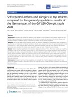
Loss of CFTR Results in high accumulation of IL-8 mRNA
CF cell lines stimulated by TNFα/IL-1β secreted much greater concentrations of IL-8
than similarly stimulated non-CF cells. We previously reported that this was associated
with increased and persistent activation of IKK and NFκB in the AS cells as compared to
S cells [2, 14]. In this study, we determined the expression of IL-8 mRNA in AS/S and
IB3-1/S9 cell lines in response to IL-1β and/or TNFα by real time PCR. The mRNA
contents were normalized for GAPDH and the results are given in relative units.
Following treatment with TNFα/IL-1β, IL-8 mRNA was significantly higher in AS cells
compared to S cells (Fig. 1A). At 3 h post stimulation, and in accordance with the high
production of IL-8 protein (Figure 1 inset), IL-8 mRNA was increased by more than 85
fold in AS cells (p=0.003), while in S cells the increase was only 33 fold (Fig. 1A) when
compared to their non-stimulated baseline controls. At 6 h after stimulation, IL-8 mRNA
was still elevated by 56 fold in the AS cells (p=0.0002), as compared to 26-fold in the S
cells when compared to their non-stimulated baseline controls. Similarly IB3-1 cells also
increased IL-8 mRNA accumulation much more than S9 cells (Fig.1B). At 3 h post
stimulation IL-8 mRNA was increased by more than 1500 fold in IB3-1 cells (p=0.0007)
while in S9 cells the increase was only 400 fold, when compared to their non-stimulated
baseline controls. At 6 h after stimulation IL-8 mRNA was still elevated by 600 fold in
the IB3-1 cells as compared to 350 fold in the S9 cells compared to baseline controls.
12
TNF-α and/or IL-1β
ββ
β causes increased activation of AP-1 in CF as compared to
control cells
Recently, there have been major advances in understanding how different signaling
pathways coordinately regulate IL-8 transcription and mRNA stabilization in response to
external stimuli. Besides its binding sites for NFκB, the IL-8 promoter also contains
binding sites for activator protein-1 (AP-1), which is not essential for baseline expression
but is required for maximal expression of IL-8 [16, 17, 24, 25]. To understand how the
two transcription factors might contribute to the tremendous increase in IL-8 protein
secretion by CF epithelial cells, we studied the activation of AP-1 as well as NFκB. As
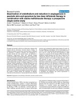
reported previously, and shown here as a positive control, unstimulated AS and S cells
have little detectable active nuclear NFκB (Figure 2A, time 0). However, stimulation
with TNFα/IL-1β increased binding of NFκB by 15 min in both cell types (Figure 2A,
2B). In AS cells, the increase in NFκB activation was significantly higher at 30 and 60
min (p=0.001 and p=0.007, respectively). The same phenomenon was observed with the
IB3 and S9 model. TNFα stimulation significantly increased NFκB binding at 30
minutes (Figure 2B). However, the NFκB levels of activation were comparable between
the IB3 (CF cells) and S9 (controls) at 60 minute, potentially related to the different
origins of the cell lines. The AS/S and IB3/S9 models from the same studies were also
evaluated for AP-1 activation post-TNFα stimulation. Baseline AS/S and IB3/S9 cells
had little or no nuclear AP-1 (Figure 2C and 2D, time 0). Incubation with TNFα/IL-1β
increased AP-1 activation significantly in both AS (p=0.002) and IB3 (p=0.05) cell lines
by 30 min Similarly, the elevated levels of AP-1 in AS cells were sustained out to 180
minutes consistent with the NFκB studies, with the IB3 comparable with the S9 cells
13
(Figures 2C and 2D). These data suggest that both NFκB and AP-1 activation is
dysregulated in CF epithelial cells, potentially contributing to the excessive IL-8 message
expression and protein secretion observed under these conditions.
TNFα- and/or IL-1β
ββ
β-induces over-activation of p38, ERK and JNK in cells with
defective CFTR expression
Because AP-1 is activated by mitogen-activated protein kinases (MAPKs) [17, 26, 27],
we sought to determine whether activation of these enzymes might be associated with
AP-1 activation in the CF cells. MAPK Phospho-p38 , JUN-N terminal protein kinase
(JNK), and the extracellular-regulated protein kinase (ERK) cascades are active by
phosphorylation through dual specificity MAPK kinases (MKK) and may all contribute
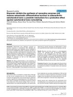
to IL-8 expression. We found no difference in baseline ERK-phosphorylation between b
AS and S cell lines (time 0, Figure 3A). TNFα/IL-1β stimulation of AS cells resulted in
ERK phosphorylation at all time points. ERK phosphorylation was significantly
increased in AS cells at 30 and 180 min (p=0.05)) (Figure 3B, solid bars), while it was
markedly reduced by 30 min and barely detectable at 60 and 180 min in the S cells
(Figure 3B, open bars). Figure 3A shows marked increases in phosphorylated p38 at in
the AS cells after 15 and 30 minutes post-stimulation with TNFα/IL-1β, which was only
modest in the S cells. A similar pattern was observed for the phosphorylation of the p54
and p46 isoform of JNK (Figures 3A and C). Phospho-p54 was significantly increased in
AS cells at 15 and 30 min (p=0.04 and p=0.01, respectively) (Figure 3C, solid pars).
Phospho-p46 was also significantly high in AS cells at 15 and 30 min (p=0.04 and
p=0.01, respectively) as compared to control S cells (Figure 3C, hatched bars). In all
cases TNFα/IL-1β stimulation did not alter the total amounts of these proteins (data not
14
shown) suggesting that the treatment change activity of the MAPKs rather than the
abundance of the proteins. In comparison to AS cells, all 3 MAPKs were significantly
elevated in IB3-1 cells. Figure 3D shows that ERK phosphorylation is very significant at
the baseline in IB3-1 cells (time 0, p=0.01) compared to S9 cells. TNFα stimulation
significantly increased phospho-ERK in IB3-1 at 30 min (p=0.04) (Figure 3D, solid bars).
Phospho-p38 was significantly increased in TNFα stimulated IB3-1 cells at 15 and 30
min (Figure 3E, p=0.003 and p=0.01, respectively) (Figure 3E, solid bars). Figure 3F,
shows a significant elevation in phospho-JNK at all-time point in IB3-1 cells at 15, 30
and 60 min (p=0.00004, p=0.01 and p=0.05 respectively) compared to control S9 cells.
Parthenolide inhibits TNFα/IL-1β-induced activation/nuclear translocation of AP-1.
Parthenolide is an anti-inflammatory drug thought to be able to suppress inflammation
through inhibiting NFκB, amongst other effects [14]. The observed differences in
MAPKs activity opened new questions about the mechanisms of parthenolide’s effects in
CF cells. Therefore, we sought to determine if the inhibitory activities of parthenolide
also extended to the MAPK-AP-1 pathway. AS and S cells were pretreated with
parthenolide then stimulated with TNFα/IL-1β as in Figure 1 and Figure 2. Nuclear
extracts were prepared and DNA-binding activity of AP-1 was measured by EMSA. For
comparison, NFκB was also assayed. The inhibition of NFκB activation by parthenolide
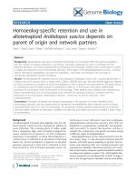
was similar to what we reported previously [14]. Parthenolide pretreatment inhibits
significantly NFκB activation in AS cells at 15, 30 and 60 min (p=0.003, p=0.003 and
p=0.004, respectively) (Fig 4A and 4B). Pretreatment with parthenolide also inhibits
significantly AP-1 activation in AS at 30 min (p=0.04) (Figure 4C and 4D).
15
Densitometry of the parthenolide inhibitor effect on NFκB and AP-1 activity in IB3-cells
with (white bars) and without (solid bars) parthenolide is shown (Figures 4E and 4F).
Parthenolide inhibited ERK1/2 and JNK phosphorylation and stabilized p38
phosphorylation
We hypothesized that parthenolide’s inhibition of AP-1 activation might be due to
inhibition of the upstream MAPKs which activate this transcription factor. Because the
p38 MAPK pathway is also believed to regulate a specific, post-transcriptional step in IL-
8 mRNA processing, we focused on determining if parthenolide alters p38-
phosphorylation [16, 17]. Since MAPKs ERK and JNK activate AP-1, we also evaluated
the effect of parthenolide on their activation. Figures 5A and 5B show that pretreatment
with parthenolide inhibited phosphorylation and activation of ERK 1 and 2 at 15 and 30
min and the inhibition was significant at 30 min (p=0.05). In contrast, parthenolide
stabilized the phosphorylation of JNK, especially p46 at 30 and 60 min (p=0.01 and
p=0.006, respectively) (Figure 5C and 5D, open bars), and the phosphorylated of JNK-
p54 was not significantly different after parthenolide pretreatment (data not shown).
Also, pre-treatment with parthenolide significantly stabilized phosphorylated p38 at 60
min (p=0.02) (Figure 5E and 5F). Parthenolide pretreatment did not alter the total amount
of all three proteins in the cells (data not shown).
In IB3-1 cells stimulated with TNFα alone, parthenolide pretreatment has the same effect
as in AS cells stimulated with TNFα/IL-1β. Parthenolide pretreatment significantly
inhibited phosphorylation and activation of ERK 1 and 2 at 30 min (p=0.04) (Figure 5G
and 5H, open bars), however pre-treatment with parthenolide stabilized phosphorylated
16
phospho-JNK at 30 min (p=0.006) (Figure 5I and 5J, open bars), and also stabilized the
phosphorylated p38 at all time points 15, 30 and 60 min after TNFα stimulation
(p=0.0001, p=0.05 and p=0.004, respectively) (Figure 5K and 5L, open bars).
Effects of parthenolide on IL-8 mRNA
Because phosphorylated p38 has been reported to stabilize IL-8 mRNA, we wanted to
determine if that mechanism was operating concurrently with the inhibition of net IL-8
secretion caused by parthenolide. Therefore, we evaluated the effect of the drug on IL-8
mRNA expression as determined by RT-PCR. ELISAs were performed with supernatants
from the same cells used for the real time PCR experiments, to confirm that parthenolide
in fact inhibited IL-8 production during each experiment. The results verified that
parthenolide inhibited IL-8 protein production in both AS and S cells (Data not shown).
Parthenolide pretreatment significantly increased the content of IL-8 mRNA in TNFα/IL-
1β stimulated AS at 3 and 6 h (p=0.001 and p=0.00009, respectively) (Figure 6A)
compared to cells treated with vehicle alone (placebo, solid bars). In contrast,
parthenolide treatment did not significantly increase mRNA in S cells (Figure 6A, inset).
These data suggest that parthenolide treatment results in stabilization of that mRNA
which is transcribed. The increased mRNA in the face of decreased protein production
suggests an additional inhibitory effect at a translational level. In IB3-1 cells stimulated
with TNFα alone, parthenolide pretreatment leads to the inhibition of TNFα-induced IL-8
mRNA accumulation (Figure 6B), whereas in control S9 cells it did not significantly
increase IL-8 mRNA accumulation (Figure 6B, inset).
17
Primary HTE cells rendered “CF like” with 20µM CFTR
inh
172 (21) were also evaluated.
Figure 6C showed that IL-8 mRNA accumulation in response to TNFα alone is not
significantly different between parthenolide and DMSO pretreated unstimulated HTE
cells (5.6±1.69 vs. 4.54±1.88 respectively), whereas Parthenolide significantly inhibits
the TNFα- induced increase in IL-8 secretion (p=0.04) (Fig 6C, inset). In a separate
experiment AS cells were pretreated with parthenolide, then one hour later stimulated
with TNFα alone or IL-1β/TNFα together. Parthenolide significantly increased IL-8
mRNA accumulation in AS cells stimulated with IL-1β/TNFα, whereas the parthenolide
effects was not observed when cells were stimulated with TNFα alone (Figure 6D). In all
cases, parthenolide significantly inhibited IL-8 protein (data not shown).
Analysis of IL-8 promoter activity after pretreatment by parthenolide and
stimulation with both TNFα/IL-1β.
Because the observation of increased IL-8 mRNA in AS cells seems paradoxical in the
face of inhibition of activation of the transcription factors AP-1 and NFκB, which are
considered the most important regulators of IL-8 gene expression, we sought to directly
investigate the influence of parthenolide on transcription independently of possible
effects on IL-8 mRNA stability or translation. AS and S cell lines were transfected with a
plasmid driving expression of the firefly luciferase reporter under control of the IL-8
promoter [23]. Firefly luciferase activity was normalized against a constitutively
expressed Renilla luciferase reporter. AS and S cell lines were pretreated with
parthenolide or DMSO, then one hour later stimulated with TNFα/IL-1β for 3 h.
Supernatants were collected and assessed by ELISA for IL-8 production and the cells
18
were assessed for IL-8 promoter activity. IL-8 promoter activity responded to TNFα/IL-
1β with 1.5-2 fold increases in luciferase expression at 3 and 6 hrs respectively in AS
cells (Figure 7). Parthenolide inhibited this stimulated firefly luciferase expression as
well as the basal expression without stimulation (Figure 7), confirming inhibition of
transcription. ELISAs verified that parthenolide inhibited the production of IL-8 protein
in response to TNFα/IL-1β (data not shown).
19
Discussion
Human bronchial epithelial cell lines with defective CFTR expression and/or
function have exaggerated IL-8 mRNA expression and protein secretion in response to
stimulation with TNF-α and IL-1β [7]. This result is in agreement with most other studies
of IL-8 mRNA in CF cells and tissues [13, 18, 28, 29]. We and others have previously
shown that this is associated with increased and prolonged activation of IKK and NFκB
in CF as compared to non-CF cells [10, 13, 14, 30]. The present results confirm that
inflammatory cytokine stimulation causes a huge increase in IL-8 production and gene
expression in CF compared with control cells. Furthermore, we show that the increased
IL-8 production is also associated with: 1) excessive and prolonged activation of AP-1 in
addition to NFκB, and 2) excessive and prolonged activation of the MAP Kinases p38,
JNK and ERK. Thus, the MAPKs/AP-1 signaling pathways as well as the IKK/NFκB
pathway shows exaggerated and prolonged activation after stimulation of cells with CF
defects. Both pathways likely contribute to the excessive amount of IL-8 mRNA and
excessive protein secretion which characterizes CF.
IL-8 is a potent chemokine which attracts neutrophils to the lung. An excess of
this chemokine is believed to make an important contribution to the excessive influx of
neutrophils that ultimately causes lung damage and death in CF patients. A large body of
evidence has shown that the increased IL-8 secretion in CF is associated with excessive
activation of IΚK and NFκB [10, 12-14, 30]. However, little is known about IL-8
regulation in the context of mitogen-activated protein kinases (MAPKs) and the
transcription factor, activator protein-1 (AP-1) in CF. Three MAPK pathways are
believed to contribute to IL-8 gene expression, the extracellular-regulated protein kinase
20
(ERK), JUN-N-terminal protein kinase (JNK), and p38 MAPK [21]. To our knowledge
there are few studies that report a higher ERK activation in CF cells [15], and no study
about the MAPK p38 or the transcription factor AP-1. Blau and colleagues however did
not demonstrate a prolonged activation of ERK, which we believe to be of major interest
in CF disease. We believe that our paper for the first time showed that all 3 MAPKs and
AP-1 are dysregulated and their activation is excessive and prolonged in CF. All of these
are activated by phosphorylation through dual-specificity MAP kinase kinases, also
referred to as MKK [16, 17, 31]. Holtmann and colleagues found that introduction of an
activating mutation in MKK6, increased p38 and, stabilized IL-8 mRNA and further
increased IL-8 protein formation induced by NFκB-inducing kinase (NIK) [16]. The
active form of the MAPK-activated protein kinase 2 (MK-2), a downstream substrate of
the p38 MAPK pathway, also induced mRNA stabilization. Negative mutants of MK-2
decreased mRNA stability [16, 32]. This data suggest that IL-1β and TNFα can induce
IL-8 production through simultaneous activation of MAPK cascades that regulate AP-1
in addition to the IKK pathway, which activates NFκB [17]. Unlike NFκB, AP-1 is not
essential for transcription of IL-8 but it is required for maximal gene expression [16, 17,
24]. Further cooperation of these two pathways resulting in maximal IL-8 secretion may
be due to activation of IKK by MAPK kinase kinase (also called MEKK1), which also
activates the three MAPKs [29]. In addition, NFκB and AP-1 modulate each other and
can function cooperatively [18, 29, 28, 33] for rapid and maximum transcriptional
activation. However, their activities are also rapidly down-regulated in normal cells so
that rigorous responses are often transient.
21
Our results clearly show that in epithelial cells with CF defect, stimulation with TNFα
alone or TNFα/IL-1β results in marked increases in the amounts of phospho-p38,
phospho-ERK, and phospho-JNK p46/p54 as compared to control cells. The time
intervals at which the phosphorylated intermediates of the different MAPKs differ from
each other, but for all three, phosphorylated forms are present for longer intervals after
stimulation in “CF-Like” cells than in similarly stimulated “Wild-Type-Like”cells. These
results correlate with, and likely account for the increased AP-1 observed at all time
intervals after stimulation in CF cells as compared to non-CF cells. Thus, the CF defect
seems to have similar effects on the MAPK/AP-1 pathway as on the IKK/NFκB pathway.
The persistent phosphorylation of all 3 MAPKs; ERK, JNK and p38 in cells with
defective CFTR, suggest that CF alters a phosphatase whose decreased activity could
explain the persistence of these enzymes. Together, simultaneous and prolonged
activation of these two transcription factors is very likely to play a major role in the
excessive and prolonged IL-8 production seen in CF cells and patients.
Parthenolide, a sesquiterpene lactone found in the medicinal plant feverfew
(Tanacetum parthenium) has powerful anti-inflammatory properties [14, 34, 35]. We
have previously showed that parthenolide inhibits inflammation in CF both in vivo and in
vitro by inhibiting IKK/NFκB pathway. Parthenolide has been considered by some to be
a specific inhibitor of the NFκB pathway [34-39]. During these studies, we wished to
determine the effects of this inhibitor on the MAPKs and AP-1 pathway, because of the
similar reactions and likely cross-talk between the two pathways which lead to formation
of AP-1 and activation of NFκB, respectively. In agreement with previous results, we
found that parthenolide inhibited secretion of IL-8 protein by CF as well as non-CF cells.
22
Our new results show that parthenolide inhibited ERK phosphorylation induced by
TNFα/IL-1β stimulation, and also inhibited AP-1 activation; but that phosphorylated p38
was preserved by this inhibitor. Since phosphorylated p38 can play an important role in
stabilization of the mRNAs for pro-inflammatory cytokines, we sought to dissect the
possible effects of parthenolide on transcriptional activation of the IL-8 promoter versus
its net effects on IL-8 mRNA in cells stimulated with TNFα/IL-1β. Results using a
luciferase reporter construct show that parthenolide inhibited transcription per se.
However, using real time PCR, we found that parthenolide pretreatment, actually
increased IL-8 mRNA in TNFα/IL-1β stimulated cells. This is most likely a consequence
of the preservation of phosphorylated p38 caused by the drug. [16, 32] since phospho-p38
is known to play an important role in modulating the effect of the AU rich sequences that
would otherwise destabilize pro-inflammatory cytokine mRNAs. In turn, the
preservation of phospho-p38 in the face of decreased phosphorylated ERK suggests the
possibility that parthenolide acts on a phosphatase which specifically targets p38 [40, 41].
The results also suggest that parthenolide inhibits translation of the IL-8 message,
accounting for the apparent paradox of finding increased mRNA in the face of decreased
protein secretion.
The differences between cell lines is probably due to the sources, however across each
model parthenolide pretreatment results in an inhibition of inflammatory signaling in
cells with CF defects.
Production of IL-8 and other pro-inflammatory cytokines is also governed by
mechanisms regulating in their mRNA half-lives. Several reports have shown that IL-8
mRNA degradation is modulated by AU-rich sequences present in the 3’UTR of IL-8
23
mRNA [23, 28, 42]. These sequences function as potent destabilizing elements that cause
rapid decay of the transcripts. Furthermore, a recently identified family of genes called
micro-inhibitory RNAs (miRNAs) has been shown to regulate protein expression mainly
at the post-transcriptional level [43], and has been shown to be dysregulated in several
inflammatory diseases including psoriasis, rheumatoid arthritis and asthma [44, 45]. The
miRNA/mRNA interaction occurs within the 3’UTR of the target mRNA and decreases
protein translation. Since our results suggest that parthenolide also inhibits translation
(see below), we speculate that this may involve stabilization of these inhibitory miRNAs,
which in turn inhibits IL-8 mRNA translation. This hypothesis would best explain our
results, which showed decreased IL-8 protein secretion despite increased message
concomitantly with stabilization of phospho-p38.
Overall, the results in three different cellular models of CF show remarkable
similarity in the ways that multiple different pathways of transcription factor signaling
are affected by the CF defect. In response to physiologic stimulation, phosphorylation
and activation of multiple MAPKs is greater and persists for longer in CF airway
epithelial cells as compared to non-CF airway epithelial cells. The termination of the
activities of MAPKs and IKK is normally insured by dual specificity phosphatases
(DUSP), also called MAP kinase phosphatase (MKP) [46, 48].
Our data showing increased activity of multiple kinase pathways may therefore suggest
that decreased expression or function of CFTR somehow decreases activity of a multi-
specific protein phosphatase [49]. Although this particular study has focused only on one
important pro-inflammatory cytokine, IL-8, it is likely that many other genes are also
over-expressed in CF as a consequence of the dysregulation of these signaling pathways.
24
Conclusions
Our results suggest that 1) in addition to IKK/NFκB pathway, MAPKs and AP-1
are also dysregulated in CF epithelial cells, and 2) the effects of a widely used inhibitor,
parthenolide, are less specific than previously believed. However, the results also suggest
that this type of compound, which simultaneously targets multiple signaling pathways,
may have beneficial effects in CF and other diseases in which multiple mechanisms that
should control inflammation are disrupted.
Abbreviations
CF: Cystic fibrosis. MAPKs: Mitogen activated protein kinases. HBEC: Human
bronchial epithelial cells. ERK: Extracellular-regulated protein kinase. JNK: Jun-N
terminal protein kinase. AP-1: Activator-protein-1. 16 HBE: human bronchial epithelial
cell line. AS: 16HBE stably transfected with antisense oligonucleotides which inhibit
expression of CFTR. IB3-1: Genetically CF Clinical sample. CFTR-Inhibitor 172:
CFTR
inh
-172
Competing interests
The authors declare that they have no competing interests.
Authors’ contributions
MB and AS participated in the conception and design of the study, TLB contributed to
the design and process of the review in the later phases of experimentation. MB, TLB
and EJ reviewed the article for important intellectual content and clarity. AS conducted
the literature search, acquisition of data analysis, interpretation of data, wrote the first