Bone Regeneration and Repair - part 3 doc
Bạn đang xem bản rút gọn của tài liệu. Xem và tải ngay bản đầy đủ của tài liệu tại đây (1.35 MB, 41 trang )
70 Bruder and Scaduto
66-kDa homolog of mammalian osteopontin (10). As the vasculature penetrates the first collar of
bone formed along the diaphysis, phagocytic cells remove the hypertrophic cartilage core and allow
its replacement by stromal and hematopoietic marrow elements. In this way, the cartilage core pre-
cisely defines the geometric boundaries of the eventual marrow cavity.
CELL LINEAGE AND THE ORIGIN OF OSTEOBLASTS
Differentiation of the totipotent zygote into developmentally restricted pluripotent stem cell popu-
lations, and the subsequent commitment and expression of specific cellular phenotypes, is believed
to be regulated by a variety of factors including inherent genomic potential, cell–cell interactions,
and environmental cues. In considering the mechanisms involved, the concept of cell lineage is fun-
damentally relevant. Our logic, in part, is based on the cellular relationships proven to exist in the
hematopoietic lineage pathways. As it is generally understood, the term lineage refers to the progres-
sion of particular cell precursors as they mature and give rise to differentiated cells, tissues, and organs.
The accurate description of such a cell lineage depends on the ability to identify a particular feature,
or collection of features, which can be traced from the differentiated cell type back through its pre-
cursors. Because the formation of specialized tissues is a progressive phenomenon, many generations
of cells lie between the stem cell and the fully differentiated phenotype, which forms the mature tissue.
Our hypothesis was that a discrete series of steps, or transitions, exists between osteoprogenitor
cells and the fully expressive osteoblast, comparable to that documented during hematopoiesis (11)
or development of the nematode Caenorhabditus elegans (12). Analysis of these lineage steps is, para-
doxically, both facilitated and complicated by the variety of tissues containing osteoblast progeni-
tors. Embryonic limb bud mesenchyme, developing and mature periosteum, and bone marrow all
contain these precursors. In addition, calvarial tissue, which is derived from neural crest, possesses a
repository of progenitor cells. Fortunately, experimental systems for analyzing each of these tissues
have been developed. In addition to dynamic studies of limb development in situ, conditions to demon-
strate differentiation of osteoblasts from isolated periosteum in vitro (13,14) and in vivo (15,16) have
been established. For example, organ culture of folded chick calvarial periosteum has become a use-
ful model for studies of bone cell differentiation. In addition, inoculating marrow cell suspensions
into diffusion chambers and implanting these chambers into athymic mouse hosts served to provide
the first evidence for osteochondral progenitors in bone marrow (17,18). While host-derived cells are
prevented from entering the chamber, nutrients and growth factors may pass freely through its pores.
In this setting, bone forms along the inner surface of the porous membrane, adjacent to external vas-
culature, and cartilage forms within the center of the chamber.
Although these anatomically distinct sources of progenitor cells all give rise to bone, the precise
sequence of cellular transitions that occurs during maturation has not been appreciated fully. That is,
do marrow-derived and periosteal osteoblasts proceed through the same developmental pathway? Does
embryonic limb bud mesenchyme give rise to osteoblasts through a different sequence than ectodermal
neural crest? And finally, how do these cellular transitions compare between embryonic and adult
sources of osteoprogenitors, both in vivo and in vitro? As a basis for answering these questions, one
must first understand that many developing eukaryotic systems have been studied using biochemical
and immunological techniques aimed at demonstrating alterations in the surface architecture of cells
as a function of stage-specific morphologies, activities, and requirements. In recent years, investigators
have used monoclonal antibody technology to generate probes that detect these alterations. This is
especially clear in the study of hematopoiesis, which now boasts over 160 cell-surface cluster designa-
tion (CD) antigens. These probes also provide the means by which to purify antigens or cells, and in
some cases, determine the function of the molecules for which they select. As an extension of this suc-
cessful logic, we sought to generate a battery of monoclonal antibody probes selective for surface anti-
gens on osteogenic cells at various stages of differentiation.
This is trial version
www.adultpdf.com
Cell Therapy for Bone Regeneration 71
THE GENERATION OF MONOCLONAL
ANTIBODIES AGAINST OSTEOGENIC CELL SURFACES
We immunized mice with a heterogeneous population of chick embryonic bone cells obtained from
the first bony collar formed in the tibia, and subsequently generated and selected for monoclonal anti-
bodies against osteogenic cell surface determinants. Supernatants from growing hybridomas were defin-
itively screened immunohistochemically against frozen sections of developing tibiae. Four unique cell
lines were cloned, referred to as SB-1, SB-2, SB-3, and SB-5, each of which reacts with a distinct set
of cells in the developing bone (19–22). Detailed morphologic analyses of the dynamic changes dur-
ing bone histogenesis document the restricted expression of specific antigens during embryogenesis.
Progenitor cells in the stacked cell layer are not stained by any of these antibodies; however, a broad
layer of cells between the surface of newly formed bone and the overlying inner cambium layer react
with SB-1 (Fig. 2C,D). By contrast, SB-3 and SB-2 appear sequentially during the maturation of cells
as they begin to secrete osteoid matrix and initiate mineralization. As a subset of these cells begins to
surround themselves with bone matrix, the SB-1 and SB-3 antigens are lost. The resulting SB-2-posi-
tive cells then express the SB-5 antigen, which is restricted to nascent and mature osteocytes. The sub-
sequent loss of SB-2 reactivity and the formation of characteristic stellate processes that react with SB-5
and extend through the bone matrix define this terminal differentiation step (Fig. 2E,F). By carefully
tracking the reactivity of discrete cell populations, these experiments not only establish the existence of
an osteoblastic lineage, but also indicate that osteocytes are derived directly from cells expressing the
SB-1, -2, and -3 antigens.
A natural progression of this effort was to identify the antigens recognized by these antibodies. One
antibody, SB-1, was observed to react with a family of cells in bone, liver, kidney, and intestine that
were identically stained by the histochemical substrate for alkaline phosphatase (APase) (20). Partial
purification of intestinal or bone APase on a Sepharose CL-6B column results in the co-elution of enzyme
activity and high-affinity antibody-binding material. Western immunoblots of bone extract show that
SB-1 reacts with a single approx 155-kDa band, which also is stained in the sodium dodecyl sulfate
(SDS)-polyacrylamide gel by APase substrate. In a similar set of immunoblot experiments, SB-1 reacts
with an intestinal APase isoenzyme whose molecular weight is approx 185 kDa. The reactive epitope
was found to be stable to SDS denaturation, not associated with the active site of the enzyme, and
dependent on disulfide bonds that impart secondary structure to the protein (23). Efforts to identify
the antigens recognized by the other antibodies have met with only limited success. Preliminary immu-
noblot data indicate that SB-5 reacts with an approx 37-kDa protein extracted from freshly isolated
osteocyte membranes; however, neither we nor Nijweide and colleagues (5,24) have yet identified a
specific antigen present on avian osteocytes. Nevertheless, it is important to emphasize that the iden-
tity of the antigens need not be known in order for these probes to remain as useful markers for char-
acterizing the lineage of osteogenic cells.
OSTEOPROGENITOR CELLS
FROM ISOLATED PERIOSTEUM AND BONE MARROW
Unlike traditional culture methods using collagenase-liberated osteoblastic cells, calvarial peri-
osteal explants form a mineralized bone tissue in 4–6 d that is virtually identical to the in vivo coun-
terpart (14). Examination of fresh explants confirmed that no mature osteoblastic cells were present,
although a discontinuous layer of SB-1-reactive preosteoblasts was evident in some regions. The inner
(cambial) surface of the tissue was folded onto itself, and the explant was then cultured at the air–
fluid interface in the presence of dexamethasone, a synthetic glucocorticoid capable of stimulating
osteoprogenitor cell differentiation. As the wave of differentiation swept through the cultured tissue,
antibody SB-1 reacted with the surface of a large family of cells associated with the developing bone.
SB-3 and SB-2 reacted with progressively smaller subsets of cells, namely, those in successively closer
This is trial version
www.adultpdf.com
72 Bruder and Scaduto
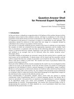
Fig. 3. Expression of osteogenic cell surface antigens in a 4-d-old periosteal culture. A H&E-stained section
from one end of the tissue fold is illustrated in (A). Bone matrix (b) containing osteocytes is evident, as is the
fibrous tissue (f) in the outer region of the explant. A broad band of cells are reactive with antibody SB-1 (B),
while a restricted population of cells reacts with SB-3 (C). A further subset of the SB-3-reactive cells is stained
by SB-2 (D), along with some cells that were recently encased in bone matrix (arrows). Morphologically recog-
nizable osteocytes are stained with SB-5 (E). Bar, 25 µm.
physical association with the newly formed and mineralizing bone (Fig. 3). Since the early events of
osteogenesis are extended over a 4-d period in this culture system, folded periosteal explants provide an
exaggerated model useful for the study of individual lineage steps. Specifically, this system allows
further dissection of the transitory stages associated with sequential acquisition of the SB-3 and SB-2
This is trial version
www.adultpdf.com
Cell Therapy for Bone Regeneration 73
antigens. Furthermore, the relatively high cellularity of the bone matrix accentuates the brief stage of
SB-2 and SB-5 co-expression prior to terminal differentiation of SB-5-positive osteocytes (25). Addi-
tional studies document that in the absence of β-glycerophosphate, which is necessary for mineral-
ization in vitro, the SB-5 antigen is not expressed despite the normal morphological appearance of
osteocytes in the developing bone (26,27). These experiments support the conclusion that expression
of the SB-5 antigen is an inducible process, is associated with bone mineralization, and that such min-
eralization is obligatory to the terminal differentiation of osteogenic cells.
The emergence of osteogenic cell-surface molecules by avian marrow–derived osteochondral pro-
genitors was similarly evaluated in diffusion chamber cultures in vivo. Fresh marrow cells from young
chick tibiae were implanted intraperitoneally in athymic mice and harvested at multiple time points
out to 60 d. Although first noted in other species (17,18,28,29), these marrow-derived avian cells also
gave rise to bone and cartilage within the chambers (30). Type I collagen was observed adjacent to the
inner surface of the membrane, and type II collagen was elaborated by chondrogenic cells in the cen-
tral portion of the chamber, where access to vascular-derived nutrients and cues was relatively reduced
(Fig. 4). Immunostaining with SB-1 revealed the expression APase-positive cells 12 d after implanta-
tion. As development progressed, the staining intensity and number of SB-1-positive cells increased.
By 20 d after implantation, antibodies SB-3 and SB-2 were observed to react with cells associated
with the developing bone. Finally, cells within the type I collagen matrix reacted with the osteocyte-
specific antibody SB-5 (Fig. 4). The morphology of these cells, with their slender pseudopodia-like
processes entering matrix-free canaliculi, is identical to that seen in embryonic chick bone and peri-
osteal explant cultures.
THE FIRST OSTEOGENIC CELL LINEAGE MODEL
The above investigations led to the creation of a lineage paradigm presented diagrammatically in
Fig. 5. The key aspects of this model describe the differentiation of APase-positive preosteoblasts
from undifferentiated progenitor cells. These preosteoblasts undergo a series of transitory osteoblast
stages, defined in part by their sequential SB-3 and SB-2 immunoreactivity, before becoming secre-
tory osteoblasts. A fraction of these cells surround themselves in matrix as SB-2/SB-5-positive osteo-
cytic osteoblasts, and terminal differentiation into an osteocyte is characterized by loss of the SB-2
antigen. That osteocytes are derived directly from secretory osteoblastic cells is now clear; however,
whether incorporation of cells into the matrix is a random event or specifically programmed to a sub-
set of cells is not yet known. Importantly, these molecular probes document that the cellular transi-
tions of the osteogenic lineage are shared by embryonic limb bud mesenchyme, by periosteal cells
from the long bone or calvarium, and by postnatal stromal cells from the marrow.
With such a lineage in mind, we have used the antibodies to isolate and purify cells at key stages
along their pathway. We employed antibody-coated magnetic bead techniques, as well as complement-
mediated cell lysis, to purify preosteoblastic populations and follow their subsequent expression of
mature phenotypic markers in vitro (31). We have also used fluorescent-activated cell sorting (FACS)
to isolate SB-5-positive osteocytes for further in vitro characterization (32). In addition, collaborators
have used these probes to establish statistical models for evaluating the effect of various hormones on
cells at specific lineage stages (33–35). Finally, these antibodies have been used to describe the differ-
entiation of scleral ossicles in the avian eye (7,36), and during osteogenesis of isolated periosteal cells
in diffusion chambers (16), on tissue culture plastic (13), and in subcutaneous implantations in athymic
mice (15).
IDENTIFICATION OF HUMAN OSTEOBLASTIC PROGENITORS
Studies of animal bone marrow–mediated osteogenesis in diffusion chambers (17,18) and ectopic
implants (37–39) served as the foundation for isolating analogous progenitor cells from humans.
Haynesworth and his colleagues first reported the isolation, cultivation, and characterization of human
This is trial version
www.adultpdf.com
74 Bruder and Scaduto
marrow–derived progenitor cells with osteochondral potential (40,41). By loading small porous hydroxy-
apatite/tricalcium phosphate (HA/TCP) cubes (3 mm per side) with culture-expanded cells, and implant-
ing the construct into athymic mice, Haynesworth demonstrated that bone and cartilage would form in
the pores of the ceramic. These cells are now known as mesenchymal stem cells (MSCs) (42), because
they have a high replicative capacity and give rise to multiple mesenchymal tissue types including
bone, cartilage, tendon, muscle, fat, and marrow stroma (43–48). We have extended these observations
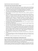
Fig. 4. (1) Toluidine-blue staining of membranous bone (B) and hyaline cartilage (C) in a diffusion chamber
inoculated with fresh chick marrow and intraperitoneally incubated for 21 d. Bone is formed predominantly along
the inner face of the membrane filter (M). (2) Type I collagen immunofluorescence shows reactivity within the
bone, and type II collagen immunofluorescence (3) resides exclusively within the cartilage. (4) Von Kossa-stained
bone (B) along the inner surface of the membrane is filled with SB-5-positive osteocytes in this 40-d sample
(5), while adjacent polygonal osteoblasts are stained by SB-1 along their surface (6). Note that SB-1 and SB-5
staining is mutually exclusive. Magnification in 1–3, ↔125. Magnification in 4–6, ↔300.
This is trial version
www.adultpdf.com
Cell Therapy for Bone Regeneration 75
to provide a detailed analysis of the surface molecules that characterize culture-expanded human
MSCs (Table 1) (49). This work stems from our effort to document the changes that occur in cell-sur-
face architecture as a function of lineage progression. The profile of cell adhesion molecules, growth
factor and cytokine receptors, and miscellaneous antigens serves to establish the unique phenotype
of these cells, and provides a basis for exploring the function of selected molecules during osteogenic,
and other, lineage progression.
Because MSCs are understood to be the source of osteoblastic cells during the processes of normal
bone growth, remodeling, and fracture repair in humans (1,4,6), we have used them as a model to study
aspects of osteogenic differentiation. When cultivated in the presence of osteogenic supplements (OSs)
(dexamethasone, ascorbic acid, and β-glycerophosphate), purified MSCs undergo a developmental
cascade defined by the acquisition of cuboidal osteoblastic morphology, transient induction of APase
activity, and deposition of a hydroxyapatite-mineralized extracellular matrix (50,51) (Fig. 6A–C).
Gene expression studies illustrate that APase is transiently increased, type I collagen is downregulated
during the late phase of osteogenesis, and osteopontin is upregulated at the late phase (49) (Fig. 6D).
Similarly, bone sialoprotein and osteocalcin (51) are upregulated late in the differentiation cascade,
while osteonectin is constitutively expressed. Additional studies detail the growth kinetics and high
replicative capacity of these cells, which do not lose their osteogenic potential following a 1 billion-
fold expansion and/or cryopreservation (52,53). We have documented that OS-treated MSCs secrete
a small-molecular-weight osteoinductive factor into their conditioned medium, which is capable of
stimulating osteogenesis in naïve cultures (54), similar to that reported for rat marrow stromal cells
directed into the osteogenic lineage (55). We have completed a comprehensive series of pulse-chase
and transient exposure experiments using dexamethasone to determine which steps of the lineage path-
way are dependent on exogenous factors, and which are supported by either (1) paracrine/autocrine fac-
Fig. 5. Diagrammatic representation of discrete cell stages that comprise the avian osteogenic cell lineage.
This is trial version
www.adultpdf.com
76 Bruder and Scaduto
tors in culture or (2) sustained lineage progression events following brief exposure to dexamethasone
(56,57). A diagrammatic representation of these results is presented in Fig. 6E.
Additional collaborations have led to insights regarding the role of BMP receptors and downstream
signaling events in osteogenesis (58–60). Recent studies of the MAP and JUN kinase signal transduc-
tion pathways establish pivotal roles for these family members in the differential commitment of human
MSCs to either the osteogenic or adipogenic lineage (61,62). Finally, studies using two-dimensional
electrophoresis of culture samples at specific time points have led to the identification of molecules,
such as α-B crystalline, that are differentially regulated during osteogenesis (63,64).
MONOCLONAL ANTIBODIES AGAINST HUMAN OSTEOGENIC CELLS
As part of characterizing the dynamic events of the differentiation process, we have generated a
number of monoclonal antibodies that react specifically with the surface of human cells during dis-
crete stages of osteogenesis. As was the case for avian-specific antibodies SB-1 through SB-5, new
probes known as SB-10, SB-20, and SB-21 have been used to localize MSCs and their progeny during
development of the fetal human skeleton (65,66). Antibody SB-10 recognizes a family of osteopro-
genitor cells present exclusively in the outer stacked cell region of the periosteum, while SB-20 and
SB-21 react with a subset of inner cambium cells expressing APase on their surface (Fig. 7). By track-
ing bone-related markers during the developmental process, we have refined our understanding of the
specific lineage transitions in osteogenesis. These data serve as a basis for our belief that sequential
acquisition and loss of specific surface molecules can be used to define positions of individual cells
within the osteogenic lineage (Fig. 8).
The antigen recognized by one of these antibodies, SB-10, was identified following its immuno-
purification from MSC plasma membrane preparations. Western blots initially demonstrated a single
approx 99-kDa-reactive band (67), which upon immunoprecipitation, purification, and peptide frag-
ment sequencing, was determined to be a cell-surface molecule known as ALCAM (68), a member of
the immunoglobulin superfamily of cell adhesion molecules (69) (Fig. 9A–C). Molecular cloning of
a full-length cDNA from a human MSC expression library confirmed nucleotide sequence identity with
ALCAM (Activated Leukocyte Cell Adhesion Molecule), and allowed us to discover homologs present
Table 1
The Cell Surface Molecular Profile of Human MSCs
Molecules present Molecules absent
Integrins
α1, α2, α3, α5, α6, αv, β1, β3, β4 β2, α4, αL
Growth factor and cytokine receptors
bFGFR, PDGFR, IL-1R, IL-3R, IL-4R, IL-6R, IL-7R, IFN-γR, EGFR-3, IL-2R
TNFIR and TNFIIR, TGFβIR and TGFβIIR
Cell adhesion molecules
ICAM-1 and -2, VCAM-1, L-selectin, LFA-3, ALCAM ICAM-3, cadherin-5, E-selectin,
P-selectin, PECAM-1
Miscellaneous antigens
Transferrin receptor, CD9, Thy-1, SH-2, SH-3, SH-4, SB-20, SB-21 CD4, CD14, CD34, CD45,
von Willebrand factor
bFGFR = basic fibroblast growth factor receptor; PDGFR = platelet-derived growth factor receptor; IL-#R = inter-
leukin-# receptor; IFN-γR = interferon gamma-receptor; TNFIR = tumor necrosis factor I receptor; TNFIIR = tumor
necrosis factor II receptor; TGFβIR = transforming growth factor beta I receptor; TGFβIIR = transforming growth
factor beta II receptor; EGFR-3 = epidermal growth factor receptor 3; ICAM = intercellular adhesion molecule; VCAM
= vascular cell adhesion molecule; LFA-3 = lymphocyte function-related antigen-3; ALCAM = activated leukocyte
cell adhesion molecule; PECAM = platelet endothelial cell adhesion molecule; CD = cluster designation.
This is trial version
www.adultpdf.com
Cell Therapy for Bone Regeneration 77
in rat, rabbit, and canine MSCs (68) (Fig. 9D–F). The addition of antibody SB-10 F
ab
fragments to MSCs
undergoing osteogenic differentiation in vitro accelerated the process, thereby implicating a role for
ALCAM during bone morphogenesis and including ALCAM in the group of cell adhesion molecules
involved in osteogenesis. Together, these results provide evidence that ALCAM plays a critical role
in the differentiation of mesenchymal tissues in multiple species across the phylogenetic tree.
Fig. 6. Osteogenic differentiation of human MSCs in vitro. Phase-contrast photomicrographs of: (A) human
MSC cultures under growth conditions display characteristic spindle-shaped morphology and uniform distribu-
tion (unstained ↔18); (B) human MSC cultures grown in the presence of osteoinductive supplements (OS) for
16 d and stained for APase and mineralized matrix. APase staining appears gray in these micrographs (originally
red) and mineralized matrix appears dark (APase and von Kossa histochemistry, ↔45). (C) Mean APase activity
and calcium deposition of MSC cultures grown in control or OS medium and harvested on d 3, 7, 13, and 16 (n =
3). The vertical bars indicate standard deviations. *p < 0.05, p < 0.005 (compared to control). (D) Expression of
characteristic osteoblast mRNAs during in vitro osteogenesis. Reverse transcriptase-polymerase chain reactions
using oligonucleotide primers specific for selected bone-related proteins were performed with RNA isolated at the
indicated times. (E) Diagrammatic representation of the stages of dexamethasone-induced osteogenic differentia-
tion of MSCs in vitro.
This is trial version
www.adultpdf.com
78 Bruder and Scaduto
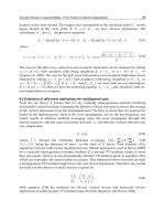
Fig. 7. Reactivity of antibodies SB-10 and SB-20 in longitudinal sections of developing human limbs. (A)
A 55-d embryonic tibia illustrates the cartilaginous core that is surrounded by a primary collar of diaphyseal
bone and a rudimentary periosteum. (Mallory-Heidenhain, ↔30). (B) High-power view of the periosteum, first
layer of bone, and underlying cartilage stained histochemically for APase (red). While the inner cambium
layer of the periosteum is intensely stained, the outer stacked cell layer (arrowheads) is free of APase activity.
This is trial version
www.adultpdf.com
Cell Therapy for Bone Regeneration 79
Fig. 7. (Continued) Phase-contrast (C) and SB-10 immunostaining (D) of a serial section to that presented
in Panel B show that the outer stacked cell layer is strongly reactive with SB-10, while the inner APase-positive
layer is negative. Panels B and D represent reciprocal staining patterns with regard to the periosteum. (Original
magnification in B–D ↔150.) (E) Phase-contrast image of the mid-diaphysis of a 62-d tibia histochemically
stained for APase activity. The stacked cell layer (arrowheads) is negative, while the inner cambium layer and
isolated chondrocytes are positive. (F) The section in panel E was also stained by antibody SB-20. Double
exposure demonstrates selected osteoblastic cells stained by SB-20 (yellow), which are a subset of the APase-
positive (red) cells in the periosteum. The stacked cell layer is not immunoreactive with SB-20. (Original magnif-
ication ↔150.) (G, H) At high power, cell surface staining on a subset of cells within the inner periosteum is appar-
ent in this 62-d embryonic femur (original magnification in E–H ↔300). (Color illustration in insert following
p. 212.)
DEVELOPMENTAL BIOLOGY APPLIED TO CLINICAL THERAPY
We have extended our basic science investigations to examine not only the role that cells of the
osteogenic lineage play in normal bone homeostasis, but also the therapeutic potential of these cells
in clinical situations requiring bone repair or bone augmentation. While autologous cancellous bone
is the current gold standard for bone grafting, a variety of problems are associated with its acquisition
including donor site morbidity, loss of function, and a limited supply (70,71). These problems have
inspired the development of alternative strategies for the repair of clinically significant bone defects.
Some of these tactics have tried to mimic portions of the natural biological sequence that occur fol-
lowing a fracture. This cascade, however, is composed of a complex series of steps including inflam-
mation, chemotaxis of progenitor cells (MSCs) to the injured site, local proliferation of MSCs to form
a repair blastema, and eventually differentiation of these cells into bone or cartilage, depending on the
mechanical stability of the site. Our approach has been to develop techniques that directly provide the
cellular machinery, namely MSCs, to the site in need of bone augmentation (1,3,49). This approach
can circumvent the early steps of bone repair, and may be particularly attractive for patients who have
fractures that are difficult to heal, or patients who have a decline in their MSC repository as a result
of age, osteoporosis, or other metabolic derangement (72–77).
Our initial efforts to design cell-based implants focused on the evaluation of a variety of delivery
vehicles. We have used a standard rat femoral gap model (78,79) to screen myriad cell-matrix combi-
nations thus far. Selection of the ideal carrier for repair of such local defects is based on several criteria:
(1) the material should foster uniform cell loading and retention; (2) the scaffold should support rapid
vascular invasion; (3) the matrix should be designed to orient the formation of new bone in anatom-
ically relevant shapes; (4) the composition of materials should be resorbed and replaced by new bone as
it is formed; (5) the material should be radiolucent to allow the new bone to be distinguished radiograph-
ically from the original implant; (6) the formulation should encourage osteoconduction with host bone;
and (7) it should possess desirable handling properties for the specific clinical indication (1,3,49).
PRECLINICAL ANIMAL MODELS OF BONE REGENERATION
One of the original preclinical studies showed that culture-expanded, syngeneic rat MSCs loaded
onto a porous HA/TCP cylinder are able to regenerate bone in a critical-sized segmental femoral defect
(80). In samples loaded with MSCs, bone formed rapidly throughout the biomatrix as a result of the
osteoblastic differentiation of the implanted cells (Fig. 10A,B). Quantitative histologic assessment of
these MSC-loaded implants demonstrated that as early as 4 wk postoperatively, bone had filled 20%
of the available pore space of the biomatrix, and by 8 wk, over 40% bone fill was achieved (Table 2).
Cell-free samples did not exceed 10% fill (osteoconduction), and even samples loaded with fresh mar-
row derived from one entire femur were not significantly better at 17% fill. Our results compare favor-
ably with other approaches described in the literature, and are strikingly better than those reported for
purified BMP in the same HA/TCP carrier (81).
This is trial version
www.adultpdf.com
80 Bruder and Scaduto
To determine the ability of purified human MSCs to heal a clinically significant bone defect, cul-
ture-expanded cells were loaded onto a HA/TCP cylinder and implanted into a segmental defect in
the femur of adult athymic (HSD:Rh-rnu/rnu) rats (82,83). Healing of bone defects was compared on
the basis of high-resolution radiography, immunohistochemistry, quantitative histomorphometry, and
biomechanical testing. The percentage bone fill with human MSCs in this study was equivalent to that
seen in euthymic rats who received syngeneic MSCs (Table 2). Immunohistochemical evaluation using
antibodies to distinguish human cells from rat cells demonstrated that tissue within the pores of the
implant during the early phase of repair was derived from donor (human) MSCs. The biomechanical
data demonstrate that torsional strength and stiffness, as measured through the implant and adjoining
diaphyseal shaft at 12 wk, were approx 40% that of intact control limbs, which is more than twice that
observed with the cell-free carrier (Fig. 10D). This result also compares favorably with the mechanical
outcome achieved in a similar study of bone repair in a primate long bone defect model, where auto-
genous bone produced only 23% of the strength of intact contralateral limbs 20 wk after implantation
(84). These studies confirm that purified, culture-expanded human MSCs can be used to regenerate
bone in a clinically significant osseous defect.
Subsequent investigations focused on advancing this technology into large animal models, and
developing prototype procedures for shipping marrow, MSCs, and autologous implants to and from
Fig. 8. Comprehensive description of the osteogenic cell lineage. Expression of selected cell surface and
extracellular matrix molecules, reported by various investigators using either monoclonal or polyclonal anti-
bodies on sections of developing bone, was used to generate this model. The beginning of each arrow reflects
the stage of differentiation when expression is first detected, while the arrowhead notes the point when expres-
sion is no longer detected. The data presented in this figure represent a collection of studies performed on
several species, including chick, pig, rat, and human. The dashed line used for antibodies SB-20 and SB-21
indicates that only a subset of cells within these stages of differentiation is immunoreactive. See original refer-
ences for additional details. 1, ref. 40; 2, ref. 104; 3, ref. 20; 4, ref. 23; 5, ref. 105; 6, ref. 106; 7, ref. 107; 8, ref.
108; 9, ref. 109; 10, ref. 110; 11, ref. 25; 12, ref. 111; 13, ref. 112; 14, ref. 113; 15, ref. 114; 16, ref. 65.
This is trial version
www.adultpdf.com
Cell Therapy for Bone Regeneration 81
Fig. 9. Identification of the SB-10 surface antigen. (A) The SB-10 antigen was immunoprecipitated, and excised from a polyacrylamide gel for lysine C-
endoproteinase digestion. (B) Recovered peptides were separated by reverse-phase high-performance liquid chromatography (HPLC). Collected peaks referred
to as K1 through K8 were subjected to N-terminal sequence analysis and found to correspond to ALCAM. (C) Control digest of a blank piece of polyacry-
lamide excised from the same gel. (D) Polymerase chain reaction (PCR) amplification of an ALCAM-specific fragment in cultured human MSCs, fetal limb,
and other tissues known to express ALCAM. (E) PCR amplification of ALCAM fragments in cultures of human, rat, rabbit, and canine MSCs. (F) Northern
blot analysis of human MSCs shows a single mRNA species approximately 6.1 kb in size, while animal ALCAM has an approximate mRNA size of 5.8 kb.
81
This is trial version
www.adultpdf.com
82 Bruder and Scaduto
distant clinical sites. Using a standardized strategy for the isolation of marrow-derived MSCs (85),
we identified conditions for effective cultivation and in vivo osteogenic differentiation of canine cells
(86). We then established a critical-sized femoral gap defect model to determine the efficacy of MSC-
based bone regeneration therapy in large dogs (87). As was done in the rodent studies, a ceramic cylin-
der was used to deliver autologous MSCs back to the site of a 21-mm-long osteoperiosteal segmental
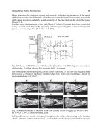
Fig. 10. MSC-mediated bone regeneration in preclinical animal studies of segmental femoral defect repair. (A)
Rat defects fitted with a MSC-loaded HA/TCP carrier form a solid osseous union with the host, and contain
substantial new bone throughout the pores by 8 wk. (B) Defects fitted with a cell-free HA/TCP carrier do not
contain bone within the pores of the implant, nor is there significant union at the interfaces, noted by the arrow-
heads. (Toluidine blue-O, ↔16.) (C) Radiographic appearance of bone healing in a 21-mm canine femoral gap
defect. Animals that did not receive an implant established a fibrous nonunion by 16 wk. Animals that received
an MSC-loaded HA/TCP cylinder regenerated a substantial amount of bone at the defect site, including a peri-
implant callus that remodeled to the size of the original bone by 16 wk. Those animals receiving cell-free implants
did not successfully heal their defects, as noted by the lack of new bone and the multiple fractures throughout the
body of the implant material. (D) Graphic results of biomechanical torsion testing performed on athymic rat
femora 12 wk following implantation with human MSC-loaded ceramics. (*p < 0.05 compared to carrier alone.)
This is trial version
www.adultpdf.com
Cell Therapy for Bone Regeneration 83
femoral defect, which was stabilized by a stainless-steel internal fixation plate with bicortical screws.
Radiographic (Fig. 10C) and histological evidence reveal an impressive periimplant callus of bone, as
well as bone throughout the pores of the entire implant by 16 wk (88,89). We attribute the formation
of this large callus to the combined action of cells delivered on the surface of the ceramic material and
the secretion of osteoinductive factors by these cells during the process of differentiation (54). Such
combined osteogenic and osteoinductive activity serve to create a mass of new bone that is derived
from the implanted cells, as well as host-derived cells that are competent to respond to secreted bone
morphogens. Importantly, none of the empty defects healed, and those animals receiving cell-free cer-
amics did not possess any periimplant callus or bone in the center of the implant region. Table 2 dem-
onstrates the similarity of bone fill between the canine studies outlined here and the previous efforts
using rat or human MSCs in rodent hosts.
PRECLINICAL ANIMAL MODELS
OF BONE MARROW-BASED BONE REGENERATION
Culture expansion of MSCs can provide an abundant supply of osteogenic cells for repair and defect
healing, but the steps necessary for expansion, and the delay between harvest and implantation are chal-
lenging to integrate into a clinical setting. An intraoperative technique that eliminates the steps of
culture expansion but provides an enriched population of osteoprogenitor cells to the graft site may
be effective in many clinical conditions.
Osteoprogenitor cells present in bone marrow are obtained by simple aspiration. We initially focused
on optimizing the osteogenic capacity of fresh, intraoperatively manipulated bone marrow. Employ-
ing our standard rat femoral defect model, we evaluated a variety of matrix carriers including ceramics,
synthetic polymers, and natural polymers. When bone marrow was combined with a porcine-derived
gelatin product (Gelfoam Upjohn, Kalamazoo, MI) and peripheral blood, the femoral defects healed
successfully; however, such defects did not heal when the same amount of marrow was implanted on
a synthetic matrix, or when a reduced amount of marrow was delivered using Gelfoam (see Fig. 11
for details). When using a similar combination of fresh bone marrow with Gelfoam in a large animal
model of bone repair, excellent results were observed in several animals, though the uniformity of
the outcome was not ideal—only six of nine animals had a solid bony bridge spanning the defect (90).
This line of investigation also highlights two important issues: (1) that there are non-MSC compo-
nents in the marrow that are important to the healing response; and (2) that the delivery matrix is criti-
cal to eventual success. We conclude this based on the observation that even the large number of purified
MSCs required to heal a long bone defect on HA/TCP is not capable of healing the defect when deliv-
ered on Gelfoam. However, successful healing is observed on Gelfoam when as little as 500 times
fewer MSCs are delivered in conjunction with other endogenous marrow-derived cells and factors.
We refer to these other non-MSC, marrow-derived cells as accessory cells. Whether accessory cell
function is paracrine in nature or mediated by cell-to-cell contact remains to be evaluated.
Table 2
Quantitative Histomorphometry of Bone Fill as a Percentage
of Available Space in Selected Models of Segmental Bone Defect Repair
Canines Athymic rats Fischer rats
(implanted with (implanted with (implanted with
Implant type autologous MSCs) human MSCs) syngeneic MSCs)
Cell-free HA/TCP 24.0 ± 15.5 29.5 ± 8.9 10.4 ± 2.4
MSC-loaded HA/TCP 39.9 ± 6.1* 46.6 ± 14.8* 43.2 ± 7.7*
*Indicates p < 0.05 compared to cell-free controls.
This is trial version
www.adultpdf.com
84 Bruder and Scaduto
Osteoprogenitors constitute significantly less than 1% of the nucleated cells in the marrow of a
healthy adult (41,53). Because these are the cells that go on to synthesize new bone, one possible way
to improve the efficacy of a bone marrow aspirate is by concentrating the endogenous osteoprogenitor
cells (91). Using simple centrifugation of fresh whole marrow, Connolly reported successful treatment
of 18 of 20 tibial nonunions via percutaneous injection of bone marrow concentrate with and without
intramedullary nailing (92).
Recent work by several investigators has focused on developing a means to intraoperatively con-
centrate osteoprogenitor cells while optimizing their clinical delivery and local retention. Ideally, this
process would combine cells participating in bone formation with a directly implantable substrate
that enhances their activity. Bone marrow cells have been shown to possess a high affinity for certain
solid substrates. For example, osteoprogenitor cells are selectively retained when marrow is filtered
through specific porous configurations of calcium phosphate or bone matrix. Using demineralized
bone matrix to capture osteoprogenitors cells, and then implant the composite graft directly, Takigami
et al. reproducibly obtained spine fusion in a canine model (93). The results of the cell-enriched graft
were significantly better than allograft alone or allograft mixed with whole marrow. Kapur and col-
leagues (94) have similarly shown, in a canine long bone defect model, the beneficial effect of selec-
tive retention on graft performance (see Fig. 12). In this study, the bone grafts were created using a
matrix consisting of a mixture of allogeneic demineralized bone fibers and undemineralized cancel-
lous bone chips (DBM-CC). The experimental group contained grafts prepared by flowing marrow
through the matrix under controlled conditions that selectively retain the osteoprogenitors. As part of
Fig. 11. Fresh marrow-based bone regeneration in preclinical animal studies of segmental femoral defect
repair. As in Fig. 10, rat segmental defects were fitted with various implants containing either culture-expanded,
purified MSCs, or fresh marrow obtained from 1–4 diaphyseal segments of syngeneic femora. MSCs on a HA/
TCP cylinder reproducibly heal defects, and such implants contain approximately 2000 times the number of
MSCs when compared to the same volume of fresh marrow from one diaphyseal segment. Purified MSCs deliv-
ered on a porcine gelatin sponge (Gelfoam) exhibit no healing, but when the same carrier is used to deliver whole
marrow from four femoral segments, solid bone bridging ensues. A dose–response effect is observed when mar-
row from only one femur is delivered in Gelfoam; these animals show modest bone formation. Animals implanted
with synthetic polylactic acid (PLA) carriers and marrow from four femora similarly showed poor bone forma-
tion. Together, these data provide insight into the importance of proper carriers, proper cellular dosing, and the
benefit of accessory cells in fresh marrow that reduce the need for large numbers of purified MSCs.
This is trial version
www.adultpdf.com
Cell Therapy for Bone Regeneration 85
the final step of graft preparation, the concentrated osteoprogenitor-graft was clotted together with
autologous platelet-rich plasma (PRP) (Con Osteoprogenitor-PRP). The control groups consisted of an
iliac crest bone graft, allogeneic DBM-CC mixture alone, or the DBM-CC mixture loaded with whole
marrow (DBM-CC-Marrow). The rate and incidence of union was assessed by radiographic analysis,
including plain films every 4 wk and CT scan upon sacrifice at 16 wk. In the Autograft and Con-Osteo-
progenitor-PRP groups, fusion was achieved in all animals (Fig. 12). In contrast, when the allogeneic
DBM-CC mixture was used alone or in combination with native bone marrow, there was an unsatis-
factory healing response, with approximately half of the canines going onto fusion.
Although the above results are promising, maximizing cell capture and concentration does not
necessarily guarantee optimal conditions for bone formation. To better approximate the ideal biolog-
ical milieu for bone formation, conditions must aim to optimize cell interaction and supply the cytokines
and growth factors involved in bone formation. Using the selective retention technique, Muschler
et al. demonstrated improved graft performance when a bone marrow clot was added to the enriched
cell matrix in a canine spine fusion model (95). Interestingly, a cellular composite that contained
twice the number of osteogenic cells was inferior to a graft containing fewer progenitors but included
the clot environment. They hypothesized that the fibrin clot may provide additional mechanical stabil-
ity, deliver osteotropic and angiogenic growth factors, and possibly replace cells that contribute to bone
formation that are excluded by the selective retention process. Toward this overall goal, a disposable,
single-use kit for the preparation of osteoprogenitor cell-enriched bone graft materials has recently
been cleared for use by the US Food and Drug Administration. The initial clinical study results in both
long bone repair (96) and spine fusion (97) are encouraging.
THE FUTURE OF CELL-BASED THERAPY
In summary, these studies establish the existence of an osteogenic cell lineage, which can be defined
by the sequential expression of specific cell surface and extracellular matrix molecules. In an effort
to refine our understanding of the specific transition steps that constitute development of the osteo-
blast phenotype, we have generated a battery of specific monoclonal antibody probes against cell sur-
Fig. 12. Summary of canine femoral gap study. Radiographic fusion was observed in all animals treated with
autograft or concentrated osteoprogenitor cells and platelet-rich plasma. The benefit of selective retention com-
pared to DBM-CC plus fresh marrow or DBM-CC alone is apparent. Each group contained at least five animals,
all sacrificed at 16 wk.
This is trial version
www.adultpdf.com
86 Bruder and Scaduto
face antigens. These markers have enabled us, in part, to unravel the cellular events and describe reg-
ulatory aspects of osteoblast differentiation in vivo and in vitro. Furthermore, the generation of such
probes has allowed us to identify progenitor and lineage-progressed cells present in animal and human
bone marrow. Techniques for the cultivation of these marrow-derived progenitors have now become
routine, and serve as the foundation for establishing cell-based therapies for the 21st century.
Based on the preclinical studies reviewed here, and an ability to manipulate and/or isolate and cul-
tivate large numbers of human osteoprogenitor cells (MSCs), some clinical therapies to achieve bone
(and other tissue) regeneration in humans are here today. Figure 13 outlines three possible paradigms
for achieving this goal, and may be generally classified on the basis of using (1) fresh autologous bone
marrow, (2) culture-expanded autologous MSCs, or (3) cryoproserved culture-expanded allogeneic
MSCs. Regardless of the cell source, this active cellular component must be combined with an appro-
priate biomaterial to form an indication-specific implant. For fresh bone marrow to be used as a routine
bone grafting substitute, we must establish techniques for reproducibly enriching the active fraction
at the bedside in the operative suite. While this approach will most certainly be effective for other-
wise healthy patients, there still may be scenarios where sufficient osteogenicity of the preparation
cannot be attained. For example, elderly patients and those with diabetes or metabolic bone disease
may have a reduction in their endogenous osteoprogenitor cache. While it is clear that the selective
retention technology can indeed serve to boost whatever the native number of progenitor cells is in
an individual, it is also true that the absolute number of cells required under various pathological con-
ditions has not yet been experimentally determined. In some compromised patients, the use of cul-
ture-expanded stem cells may be required, providing the effect of compensating for the lack of other
natural processes in new tissue synthesis. Following aspiration of a small amount (10–20 mL) of mar-
row from the iliac crest, MSCs are isolated and expanded in culture. Even in these skeletally chal-
lenged patients, the rare MSCs can be isolated, cryopreserved, and culture-expanded over 1 billion-
fold without a loss in their osteogenic potential (53), thus restoring or enhancing a patient’s ability to
heal tissue defects. Specific and varied MSC-matrix formulations for the regeneration or augmenta-
tion of bone in selected circumstances, such as craniofacial reconstruction, spine fusion, long bone
repair, and prosthetic implant fixation, will be required. As an example of this strategy, one European
investigator group has already shown that long bone defects could be repaired by combining autolo-
gous expanded MSCs with HA scaffolds (98). Bone defects of the tibia, ulna, and humerus varying in
size from 4 to 7 cm were successfully treated in three patients by implanting expanded MSCs on a
HA scaffold stabilized with external fixation. All three patients were noted to have callus formation and
integration at the host–graft interface by 2 mo and recovered full limb function between 6 and 12 mo.
These results are indeed encouraging from an outcome perspective; however, the logistics and costs
associated with such therapy are too burdensome to support a broad commercialization effort. In addi-
tion, one of the pioneers in this field has been evaluating the influence of culture-expanded cells on the
interfacial surface of total joint prostheses prior to their implantation in the bony host region (Dr. Hajime
Ohgushi, National Institute of Advanced Industrial Science and Technology, unpublished results).
The principal advantage that all cell-based techniques offer over other bone-regeneration strategies
is the direct delivery of the cellular machinery required for bone formation. In the future, we may be
able to establish universal donor cell banks offering validated materials that do not elicit an immune
response when implanted in allogeneic hosts. Recently, culture-expanded allogeneic canine MSCs
from animals with major DLA mismatches were shown to regenerate bone in segmental defects with-
out stimulating an immune response in vivo (99). Although human MSCs do not overtly express Class
II MHC antigens or other costimulatory molecules such as B-7, the precise mechanism by which allo-
graft rejection is avoided remains mysterious at present. It is therefore possible that cryopreserved
MSCs, like other allogeneic graft material, could eventually be stored in hospital freezers around the
world, ready for immediate use by surgeons seeking osteogenic bone graft materials. The possibility
of even further enhancement of such cells is suggested by a recent report in which investigators used
allogeneic MSCs genetically engineered to produce BMP-2 to heal segmental defects in rats (100).
This is trial version
www.adultpdf.com
Cell Therapy for Bone Regeneration 87
Fig. 13. Diagrammatic representation of clinical strategies for MSC-based bone regeneration.
87
This is trial version
www.adultpdf.com
88 Bruder and Scaduto
It may also be possible to further expedite the healing process by directing culture-expanded MSCs
to enter the osteogenic lineage prior to implantation, thus decreasing the in situ interval between
surgical delivery and their biosynthetic activity as secretory osteoblasts. Yoshikawa (101) and Ishuag-
Riley (102) have set the stage for this approach by showing that, following implantation in syngeneic
rat hosts, rat marrow stromal cells directed into the osteogenic lineage in vitro form a greater amount
of bone faster than undifferentiated stromal cells. With this in mind, other investigators have demon-
strated that modifications of the ceramic carrier itself can also induce osteogenic differention of cul-
tured MSCs (103).
Continuing studies of the regulatory pathways and transitions comprising osteogenic lineage pro-
gression will serve to guide our cellular treatment protocols and define the precise stage of cells that
are implanted in patients for therapeutic purposes.
ACKNOWLEDGMENTS
Portions of this work were funded by research grants from the National Institutes of Health, The
Arthritis Foundation, The Orthopaedic Research and Education Foundation, Case Western Reserve
University, Osiris Therapeutics, Inc., and DePuy, Inc.
REFERENCES
1. Bruder, S. P., Fink, D. J., and Caplan, A. I. (1994) Mesenchymal stem cells in bone formation, bone repair, and skel-
etal regeneration therapy. J. Cell Biochem. 56(3), 283–294.
2. Caplan, A. I. and Pechak, D. (1987) The cellular and molecular biology of bone formation, in Bone and Mineral
Research, vol. 5 (Peck, W. A., ed.), Elsevier, New York, pp. 117–183.
3. Caplan, A. I. and Bruder, S. P. (1997) Cell and molecular engineering of bone regeneration, in Textbook of Tissue
Engineering (Lanza, R., Langer, R., and Chick, W., eds.), R.G. Landes, Georgetown, pp. 603–618.
4. Hollinger, J., Buck, D. C., and Bruder, S. P. (1998) Biology of bone healing: its impact on clinical therapy, in
Applications of Tissue Engineering in Dentistry (Lynch, S., ed.), Quintessence, Chicago, pp. 17–53.
5. Nijweide, P. J., Van der Plas, A., and Olthof, A. A. (1988) Osteoblastic differentiation, in Cell and Molecular Biology
of Vertebrate Hard Tissues, CIBA Foundation Symposium 136 (Evered, D. and Harnett, S., eds.), John Wiley and
Sons, New York, pp. 61–72.
6. Owen, M. (1985) Lineage of osteogenic cells and their relationship to the stromal system, in Bone and Mineral, 3
(Peck, W. A., ed.), Elsevier, Amsterdam, pp. 1–25.
7. Watanabe, K., Bruder, S. P., and Caplan, A. I. (1994) Transient expression of type II collagen and tissue mobilization
during development of the scleral ossicle, a membranous bone, in the chick embryo. Dev. Dyn. 200, 212–226.
8. Bruder, S. P. (1987) Rethinking embryonic bone formation. JAMA 257(17), Pulse 3–7.
9. Bruder, S. P. and Caplan, A. I. (1989) Cellular and molecular events during embryonic bone development. Conn. Tiss.
Res. 20, 65–71.
10. Bruder, S. P., Caplan, A. I., Gotoh, Y., Gerstenfeld, L. C., and Glimcher, M. J. (1991) Immunohistochemical local-
ization of a ~66kD glycosylated phosphoprotein during development of the embryonic chick tibia. Calcif. Tissue Int.
48, 429–437.
11. Ogawa, M., Porter, P., and Nakahata, T. (1983) Renewal and commitment to differentiation of hemopoietic stem cells.
Blood 61, 823–829.
12. Sulston, J., Schierenberg, E., White, J., and Thomson, J. (1983) The embryonic cell lineage of the nematode Caeno-
rhabditis elegans. Dev. Biol. 110, 64–119.
13. Nakahara, H., Dennis, J. E., Bruder, S. P., Haynesworth, S. E., Lennon, D. P., and Caplan, A. I. (1991) In vitro
differentiation of bone and hypertrophic cartilage from periosteal-derived cells. Exp. Cell Res. 195, 492–503.
14. Tenenbaum, H. C. and Heersche, J. N. M. (1985) Dexamethasone stimulates osteogenesis in chick periosteum in vitro.
Endocrinology 117, 2211–2217.
15. Nakahara, H., Bruder, S. P., Goldberg, V. M., and Caplan, A. I. (1990) In vivo osteo-chondrogenic potential of
cultured cells derived from the periosteum. Clin. Orthop. Rel. Res. 259, 223–232.
16. Nakahara, H., Bruder, S. P., Haynesworth, S. E., et al. (1990) Bone and cartilage formation in diffusion chambers by
subcultured cells derived from the periosteum. Bone 11, 181–188.
17. Ashton, B., Allen, T., Howlett, C., Eaglesome, C., Hattori, A., and Owen, M. (1980) Formation of bone and cartilage
by bone marrow stromal cells in diffusion chambers in vivo. Clin. Orthop. Rel. Res. 151, 294–307.
18. Friedenstein, A. J. (1976) Precursor cells of mechanocytes. Int. Rev. Cytol. 47, 327–355.
19. Bruder, S. P. and Caplan, A. I. (1989) Discrete stages within the osteogenic lineage are revealed by alterations in the
cell surface architecture of embryonic bone cells. Connect. Tissue Res. 20, 73–79.
This is trial version
www.adultpdf.com
Cell Therapy for Bone Regeneration 89
20. Bruder, S. P. and Caplan, A. I. (1989) First bone formation and the dissection of an osteogenic lineage in the embry-
onic chick tibia is revealed by monoclonal antibodies against osteoblasts. Bone 10, 359–375.
21. Bruder, S. P. and Caplan, A. I. (1989) Monoclonal antibodies against osteoblasts reveal cell surface alterations cor-
relating to discrete stages of differentiation. Trans. Orthop. Res. Soc. 14, 61.
22. Bruder, S. P. and Caplan, A. I. (1990) Terminal differentiation of osteogenic cells in the embryonic chick tibia is
revealed by a monoclonal antibody against osteocytes. Bone 11, 189–198.
23. Bruder, S. P. and Caplan, A. I. (1990) A monoclonal antibody against the surface of osteoblasts recognizes alkaline
phosphatase in bone, liver, kidney, and intestine. Bone 11, 133–139.
24. Nijweide, P. and Mulder, R. (1986) Identification of osteocytes in osteoblast-like cell cultures using a monoclonal
antibody specifically directed against osteocytes. Histochemistry 84, 342–347.
25. Bruder, S. P. and Caplan, A. I. (1990) Osteogenic cell lineage analysis is facilitated by organ cultures of embryonic
chick periosteum. Dev. Biol. 141, 319–329.
26. Bruder, S. P., Nakamura, O., and Caplan, A. I. (1991) Terminal osteogenic cell differentiation in culture requires
beta-glycerol phosphate. Trans. Orthop. Res. Soc. 16, 58.
27. Chung, C H., Bruder, S. P., and Shapiro, I. M. (1997) Beta-glycerophosphate induces osteocyte differentiation in
chick osteoblast cultures. Dent. Res. 76, 341 (#2621).
28. Bab, I., Passi-Even, L., Gazit, D., et al. (1988) Osteogenesis in in vivo diffusion chamber cultures of human marrow
cells. Bone Miner. 4, 373–386.
29. Johnson, K. A., Howlett, C. R., Bellenge, C. R., and Armati-Guslon, P. (1988) Osteogenesis by canine and rabbit bone
marrow in diffusion chambers. Calcif. Tissue Int. 42, 113–118.
30. Bruder, S. P., Gazit, D., Passi-Even, L., Bab, I., and Caplan, A. I. (1990) Osteochondral differentiation and the emer-
gence of osteogenic cell-specific markers by bone marrow cells in diffusion chambers. Bone Miner. 11, 141–151.
31. Bruder, S. P. and Caplan, A. I. (1989) Isolation of osteogenic cells at distinct stages of differentiation is facilitated by
monoclonal antibodies against osteogenic cells. J. Bone Miner. Res. 4, S128.
32. Kaysinger, K. K., Ramp, W. K., and Bruder, S. P. (1996) Low pH stimulates the activity of an osteocyte-enriched
population. Trans. Orthop. Res. Soc. 21, 105.
33. Broess, M., Schaffer, J. L., Davidovitch, M., et al. (1995) Differential phenotypic expression and responsiveness
during osteoblast lineage progression. Trans. Orthop. Res. Soc. 20, 487.
34. Gerstenfeld, L. C., Broess, M., Bruder, S. P., Caplan, A. I., and Landis, W. J. (1992) Regulation of osteoblast extra-
cellular matrix formation: post-translational regulation of extracellular matrix deposition and relationship between
embryonic development and hormonal response, in The Chemistry and Biology of Mineralized Tissues (Slavkin, H.
and Price, P., eds), Elsevier Science, New York, pp. 287–295.
35. Gerstenfeld, L. C., Zurakowski, D., Schaffer, J. L., et al. (1996) Variable hormone responsiveness of osteoblast
populations solated at different stages of embryogenesis and its relationship to the osteogenic lineage. Endocrinology
137(9), 3957–3968.
36. Bruder, S. P., Watanabe, K., and Caplan, A. I. (1994) Bone cell differentiation and matrix morphogenesis in the
scleral ossicle occurs via transient epithelial interaction and expression of type II collagen.
J. Bone Miner. Res. 9, S-236.
37. Dennis, J. E., Haynesworth, S. E., Young, R. G., and Caplan, A. I. (1992) Osteogenesis in marrow-derived mesenchy-
mal cell porous ceramic composites transplanted subcutaneously: effect of fibronectin and laminin on cell retention
and rate of osteogenic expression. Cell Transplant 1, 23–32.
38. Goshima, J., Goldberg, V. M., and Caplan, A. I. (1991) The origin of bone formed in composite grafts of porous
calcium phosphate ceramic loaded with marrow cells. Clin. Orthop. Rel. Res. 269, 274–283.
39. Goshima, J., Goldberg, V. M., and Caplan, A. I. (1991) The osteogenic potential of culture-expanded rat marrow
mesenchymal cells assayed in vivo in calcium phosphate ceramic blocks. Clin. Orthop. Rel. Res. 262, 298–311.
40. Haynesworth, S. E., Baber, M. A., and Caplan, A. I. (1992) Cell surface antigens on human marrow-derived mesen-
chymal cells are detected by monoclonal antibodies. Bone 13, 69–80.
41. Haynesworth, S. E., Goshima, J., Goldberg, V. M., and Caplan, A. I. (1992) Characterization of cells with osteogenic
potential from human marrow. Bone 13, 81–88.
42. Caplan, A. I. (1991) Mesenchymal stem cells. J. Orthop. Res. 9, 641–650.
43. Johnstone, B., Hering, T. M., Caplan, A. I., Goldberg, V. M., and Yoo, J. U. (1998) In vitro chondrogenesis of bone
marrow-derived mesenchymal progenitor cells. Exp. Cell Res. 238, 265–272.
44. Majumdar, M. K., Thiede, M. A., Mosca, J. D., Moorman, M. K., and Gerson, S. L. (1998) Phenotypic and functional
comparison of cultures of marrow-derived mesenchymal stem cells (MSCs) and stromal cells. J. Cell Physiol. 176,
57–66.
45. Pittenger, M. F., Mackay, A. M., Beck, S. C., et al. (1999) Multilineage potential of adult human mesenchymal stem
cells. Science 284, 143–147.
46. Saito, T., Dennis, J. E., Lennon, D. P., Young, R. G., and Caplan, A. I. (1995) Myogenic expression of mesenchymal
stem cells within myotubes of mdx mice in vitro and in vivo. Tissue Eng. 1(4), 327–343.
47. Wakitani, S., Saito, T., and Caplan, A. I. (1995) Myogenic cells derived from rat bone marrow mesenchymal stem
cells exposed to 5-azacytidine. Muscle Nerve 18, 1417–1426.
This is trial version
www.adultpdf.com
90 Bruder and Scaduto
48. Young, R. G., Butler, D. L., Weber, W., Caplan, A. I., Gordon, S. L., and Fink, D. J. (1998) The use of mesenchymal
stem cells in a collagen matrix for Achilles tendon repair. J. Orthop. Res. 16, 406–413.
49. Bruder, S. P., Jaiswal, N., Ricalton, N. S., Mosca, J. D., Kraus, K. H., and Kadiyala, S. (1998) Mesenchymal stem
cells in osteobiology and applied bone regeneration. Clin. Orthop. Rel. Res. 355S, 247–256.
50. Bruder, S. P., Eames, B. F., and Haynesworth, S. E. (1995) Osteogenic induction of purified human mesenchymal
stem cells in vitro: quantitative assessment of the osteoblastic phenotype. Trans. Orthop. Res. Soc. 20, 464.
51. Jaiswal, N., Haynesworth, S. E., Caplan, A. I., and Bruder, S. P. (1997) Osteogenic differentiation of purified, cul-
ture-expanded human mesenchymal stem cells in vitro. J. Cell. Biochem. 64(2), 295–312.
52. Bruder, S. P. and Jaiswal, N. (1996) The osteogenic potential of human mesenchymal stem cells is not diminished
after one billion-fold expansion in vitro. Trans. Orthop. Res. Soc. 21, 580.
53. Bruder, S. P., Jaiswal, N., and Haynesworth, S. E. (1997) Growth kinetics, self-renewal and the osteogenic potential
of purified human mesenchymal stem cells during extensive subcultivation and following cryopreservation. J. Cell
Biochem. 64(2), 278–294.
54. Jaiswal, N. and Bruder, S. P. (1997) Human osteoblastic cells secrete paracrine factors which regulate differentiation
of osteogenic precursors in marrow. Trans. Orthop. Res. Soc. 22, 524.
55. Hughes, F. and McCulloch, C. A. G. (1991) Stimulation of the differentiation of osteogenic rat bone marrow stromal
cells by osteoblast cultures. Lab. Invest. 64, 617–622.
56. Bruder, S. P. and Jaiswal, N. (1995) Transient exposure of human mesenchymal stem cells to dexamethasone is capa-
ble of inducing sustained osteogenic lineage progression. J. Bone Miner. Res. 10, S-41.
57. Jaiswal, N. and Bruder, S. P. (1996) The pleiotropic effects of dexamethasone on osteoblast differentiation depend
on the developmental state of the responding cells. J. Bone Miner. Res. 11, S-259.
58. Connolly, T., Bruder, S. P., and Kerrigan, L. (1996) Dexamethasone induces the expression of a type I BMP receptor
in pluripotential mesenchymal progenitor cells. Trans. Orthop. Res. Soc. 21, 180.
59. Kerrigan, L., Murphy, J. M., Connolly, T. J., and Bruder, S. P. Human mesenchymal stem cells are responsive to BMP
receptor-mediated signaling. Submitted.
60. Kerrigan, L. A., Murphy, J. M., Bruder, S. P., and Connolly, T. J. (1997) Bone morphogenic protein (BMP) receptor
expression in human mesenchymal stem cells. Trans. Orthop. Res. Soc. 22, 225.
61. Jaiswal, R. K., Jaiswal, N., Bruder, S. P., Marshak, D. R., and Pittenger, M. F. (2000) Inverse regulation of osteogenic
and adipogenic differentiation of human mesenchymal stem cells by MAP kinases. J. Biol. Chem. 275(13), 9645–9652.
62. Jaiswal, R. K., Jaiswal, N., Bruder, S. P., Marshak, D. R., and Pittenger, M. F. (1997) Differential regulation of MAP
kinases during osteogenic differentiation of human mesenchymal stem cells. Mol. Biol. Cell 8S, 247a (1432).
63. Pittenger, M. F., Beck, S., Bruder, S. P., Mackay, A. M., and Kobayashi, R. (1996) Alpha B crystalline is expressed
during osteogenic differentiation of human mesenchymal stem cells. Mol. Biol. Cell 7S, 583a (3390).
64. Pittenger, M. F., Bruder, S. P., and Kobayashi, R. (1996) Identification of proteins involved in osteogenesis of human
mesenchymal stem cells by high resolution 2D gel electrophoresis. Trans. Orthop. Res. Soc. 21, 111.
65. Bruder, S. P., Horowitz, M. C., Mosca, J. D., and Haynesworth, S. E. (1997) Monoclonal antibodies reactive with
human osteogenic cell surface antigens. Bone 21(3), 225–235.
66. Bruder, S. P., Lawrence, E. G., and Haynesworth, S. E. (1995) The generation of monoclonal antibodies against human
osteogenic cells reveals embryonic bone formation in vivo and differentiation of purified mesenchymal stem cells in
vitro. Trans. Orthop. Res. Soc. 20, 8.
67. Bruder, S. P., Barry, F. P., and Lawrence, E. G. (1996) Identification and characterization of a cell surface differen-
tiation antigen on human osteoprogenitor cells. Trans. Orthop. Res. Soc. 21, 574.
68. Bruder, S. P., Ricalton, N. S., Boynton, R., et al. (1998) Mesenchymal stem cell surface antigen SB-10 corresponds
to activated leukocyte cell adhesion molecule and is involved in osteogenic differentiation. J. Bone Miner. Res. 13(4),
655–663.
69. Bowen, M. A., Patel, D. D., Li, X., et al. (1995) Cloning, mapping, and characterization of activated leukocyte-cell
adhesion molecule (ALCAM), a CD6 ligand. J. Exp. Med. 181, 2213–2220.
70. Laurie, S. W. S., Kaban, L. B., Mulliken, J. B., and Murray, J. E. (1984) Donor-site morbidity after harvesting rib and
iliac bone. Plast. Reconstr. Surg. 73, 33–938.
71. Younger, E. M. and Chapman, M. W. (1989) Morbidity at bone graft donor sites. J. Orthop. Trauma 3, 192–195.
72. Bergman, R. J., Gazit, D., Kahn, A. J., Gruber, H., McDougall, S., and Hahn, T. J. (1996) Age-related changes in
osteogenic stem cell in mice. J. Bone Miner. Res. 11, 568–577.
73. Inoue, K., Ohgushi, H., Yoshikawa, T., et al. (1997) The effect of aging on bone formation in porous hydroxyapatite:
biochemical and histological analysis. J. Bone Miner. Res. 12(6), 989–994.
74. Kahn, A., Gibbons, R., Perkins, S., and Gazit, D. (1995) Age-related bone loss: a hypothesis and initial assessment in
mice. Clin. Orthop. Rel. Res. 313, 69–75.
75. Quarto, R., Thomas, D., and Liang, T. (1995) Bone progenitor cell deficits and the age-associated decline in bone repair
capacity. Calcif. Tissue Int. 56, 123–129.
76. Tabuchi, C., Simmon, D. J., Fausto, A., Russell, J., Binderman, I., and Avioli, L. (1986) Bone deficit in ovariectomized
rats. J. Clin. Invest. 78, 637–642.
This is trial version
www.adultpdf.com
Cell Therapy for Bone Regeneration 91
77. Tsuji, T., Hughes, F. J., McCulloch, C. A., and Melcher, A. H. (1990) Effect of donor age on osteogenic cells of rat
bone marrow in vitro. Mech. Ageing Dev. 51, 121–132.
78. Einhorn, T. A., Lane, J. M., Burstein, A. H., Kopman, C. R., and Vigorita, V. J. (1984) The healing of segmental bone
defects induced by demineralized bone matrix. J. Bone Joint Surg. 66, 274–279.
79. Takagi, K. and Urist, M. R. (1982) The role of bone marrow in bone morphogenetic protein-induced repair of femoral
massive diaphyseal defects. Clin. Orthop. Rel. Res. 171, 224–231.
80. Kadiyala, S., Jaiswal, N., and Bruder, S. P. (1997) Culture-expanded, bone marrow-derived mesenchymal stem cells
can regenerate a critical-sized segmental bone defect. Tissue Eng. 3, 173–185.
81. Stevenson, S., Cunningham, N., Toth, J., Davy, D., and Reddi, A. H. (1994) The effect of osteogenin (a bone morpho-
genetic protein) on the formation of bone in orthotopic segmental defects in rats. J. Bone Joint Surg. 76, 1676–1687.
82. Bruder, S. P., Kurth, A. A., Shea, M., Hayes, W. C., and Kadiyala, S. (1997) Quantitative parameters of human mes-
enchymal stem cell-mediated bone regeneration in an orthotopic site. Trans. Orthop. Res. Soc. 22, 250.
83. Bruder, S. P., Kurth, A. A., Shea, M., Hayes, W. C., Jaiswal, N., and Kadiyala, S. (1998) Bone regeneration by implan-
tation of purified, culture-expanded human mesenchymal stem cells. J. Orthop. Res. 16(2), 155–162.
84. Cook, S. D., Wolfe, M. W., Salkeld, S. L., and Rueger, D. C. (1995) Effect of recombinant human osteogenic protein-
1 on healing of segmental defects in non-human primates. J. Bone Joint Surg. 77A, 734–750.
85. Lennon, D. P., Haynesworth, S. E., Bruder, S. P., Jaiswal, N., and Caplan, A. I. (1996) Development of a serum
screen for mesenchymal progenitor cells from bone marrow. In Vitro Cell Dev. Biol. 32, 602–611.
86. Kadiyala, S., Young, R. G., Thiede, M. A., and Bruder, S. P. (1997) Culture-expanded canine mesenchymal stem
cells possess osteochondrogenic potential in vivo and in vitro. Cell Transplant 6(2), 125–134.
87. Kraus, K. H., Kadiyala, S., Wotton, H. M., et al. (1999) Critically sized osteo-periosteal femoral defects: a dog model.
J. Invest. Surg. 12, 115–124.
88. Bruder, S. P., Kraus, K. H., Goldberg, V. M., and Kadiyala, S. (1998) Critical-sized canine segmental femoral defects
are healed by autologous mesenchymal stem cell therapy. Trans. Orthop. Res. Soc. 23, 147.
89. Bruder, S. P., Kraus, K. H., Goldberg, V. M., and Kadiyala, S. (1998) The effect of implants loaded with autologous
mesenchymal stem cells on the healing of canine segmental bone defects. J. Bone Joint Surg. 80A, 985–996.
90. Bruder, S. P. (2004) A bench to bedside case study—the development pathway of an intraoperative, autologous pro-
genitor cell preparation kit, in Tissue Engineering in Musculoskeletal Clinical Practice (Sandell, L. J. and Grodzinsky,
A. J., eds.), AAOS Press, Chicago, IL, pp. 377–387.
91. Connolly, J. F., Guse, R., Lippiello, L., and Dehne, R. (1989) Development of an osteogenic bone-marrow prepara-
tion. J. Bone Joint Surg. 71A, 684–691.
92. Connolly, J. F., Guse, R., Lippiello, L., and Dehne, R. (1991) Autologous marrow injection as a substitute for opera-
tive grafting of tibial nonunions. Clin. Orthop. Rel. Res. 266, 259–270.
93. Takigami, H., Muschler, G. F., Matsukura, Y., et al. (2002) Spine fusion using allograft bone matrix enriched in bone
marrow cells and connective tissue progenitors. J. Orthop. Res. Soc. 27, 807.
94. Kapur, T., Kadiyala, S., Urbahns, D. J., and Bruder, S. P. (2003) Autologous growth factors and progenitor cells as
effective components in bone grafting products for spine, in
Advances in Spinal Fusion: Molecular Science, Biomech-
anics, and Clinical Management (Lewandrowski, K. U., Wise, D. L., Trantolo, D. J., Yaszemski, M. J., and White,
A. A., eds.), Cambridge Scientific, Cambridge, MA, pp. 381–395.
95. Muschler, G. F., Nitto, H., Matsukura, Y., et al. (2003) Spine fusion using cell matrix composites enriched in bone
marrow-derived cells. Clin. Orthop. Rel. Res. 407, 102–118.
96. Takigami, H., Matsukura, Y., and Muschler, G. F. (2002) Retrospective comparison of bone grafting techniques
using cellular and acellular grafts. Assoc. Bone Joint Surg. Annu. Meet. 54, 74–75.
97. Lieberman, I. (2003) Clinical use of bone marrow-derived osteoprogenitor cells in orthopaedic surgery. Trans. Orthop.
Res. Soc. 28, 28.
98. Quarto, R., Mastrogiacomo, M., Cancedda, R., et al. (2001) Repair of large bone defects with the use of autologous
bone marrow stromal cells. N. Engl. J. Med. 344, 385–386.
99. Arinzeh, T. L., Peter, S. J., Archambault, M. P., et al. (2003) Allogeneic mesenchymal stem cells regenerate bone in
a critical-sized canine segmental defect. J. Bone Joint Surg. 85, 1927–1935.
100. Tsuchida, H., Hashimoto, J., Crawford, E., Manske, P., and Lou, J. (2003) Engineered allogeneic mesenchymal stem
cells repair femoral segmental defect in rats. J. Orthop. Res. 21, 44–53.
101. Yoshikawa, T., Ohgushi, H., and Tamai, S. (1996) Immediate bone forming capability of prefabricated osteogenic
hydroxyapatite. J. Biomed. Mater. Res. 32, 481–492.
102. Ishuag-Riley, S., Crane, G. M., Gurlek, A., et al. (1997) Ectopic bone formation by marrow stromal osteoblast trans-
plantation using poly (DL-lactic-coglycolic acid) foams implanted into the rat mesentery. J. Biomed. Mater. Res. 36,
1–8.
103. Ikeuchi, M., Ito, A., Dohi, Y., et al. (2003) Osteogenic differentiation of cultured rat and human bone marrow cells
on the surface of zinc-releasing calcium phosphate ceramics. J. Biomed. Mater. Res. 67A(4), 1115–1122.
104. Stewart, K., Screen, J., Jefferiss, C. M., Walsh, S., and Beresford, J. (1996) Co-expression of the STRO-1 antigen and
alkaline phosphatase in cultures of human bone and marrow cells. J. Bone Miner. Res. 11, S142 (abstr.).
This is trial version
www.adultpdf.com
92 Bruder and Scaduto
105. Turksen, K. and Aubin, J. E. (1991) Positive and negative immunoselection for enrichment of two classes of osteo-
progenitor cells. J. Cell Biol. 114, 373–384.
106. Turksen, K., Bhargava, U., Moe, H. K., and Aubin, J. E. (1992) Isolation of monoclonal antibodies recognizing rat
bone-associated molecules in vitro and in vivo. J. Histochem. Cytochem. 40, 1339–1352.
107. Yamaguchi, A. and Kahn, A. J. (1993) Monoclonal antibodies that recognize antigens in human osteosarcoma cells
and normal fetal osteoblasts. Bone Miner. 22, 165–175.
108. Bianco, P., Silvestrini, G., Termine, J. D., and Bonucci, E. (1988) Immunohistochemical localization of osteonectin
in developing human and calf bone using monoclonal antibodies. Calcif. Tissue Int. 43, 155–161.
109. Aubin, J. E., Gupta, A. K., Bhargava, U., and Turksen, K. (1996) Expression and regulation of galectin 3 in rat osteo-
blastic cells. J. Cell Physiol. 169, 468–480.
110. Chen, J. K., Zhang, Q., McCulloch, A. G., and Sodek, J. (1991) Immunohistochemical localization of bone sialo-
protein (BSP) in fetal porcine bone tissues: comparisons with secreted phosphoprotein 1 (SPP1, osteopontin) and SPARC
(osteonectin). Histochem. J. 23, 281–289.
111. Mark, M. P., Prince, C. W., Oosawa, T., Gay, S., Bronckers, A. L., and Butler, W. T. (1987) Immunohistochemical
demonstration of a 44 kDa phosphoprotein in developing rat bones. J. Histochem. Cytochem. 35, 707–715.
112. Bronckers, A. L., Gay, S., Finkelman, R. D., and Butler, W. T. (1987) Developmental appearance of Gla proteins
(osteocalcin) and alkaline phosphatase in tooth germs and bones of the rat. Bone Miner. 2, 361–373.
113. Mark, M. P., Prince, C. W., Gay, S., et al. (1987) A comparative immunocytochemical study on the subcellular distri-
butions of a 44 kDa bone phosphoprotein and bone gamma-carboxyglutamic acid (gla)-containing protein in osteo-
blasts. J. Bone Miner. Res. 2, 337–346.
114. Wetterwald, A., Hofstetter, W., Cecchini, M. G., et al. (1996) Characterization and cloning of the E11 antigen, a marker
expressed by rat osteoblasts and osteocytes. Bone 18(2), 125–132.
This is trial version
www.adultpdf.com
Biology of the Vascularized Fibular Graft 93
93
From: Bone Regeneration and Repair: Biology and Clinical Applications
Edited by: J. R. Lieberman and G. E. Friedlaender © Humana Press Inc., Totowa, NJ
6
Biology of the Vascularized Fibular Graft
Elizabeth Joneschild, MD and James R. Urbaniak, MD
BONE GRAFTING
Introduction
In the practice of orthopedics, bone grafting is a common procedure used to enhance the regenera-
tion of bone and lead to the restoration of skeletal integrity. Bony regeneration is needed to recon-
struct a wide variety of traumatic, developmental, degenerative, and neoplastic disorders that affect
the skeletal system. The source of bone for grafting has evolved over the past two centuries to include
autogenous cancellous or cortical, allogenic frozen, freeze-dried, or processed cortical, corticocan-
cellous, and cancellous grafts, and demineralized bone matrix. Recently, synthetic or engineered bone
graft substitutes have also been approved for use. Although this chapter concentrates on the autogenous
vascularized fibular graft, a brief review of the history and basic science of bone grafting will serve
as an introduction.
History
Historically, isolated cases of clinical bone grafting were described as early as 1668, when the Dutch
surgeon Job van Meerkeren inserted a portion of a dog’s skull to repair a soldier’s cranium (1,2).
Further work by a fellow Dutch scientist, Antonius De Heyde, helped define the process of osteogene-
sis. He concluded, after his experimental observations made on frogs, that callus forms by calcifica-
tion of the blood clot around the broken bone ends (3). Two centuries later, the Frenchman L. Ollier
published a classic paper entitled Triate experimental et elinique de la regeneration des os, in which
he showed that autographs can be viable. Ollier also recognized that separate living bone fragments
without periosteum could live and grow in a suitable environment (4).
The motivation for the clinical use of bone grafting came from the simultaneous work on bone
transplantation in the late 19th century by Barth in Germany and Curtis in the United States. Working
independently, both published their work on bone transplantation. Barth described, schleichenden
Ersatz, the absorption of dead tissue of the bone graft and formation of new bone, which grew into the
graft from the surrounding living bone (5). Curtis noted that the haversian canals, moreover, afford
easy avenues for the growth of granulation tissue, and….ossification so soon takes place in the tissues
which replace the bone graft as it is absorbed (6). Phemeister later termed this process creeping sub-
stitution. He described the penetration of newly formed bone directly into the old bone, a process that
required the simultaneous removal of the necrotic trabeculae of the devascularized bone and subse-
quent deposition of new bone (7,8).
In 1820, Philips von Walter, a German surgeon, described the first clinical autograft procedure in
which he replaced surgically removed parts of a skull after trepanotomy (9). In 1880, William Macewen,
from Scotland, described the allographic transplant of a tibia from a child with rickets to reconstruct an
infected humerus in a 4-yr-old child (10). However, it was not until after the publication of F. H. Albee’s
This is trial version
www.adultpdf.com
94 Joneschild and Urbaniak
book, Bone graft surgery, in the United States in 1915, that bone grafting was understood and became
a commonly used surgical procedure (11).
Basic Science of Bone Grafting:
Osteoconduction, Osteoinduction, and Osteogenesis
Bone grafts are used to promote healing in various situations of bone loss. The principal indications
for the use of bone graft include the need to fill bony defects and to enhance new bone formation. Many
types of bone grafts are used, and the choice of bone graft is often tailored to the clinicial situation.
Autogenous fresh cancellous and cortical bone (vascularized vs nonvascularized) are most often used,
but other graft materials include allogenic fresh frozen, freeze-dried, or processed cortical, cortico-
cancellous, and cancellous grafts. Synthetic or engineered bone graft substitutes are the latest addition
of materials used to enhance healing of bony segments.
Each of these grafts has various capacities to provide active bone formation, to induce bone for-
mation by cells of the surrounding soft tissue, and to serve as a substrate for bone formation. The bio-
logical activity of any graft is multifactorial. Its activity represents the sum of its inherent biological
activity, its capacity to activate surrounding host tissues, and its osteoinductive capacity, which is
mediated by bioactive factors within the matrix. Finally, its ability to support the ingrowth of osteo-
genic host tissue by its osteoconductive framework also plays a role in its behavior (12).
Autogenous bone grafting is currently considered the best graft material because it provides the
three elements required for bone regeneration: osteoconduction, osteoinduction, and osteogenic cells.
Osteoconduction pertains to the porous, three-dimensional architecture of cancellous bone that allows
for rapid ingrowth of sprouting capillaries, perivascular tissue, and osteoprogenitor cells from the recip-
ient host bed into the three-dimensional structure of an implant or graft (13,14). The structure functions
as a trellis, or scaffold, for the ingrowth of new host bone (15). Osteoconduction is an ordered process
following predictable spatial patterns determined by the geometry of the graft, the vascular supply from
the surrounding soft tissue, and the mechanical environment of the graft (12). The bone graft serves as
a surface on which cells attach and differentiate. Because of the graft’s three-dimensional structure,
it is able to support the growth, vascularization, and remodeling of bone.
Consequently, the mechanical environment of the graft site is paramount. Bone grafts are remodeled,
according to Wolff’s law, in response to the same mechanical stimuli as normal bone (16). In addition
to noting the clear relationship between bone structure and loading, Wolff made the critical observa-
tion that living bone adapts to alterations in loads by changing its structure in accordance with math-
ematical laws (17,18). Therefore, increased motion at the interface of grafted cortical bone and host
soft tissue will hinder or possibly prevent revascularization (8). The healing of a bone graft is strongly
influenced by the environment into which it is placed.
Within its matrix, cancellous bone contains growth factors that promote osteoinduction, a process
that supports the mitogenesis of undifferentiated perivascular mesenchymal cells (14,19). This cas-
cade leads to the formation of osteoprogenitor cells with the capacity to form new bone. The osteoin-
ductive capacity of living graft cells is related to its production of osteoindutive factors, including bone
morphogenic proteins (BMPs), TGF-β, IGF-1, IGF-II, aFGFs, interleukins, and granulocyte colony-
stimulating factors. In addition, the osteoinductive capacity of a specific molecule may be potentiated
by other factors that influence cellular responses, such as those that enhance cellular proliferation,
migration, attachment to extracellular matrix molecules, and differentiation (13). All of these factors
influence the differentiation of mesenchymal cells into bone-forming cells.
Finally, all living periosteal cells and other osteoblasts transplanted with the graft are osteogenic.
These cells, if handled properly, can survive to produce new bone (20). Cancellous bone, with its
large surface area covered with quiescent lining cells or active osteoblasts, has the potential for more
graft-originated new bone formation than does cortical bone (12). Osteogenesis of graft origin occurs
independently of the host bed, except that diffusion from the host is required for the cells to remain
This is trial version
www.adultpdf.com