Ear Surgery - part 1 ppsx
Bạn đang xem bản rút gọn của tài liệu. Xem và tải ngay bản đầy đủ của tài liệu tại đây (1.13 MB, 13 trang )
Richard R. Gacek
Ear Surgery
Richard R. Gacek
Ear Surgery
With 186 Figures, 1 Table and 6 DVDs
123
Richard R. Gacek, MD
University of Massachusetts Medical Center
Department of Otolaryngology
Head and Neck Surgery
55 Lake Avenue North
Worcester MA 01655
USA
ISBN 978-3-540-77411-2 e-ISBN 978-3-540-77412-9
DOI 10.1007/978-3-540-77412-9
Library of Congress Control Number: 2007942202
© 2008 Springer-Verlag Berlin Heidelberg
is work is subject to copyright. All rights are reserved, whether the whole or part of the mate-
rial is concerned, specically the rights of translation, reprinting, reuse of illustrations, recitation,
broadcasting, reproduction on microlm or in any other way, and storage in data banks. Dupli-
cation of this publication or parts thereof is permitted only under the provisions of the German
Copyright Law of September 9, 1965, in its current version, and permission for use must always be
obtained from Springer. Violations are liable to prosecution under the German Copyright Law.
e use of general descriptive names, registered names, trademarks, etc. in this publication does
not imply, even in the absence of a specic statement, that such names are exempt from the rel-
evant protective laws and regulations and therefore free for general use.
Product liability: the publishers cannot guarantee the accuracy of any information about dosage
and application contained in this book. In every individual case the user must check such infor-
mation by consulting the relevant literature.
Cover design: Frido Steinen-Broo, eStudio Calamar, Spain
Production & Typesetting: le-tex publishing services oHG, Leipzig, Germany
Printed on acid-free paper
9 8 7 6 5 4 3 2 1
springer.com
e author is grateful to Linda Barnes for excellent secretarial assistance in manuscript
preparation. e professional expertise in DVD production by omas Delaney, Luigi
Piarulli, and Tony Maciag is much appreciated.
Acknowledgement
Otologic procedures that endure are based on a detailed knowledge of the anatomy, physi-
ology, and pathology of the temporal bone. Several excellent texts on surgery of the tem-
poral bone are available, which comprehensibly describe surgical techniques and instru-
mentation in otologic surgery. Pictorials used in these renditions live up to the adage that
a “picture is worth a thousand words.” Building on that principle, videos of otologic sur-
gery and pathology can complete the presentation of temporal bone surgery. is mode of
illustration can convey subtleties such as the use of instruments and the management of
adverse events during surgery. e present book uses narrated and edited surgical clips to
illustrate this perspective of otologic practice.
Each chapter begins with a basic text to introduce a particular area of pathology re-
sponsible for clinical symptoms. Knowledge of the microscopic anatomy and pathology in
the temporal bone provides the surgeon with an incomparable ability to manage success-
fully expected as well as unexpected problems encountered during otologic surgery. Photo-
micrographs are utilized extensively in this book to illustrate this dimension of surgery on
the temporal bone. e book is intentionally not comprehensive, but a brief description of
major otologic procedures and their indications. e emphasis on video description and
histopathology is intended for surgeons in training as well as those beginning practice.
Richard R. Gacek, M.D.
University of Massachusetts Medical School
Worcester, Massachusetts
December 2007
Preface
1 Otosclerosis Surgery Complications
1.1 Preoperative Phase . . . . . . . . . . . . . . . . . . . . . . . . . . . . . . . . . . . . . . . . . . . . . . . . 1
1.2 Operative Phase
. . . . . . . . . . . . . . . . . . . . . . . . . . . . . . . . . . . . . . . . . . . . . . . . . . . 4
1.3 Postoperative Phase
. . . . . . . . . . . . . . . . . . . . . . . . . . . . . . . . . . . . . . . . . . . . . . . . 5
References
. . . . . . . . . . . . . . . . . . . . . . . . . . . . . . . . . . . . . . . . . . . . . . . . . . . . . . . 12
2 Tympanoplasty/Ossiculoplasty
2.1 Evaluation of the Patient . . . . . . . . . . . . . . . . . . . . . . . . . . . . . . . . . . . . . . . . . . . . 13
2.2 Eustachian Tube Function
. . . . . . . . . . . . . . . . . . . . . . . . . . . . . . . . . . . . . . . . . . 13
2.3 Control of Disease
. . . . . . . . . . . . . . . . . . . . . . . . . . . . . . . . . . . . . . . . . . . . . . . . . 13
2.4 Repair of the Sound-Conduction Mechanism
. . . . . . . . . . . . . . . . . . . . . . . . . . 16
2.5 Postoperative Care
. . . . . . . . . . . . . . . . . . . . . . . . . . . . . . . . . . . . . . . . . . . . . . . . . 24
References
. . . . . . . . . . . . . . . . . . . . . . . . . . . . . . . . . . . . . . . . . . . . . . . . . . . . . . . 26
3 Surgery for Chronic Otitis Media
References . . . . . . . . . . . . . . . . . . . . . . . . . . . . . . . . . . . . . . . . . . . . . . . . . . . . . . . 31
4 Complications of Chronic Otitis Media
4.1 Extracranial Complication . . . . . . . . . . . . . . . . . . . . . . . . . . . . . . . . . . . . . . . . . . 33
4.1.1 Labyrinthitis
. . . . . . . . . . . . . . . . . . . . . . . . . . . . . . . . . . . . . . . . . . . . . . . . . . . . . . 33
4.1.2 Facial Paralysis
. . . . . . . . . . . . . . . . . . . . . . . . . . . . . . . . . . . . . . . . . . . . . . . . . . . . 38
4.2 Intracranial Complications
. . . . . . . . . . . . . . . . . . . . . . . . . . . . . . . . . . . . . . . . . 39
4.2.1 Intradural Extension of Cholesteatoma
. . . . . . . . . . . . . . . . . . . . . . . . . . . . . . . 39
4.2.2 Meningitis
. . . . . . . . . . . . . . . . . . . . . . . . . . . . . . . . . . . . . . . . . . . . . . . . . . . . . . . 40
4.2.3 Brain Abscess
. . . . . . . . . . . . . . . . . . . . . . . . . . . . . . . . . . . . . . . . . . . . . . . . . . . . . 40
4.2.4 Lateral Sinus rombosis
. . . . . . . . . . . . . . . . . . . . . . . . . . . . . . . . . . . . . . . . . . . 40
References
. . . . . . . . . . . . . . . . . . . . . . . . . . . . . . . . . . . . . . . . . . . . . . . . . . . . . . . 42
5 Petrous Apex Lesions
5.1 Diagnosis . . . . . . . . . . . . . . . . . . . . . . . . . . . . . . . . . . . . . . . . . . . . . . . . . . . . . . . . 43
5.2 Management
. . . . . . . . . . . . . . . . . . . . . . . . . . . . . . . . . . . . . . . . . . . . . . . . . . . . . 44
5.2.1 Solid Tumors
. . . . . . . . . . . . . . . . . . . . . . . . . . . . . . . . . . . . . . . . . . . . . . . . . . . . . 44
5.2.2 Cystic Lesions
. . . . . . . . . . . . . . . . . . . . . . . . . . . . . . . . . . . . . . . . . . . . . . . . . . . . 45
5.2.3 Petrositis
. . . . . . . . . . . . . . . . . . . . . . . . . . . . . . . . . . . . . . . . . . . . . . . . . . . . . . . . . 46
Contents
5.2.4 Congenital Epidermoid Cyst . . . . . . . . . . . . . . . . . . . . . . . . . . . . . . . . . . . . . . . . 48
5.2.5 Cholesterol Granuloma
(Mucocele, Cholesterol Cyst) . . . . . . . . . . . . . . . . . . . 49
References
. . . . . . . . . . . . . . . . . . . . . . . . . . . . . . . . . . . . . . . . . . . . . . . . . . . . . . . 53
6 Cholesteatoma
6.1 Acquired Cholesteatoma . . . . . . . . . . . . . . . . . . . . . . . . . . . . . . . . . . . . . . . . . . . 55
6.2 Congenital Cholesteatoma
. . . . . . . . . . . . . . . . . . . . . . . . . . . . . . . . . . . . . . . . . . 56
References
. . . . . . . . . . . . . . . . . . . . . . . . . . . . . . . . . . . . . . . . . . . . . . . . . . . . . . . 60
7 External Auditory Canal Lesions
7.1 Bony Lesions . . . . . . . . . . . . . . . . . . . . . . . . . . . . . . . . . . . . . . . . . . . . . . . . . . . . . 61
7.2 Congenital Aural Atresia
. . . . . . . . . . . . . . . . . . . . . . . . . . . . . . . . . . . . . . . . . . . 62
7.3 Stenosing Chronic External Otitis
. . . . . . . . . . . . . . . . . . . . . . . . . . . . . . . . . . . . 63
7.4 Necrotizing External Otitis
. . . . . . . . . . . . . . . . . . . . . . . . . . . . . . . . . . . . . . . . . 63
References
. . . . . . . . . . . . . . . . . . . . . . . . . . . . . . . . . . . . . . . . . . . . . . . . . . . . . . . 64
8 Spontaneous Cerebral Spinal Fluid Otorrhea
References . . . . . . . . . . . . . . . . . . . . . . . . . . . . . . . . . . . . . . . . . . . . . . . . . . . . . . . 74
9 Facial Nerve Surgery
9.1 Anatomy of the Facial Nerve . . . . . . . . . . . . . . . . . . . . . . . . . . . . . . . . . . . . . . . . 77
9.1.1 Organization of the Facial Nerve
. . . . . . . . . . . . . . . . . . . . . . . . . . . . . . . . . . . . . 77
9.1.2 Sheath of the Facial Nerve
. . . . . . . . . . . . . . . . . . . . . . . . . . . . . . . . . . . . . . . . . . 81
9.2 Surgery of the Facial Nerve
. . . . . . . . . . . . . . . . . . . . . . . . . . . . . . . . . . . . . . . . . 83
9.2.1 Idiopathic Facial Paralysis
(Bell’s Palsy) . . . . . . . . . . . . . . . . . . . . . . . . . . . . . . . 83
9.2.2 Chronic Otitis Media
. . . . . . . . . . . . . . . . . . . . . . . . . . . . . . . . . . . . . . . . . . . . . . 83
9.2.3 Trauma: Temporal Bone
. . . . . . . . . . . . . . . . . . . . . . . . . . . . . . . . . . . . . . . . . . . . 85
9.2.3.1 Longitudinal Fracture
. . . . . . . . . . . . . . . . . . . . . . . . . . . . . . . . . . . . . . . . . . . . . . 85
9.2.3.2 Transverse Fracture
. . . . . . . . . . . . . . . . . . . . . . . . . . . . . . . . . . . . . . . . . . . . . . . . 85
9.2.4 Neoplasia
. . . . . . . . . . . . . . . . . . . . . . . . . . . . . . . . . . . . . . . . . . . . . . . . . . . . . . . . 86
9.2.5 Pseudotumor of the Facial Nerve
. . . . . . . . . . . . . . . . . . . . . . . . . . . . . . . . . . . . 86
References
. . . . . . . . . . . . . . . . . . . . . . . . . . . . . . . . . . . . . . . . . . . . . . . . . . . . . . . 87
10 Surgery for Vertigo
10.1 Antiviral erapy . . . . . . . . . . . . . . . . . . . . . . . . . . . . . . . . . . . . . . . . . . . . . . . . . . 89
10.2 Vestibular Neurectomy
. . . . . . . . . . . . . . . . . . . . . . . . . . . . . . . . . . . . . . . . . . . . . 93
10.3 Labyrinthectomy
. . . . . . . . . . . . . . . . . . . . . . . . . . . . . . . . . . . . . . . . . . . . . . . . . . 95
10.4 Singular Neurectomy
. . . . . . . . . . . . . . . . . . . . . . . . . . . . . . . . . . . . . . . . . . . . . . 95
10.5 Endolymphatic Sac Decompression
. . . . . . . . . . . . . . . . . . . . . . . . . . . . . . . . . . 97
References
. . . . . . . . . . . . . . . . . . . . . . . . . . . . . . . . . . . . . . . . . . . . . . . . . . . . . . . 98
ContentsX
11 Tumor Surgery
11.1 Internal Auditory Canal and Cerebellopontine Angle . . . . . . . . . . . . . . . . . . 99
11.2 Intralabyrinthine Vestibular/Cochlear Schwannoma
. . . . . . . . . . . . . . . . . . . 102
11.3 Benign Tumors of the Middle Ear and Mastoid
. . . . . . . . . . . . . . . . . . . . . . . . 102
11.4 Malignant Tumors of the TB
. . . . . . . . . . . . . . . . . . . . . . . . . . . . . . . . . . . . . . . 105
11.5 Pseudoepithelial Hyperplasia
of External Ear Canal . . . . . . . . . . . . . . . . . . . . 108
References
. . . . . . . . . . . . . . . . . . . . . . . . . . . . . . . . . . . . . . . . . . . . . . . . . . . . . . 109
12 Cochlear Implant Surgery
12.1 Surgery for Cochlear Implantation . . . . . . . . . . . . . . . . . . . . . . . . . . . . . . . . . . 113
12.2 Transcanal Approach to Round Window Niche (Veria Operation)
. . . . . . . 114
12.3 Cochlear Implantation in Canal Wall Down Mastoidectomy
. . . . . . . . . . . . 114
References
. . . . . . . . . . . . . . . . . . . . . . . . . . . . . . . . . . . . . . . . . . . . . . . . . . . . . . 115
13 Differential Diagnosis of Unilateral Serous Otitis Media
13.1 Level 1 . . . . . . . . . . . . . . . . . . . . . . . . . . . . . . . . . . . . . . . . . . . . . . . . . . . . . . . . . . 118
13.2 Level 2
. . . . . . . . . . . . . . . . . . . . . . . . . . . . . . . . . . . . . . . . . . . . . . . . . . . . . . . . . . 118
13.3 Level 3
. . . . . . . . . . . . . . . . . . . . . . . . . . . . . . . . . . . . . . . . . . . . . . . . . . . . . . . . . . 118
13.4 Level 4
. . . . . . . . . . . . . . . . . . . . . . . . . . . . . . . . . . . . . . . . . . . . . . . . . . . . . . . . . . 119
13.5 Level 5
. . . . . . . . . . . . . . . . . . . . . . . . . . . . . . . . . . . . . . . . . . . . . . . . . . . . . . . . . . 119
References
. . . . . . . . . . . . . . . . . . . . . . . . . . . . . . . . . . . . . . . . . . . . . . . . . . . . . . 120
Subject Index . . . . . . . . . . . . . . . . . . . . . . . . . . . . . . . . . . . . . . . . . . . . . . . . . . . . . . . . . 121
Contents XI
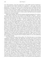
e surgical treatment for the conductive hearing
loss in otosclerosis over the past 50 years required re-
placement of the stapedial footplate, with a prosthesis
anchored to the long process of the incus. Although
total stapedectomy with tissue-wire replacement had
been the initial choice for this procedure [11], the pre-
ferred choice is a small fenestra stapedotomy, limiting
exposure of the vestibule, which accepts a piston like
prosthesis [4, 7, 12]. Several varieties of prostheses
and techniques exist for fenestrating the stapes foot-
plate. e goal is to atraumatically create a fenestra in
the footplate and replace the crural arch with a pis-
ton prosthesis of appropriate size and length for the
fenestra. e universal employment of this procedure
for over 50 years has been associated with one of the
most predictable and successful hearing levels in all
surgery. However, some minor and a few major com-
plications may result during evaluation of a patient
preoperatively, the conduct of the surgical procedure,
and in the postoperative period.
is chapter focuses on adverse events that may
occur intraoperatively and perioperatively in surgery
for otosclerosis. e discussion is followed by a video-
tape of the stapedotomy procedure and some of the
complications described in the text.
1.1 Preoperative Phase
Preoperative evaluation concerns the patient’s age,
medical status, and expectations. e hearing loss in
otosclerosis usually is brought to the attention of the
otologist in patients from the second to the fourth or
h decade, when the progressive loss has stabilized,
and the patient is able to give informed consent [6].
Patients in the second decade of life are encouraged
to delay operative intervention until the beginning or
middle of the third decade, allowing for a slowing in
the activity of the otosclerotic bone and its tendency
for regeneration. However, younger patients with a
disabling magnitude of conductive hearing loss or
aversion to the use of amplication may be acceptable
candidates for surgery. e upper end of the age scale
is more arbitrary. Since the surgical procedure may
be performed under local anesthesia with sedation, it
can be safely employed in the older patient. An asso-
ciated existing sensorineural hearing loss component
may limit the restoration of hearing even in the best
surgical result, leaving the patient still dependent on
amplication. However, patients with a severe, mixed
hearing loss pattern receiving limited improvement
with maximal electronic amplication may benet
from elimination of the conductive component by suc-
cessful stapedotomy. Such patients are uncommon but
do represent an exception to the rule.
Although 10–15% of clinical otosclerosis presents
with a unilateral conductive loss [6], this audiomet-
ric pattern should raise suspicion of a cause other
than otosclerosis. Fixation of the malleus head in the
attic typically presents with a predominant low-fre-
quency conductive hearing loss [5]. Mobility of the
manubrium can be assessed by pneumatic otoscopy
or palpation with an instrument. e possibility of a
“shadow” threshold curve caused by transmitted bone
conduction to an inadequately masked contralateral
normal ear should also receive serious consideration
in the assessment of a unilateral hearing loss. e use
of 100+ decibels (dB) white noise masking delivered
by a Bárány noise box to the contralateral ear while
Core Messages
• Conrm audiometric results with tuning fork
(512 cycles per second) and speech reception
using Barany masker in contralateral ear.
• Manage anatomical and pathologic condi-
tions of the external ear canal before the
stapedotomy.
• In unilateral conductive hearing loss, con-
sider malleus and/or incus xation.
• Stapedotomy is preferred to stapedectomy in
otosclerosis surgery.
• Prosthesis length must be carefully assessed.
• In sensorineural hearing loss aer stapedot-
omy suspect reparative granuloma.
Z
Otosclerosis Surgery Complications
speech reception is tested in the aected ear will eec-
tively identify an unsuspected “dead” ear.
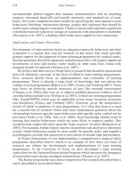
Coexistent retrolabyrinthine or labyrinthine dis-
ease may exist in patients with atypical symptoms
and clinical ndings. A conductive hearing loss with
a sensorineural component and discrimination score
that is signicantly lower than that of the contralateral
ear should raise the suspicion of a retrocochlear lesion
(i.e., acoustic neuroma), while severe vertigo associ-
ated with a low-frequency sensorineural hearing loss
suggests endolymphatic hydrops, which would be de-
compressed at stapedotomy, leading to sensorineural
hearing loss postoperatively (Fig. 1.1).
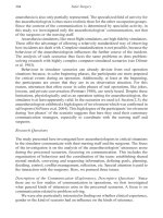
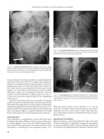
Local conditions in the ear canal may adversely
aect the performance of the stapedotomy proce-
dure. Small exostoses on the posterior canal wall can
be removed by curettage aer elevation of the tym-
panomeatal ap, permitting completion of the stape-
dotomy procedure (Fig. 1.2). However, if the exostoses
are large enough to require canaloplasty with a motor-
ized drill, then the stapedotomy should be performed
as a staged procedure.
Fig. 1.1 This photomicrograph
illustrates the vulnerability of a
dilated saccule(s) to fenestration of
the stapes footplate (FP)
.
Fig. 1.2 A small exostosis such
as this (arrow) on the posterior ear
canal wall can be removed with
curettage to allow exposure of the
middle ear. TM tympanic mem-
brane, CT chorda tympani nerve
.
1
Chapter • Otosclerosis Surgery Complications
e presence of external otitis should be control-
led medically prior to performing the surgery in or-
der to avoid contamination of the middle and inner
ear. If the external otitis is chronic, and not respon-
sive to chemotherapeutic drugs, then resection of the
infected skin with replacement by split thickness skin
gras, followed by a suciently long waiting period for
healing, should precede the stapedotomy. Anatomical
anomalies such as a dehiscent jugular bulb adjacent to
the eardrum inferiorly (Fig. 1.3) should be recognized
by preoperative otoscopy as a vascular blush in the hy-
potympanum [9]. Avoidance of such anatomical vari-
ants during ap elevation is mandatory.
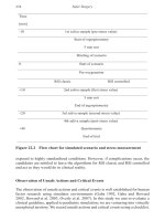
Recognition of a descending bone conduction
curve in the ear with a conductive loss should be care-
fully evaluated in anticipation of the postoperative
result (Fig. 1.4). Tilting the audiogram by closing the
air bone gap may result in a decreased discrimination
score, without injury to the sensory or neural elements
in the cochlea. e patient should be aware of this pos-
sible loss of word discrimination before the stapedot-
omy procedure.
Fig. 1.3 A large partially
dehiscent jugular bulb (J) could
be injured during elevation
of the tympanic annulus (T).
F facial nerve
.
Fig. 1.4 Closure of this air-bone
gap with stapedectomy could
result in a loss in speech discrimi-
nation because of the descending
bone conduction curve
.
. Preoperative Phase
1.2 Operative Phase
e following group of complications may occur and
be recognized intraoperatively.
Tears of the tympanic membrane occur because of
either a thin atrophic tympanic membrane or inatten-
tion to elevation of the brous annulus from its sul-
cus when raising a tympanomeatal ap. Simple tears
without a loss of tympanic membrane tissue may be
reapproximated by advancing the tympanomeatal ap
when it is returned to its anatomical position. Gelfoam
may be used in the middle ear for temporary support.
A large defect in the drum that cannot be closed by
meatal ap advancement should be repaired with adi-
pose tissue from the earlobe.
e chorda tympani nerve should be preserved
when curetting the posterior/superior canal wall.
However, in a small number of cases, probably less
than 20%, the chorda tympani nerve may be stretched
or dried out in order to achieve adequate exposure of
the oval window. Resection of the nerve segment will
avoid aberrant neural regeneration responsible for a
troublesome taste response postoperatively.
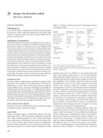
Associated xation of the malleus or incus should
be suspected in middle ear exploration [5]. It is rou-
tine during any stapedectomy procedure that all os-
sicles be individually palpated for mobility [6]. Pal-
pation of the malleus by delicate displacement of
the manubrium and of the incus by displacement of
its long process aer removal of the stapedial arch
is a routine step in the procedure. Malleus ankylosis
may be congenital or acquired and be obscured from
visualization because of its location in the epitympa-
num (Fig. 1.5). Fixation of the incus may be caused
by ossication of the posterior incudal ligaments, in
the incudal recess (Fig. 1.6). Unrecognized ossicular
xation may be responsible for failure to close the air
bone gap postoperatively.
Rarely, the incus may be dislocated during the sta-
pedectomy procedure. e initial maneuver is to re-
place the incus into its anatomical position, relying on
healing of the ligaments to retain it [6]. However, if
the dislocation is severe, and the incus does not retain
its relocated position, then malleus attachment for the
prosthesis is the most reliable solution for a satisfac-
tory result. Occasionally pneumatization of the long
process of the incus may be responsible for fracture af-
ter crimping of the wire prosthesis. is event requires
that an appropriately long new prosthesis be applied to
the manubrium of the malleus.
e critical part of the stapedotomy procedure
concerns fenestration of the ankylosed footplate. e
accompanying gures demonstrate some of the ana-
tomical variations in oval window pathology that aect
the surgical technique. In the case of a thin footplate
in an oval window niche with overhanging bone (Fig.
1.7), removal of the overhanging bone with a rotating
burr will provide complete visualization of the annu-
lar ligament. Such overhanging bone may compromise
the ability to retrieve a oating or depressed footplate.
A thick footplate with marginal xation will require
careful pressure with the drill to avoid a oating foot-
Fig. 1.5 Anterior malleus head ankylosis may be congenital
(arrow). I incus body
. Fig. 1.6 Fixation of the short process of incus (I) may be
acquired by calcication in its ligaments (arrow). * air cell in the
incus, L normal ligament
.
1
Chapter • Otosclerosis Surgery Complications