Ear Surgery - part 4 ppsx
Bạn đang xem bản rút gọn của tài liệu. Xem và tải ngay bản đầy đủ của tài liệu tại đây (855.87 KB, 13 trang )
e most serious complications of untreated COM and
mastoiditis are labyrinthitis, facial paralysis, and vari-
ous intracranial complications. e main intracranial
complications are meningitis, brain abscess, subdural
or epidural abscess, and lateral/sigmoid sinus throm-
bophlebitis.
4.1 Extracranial Complication
4.1.1 Labyrinthitis
Labyrinthitis or invasion of the perilymphatic space
by chronic middle ear and mastoid disease occur over
three pathways. ese are (1) stulization of the bony
labyrinth, (2) round window, and (3) oval window.
e most common of the pathways is bony erosion of
the labyrinthine capsule, usually of the semicircular
canals but less oen of the bony wall of the cochlea.
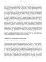
is bone erosion occurs as a result of the eects of
pressure from an enlarging cholesteatoma and/or the
chemical breakdown of collagen by collagenase en-
zymes in the cholesteatoma membrane [1] (Fig. 4.1).
Core Messages
• e most common extracranial complica-
tions of chronic otitis media (COM) are laby-
rinthitis and facial nerve paralysis.
• Labyrinthitis associated with COM occurs
through stulization of the otic capsule or in-
vasion through the oval and round windows.
• Removal of cholesteatoma membrane from
the endosteal membrane is recommended.
• Repair of oval or round window defects with
so tissue is an eective technique to pre-
serve labyrinth function.
• Facial nerve paralysis in COM requires early
surgery to remove chronic disease and pre-
serve facial function.
• Intracranial complications from COM in-
clude meningitis, brain abscess, epidural/
subdural abscess, and sigmoid sinus throm-
bophlebitis.
• Meningitis and brain abscess from COM
may occur through a preformed pathway or
by retrograde thrombophlebitis.
Z
Complications of Chronic Otitis Media
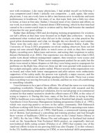
Fig. 4.1 A schematic demonstration of stages in bone erosion by cholesteatoma. BL bony labyrinth, EB endosteal bone, M mem-
branous canal, EM endosteal membrane, P perilymph
.
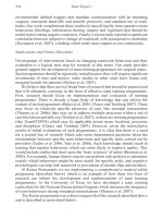
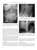
Fig. 4.2 Axial CT of a temporal bone with large cholesteato-
ma of the mastoid and erosion of the lateral canal (arrows)
.
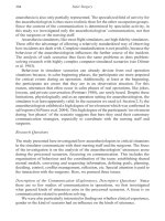
Fig. 4.3 Surgical technique for
removal of cholesteatoma mem-
brane from lateral canal stula
.
4
Chapter • Complications of Chronic Otitis Media
When bony stulization over the vestibular labyrinth
occurs, recurrent vertigo on compression of air in the
ear canal is the signal symptom heralding its pres-
ence. Conrmation by the stula test with or without
CT (Fig. 4.2) will provide some guidance as to which
of the semicircular canals is involved. A horizontally
directed nystagmus in the stula test indicates the lat-
eral canal as the site of the stula; a vertical nystagmus
points to the posterior canal as the site of involvement,
and a vertical/rotatory nystagmus identies the supe-
rior canal as the site of stulization. If uncontrolled,
such bony stulae will eventually lead to inamma-
tory invasion of the labyrinth with loss of vestibular
and auditory function, emphasizing the need for early
surgical correction.
e technique for surgical management of a bony
stula consists of sharp dissection of the cholesteatoma
membrane from the endosteal membrane in the area
of bone defect (Fig. 4.3). e guidelines for removal
of cholesteatoma membrane depend on the surgeon’s
experience, the size and location of the stula, and
the function of the involved ear and uninvolved ears
[2]. Small stulae (2 mm or less) of the bony semi-
circular canal lend themselves to safe removal of the
lining membrane, which does not adhere to the un-
derlying endosteal membrane (Fig. 4.4). Bony stulae
larger than 2 mm are managed on an individual basis.
Adherence of the matrix to the underlying endosteal
membrane at surgery is determined by sharp dissec-
tion (Fig. 4.5). Aer the cholesteatoma membrane has
been removed from the endosteal membrane, the bony
defect may be covered by a precisely sculpted cortical
bone gra held in place by a temporalis fascia free gra.
is will provide immediate relief of pressure-induced
symptoms of vertigo. Alternatively, if vestibular symp-
toms are not severe, a temporalis fascia gra may be
laid over the stula allowing eventual periosteal bone
regeneration to obliterate the bony defect. e removal
of cholesteatoma from a stula in an only hearing ear
is dependent on these guidelines, and if the foregoing
previous factors cannot provide guidance, it is best to
leave the matrix undisturbed in the only hearing ear.
In rare instances where cholesteatoma membrane in-
vades the bony semicircular canal and removal is not
possible without disruption of the membranous con-
tents, enlarging the bony stula to allow eradication
of cholesteatoma matrix is necessary. Obliteration of
the defect with bone wax may preserve residual laby-
rinthine function by sealing o the endolymphatic and
perilymphatic compartments [2].
Fistulization of the otic capsule surrounding the
cochlea [2] is usually located over the promontory
and basal turn (Fig. 4.6). A sensorineural hearing loss
should alert the surgeon to its presence, which may be
conrmed on CT of the temporal bone. Even atrau-
matic removal of cholesteatoma matrix from a coch-
lear stula will result in profound loss of function [2,
9]. erefore, removal is not recommended, and the
residual cholesteatoma membrane is exteriorized sur-
rounded by a fascial gra.
It is possible for recurrent cholesteatoma to in-
vade the cochlea and the internal auditory canal. is
unusual extension of cholesteatoma is associated with
a severe sensorineural hearing loss in combination
with additional neural decits in the temporal bone
(e.g., facial paralysis). A case illustrated in the video
Fig. 4.4 Histopathology of a small stula (arrowhead) of the
lateral canal. C cholesteatoma membrane
.
Fig. 4.5 A large bony stula (arrowhead) of the lateral canal
covered by cholesteatoma membrane is illustrated in this phot-
omicrograph. S superior canal ampulla
.
. Extracranial Complication
presented with facial paralysis along with loss of laby-
rinthine function. Imaging studies demonstrated ero-
sion of the internal auditory canal and cochlea. Chole-
steatoma membrane had extended in to the internal
auditory canal by way of the fallopian canal in its
tympanic and labyrinthine segments. Resection of the
seventh and eighth nerves was necessary for control of
cholesteatoma. A hypoglossal-to-facial nerve anasto-
mosis was used to rehabilitate the facial muscles.
Labyrinthine extension from inammatory mid-
dle ear disease may also occur through the round and
oval windows [12]. e round window membrane
forming the sole membranous barrier between middle
ear and the labyrinthine space is the next most vul-
nerable location for the spread of toxic inammatory
changes (Fig. 4.7). e anatomy of the round window
niche may have bearing on the severity of inamma-
tory tissue response on the membrane. Niches with a
narrow bony aperture may isolate inamed middle ear
mucosa with an increased potential for creating a de-
structive eect on the membrane (Fig. 4.8). Auditory
decits out of proportion to function of the contral-
ateral ear are a clinical indication of invasion through
this site. e auditory threshold decit may assume
various patterns but characteristic is a loss in speech
discrimination.
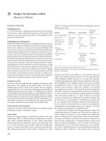
Fig. 4.6 Erosion of bone over the cochlea by chronic ostei-
tis is indicated by the arrowhead. The endosteal bone layer is
intact
. Fig. 4.7 Extension of chronic infection through the round
window membrane (arrow) is often associated with inamma-
tory changes locked in a small round window niche (*)
.
Fig. 4.8 Chronic inammatory
tissue may be sequestered in a
round window niche with a nar-
row aperture (*). R round window
membrane
.
4
Chapter • Complications of Chronic Otitis Media
Adequate removal of inammatory tissue and
cholesteatoma requires exposure of the round window
niche and adjacent sinus tympani. A reliable surgical
step is to identify the descending (mastoid) segment
of the facial nerve, followed by removal of the bony
oor of the facial recess (Fig. 4.9). A small diamond
burr can then be used to remove the overhang of the
round window niche suciently to allow sharp dis-
section of pathological tissue from the round window
membrane [3].
Fig. 4.9 Exposure of the round
window niche (RWN) for removal
of disease requires identication
of the facial nerve (a), followed
by removal of bone in the facial
recess (b). LC bone of lateral semi-
circular canal
.
. Extracranial Complication
Less common is invasion through the oval window
where the footplate forms a thin bony barrier between
the middle ear and labyrinthine uids (Fig. 4.10).
However, with chronic inammatory disease, decalci-
cation, and erosion of the footplate may allow the in-
ammatory process to aect the perilymphatic space
in the vestibule. Since the vestibular sense organs are
nearby, dizziness of varying severity and forms, rather
than an auditory decit, is a common early clinical
indicator of extension through this pathway. Control
of extension of disease through these natural windows
is accomplished with adipose tissue gra replacement
of the footplate (modied type V tympanoplasty) and
with tissue repair of round window membrane defects.
e eect of inammation in the perilymphatic space
(labyrinthitis) is generally divided into two stages, se-
rous labyrinthitis and suppurative (destructive) laby-
rinthitis [14]. e two forms are dened by the re-
covery of function (serous) or by the loss of function
(suppurative). An intermediate stage (serobrinous)
has been suggested for partial loss of function and an
end stage type included which refers to a histological
change resulting from labyrinthine suppuration (laby-
rinthitis ossicans) (Fig. 4.11).
Serous labyrinthitis can be eradicated by timely
surgery to remove the source of the inammation
(bony stula, round or oval window infection) while
suppurative labyrinthitis must be exenterated surgi-
cally together with sense organs to prevent spread to
the subarachnoid space and promote central compen-
sation for vestibular ablation. e techniques for this
surgical intervention are described elsewhere.
4.1.2 Facial Paralysis
Facial paralysis is a serious neural complication of
chronic middle ear and mastoid disease, particularly
from cholesteatoma [6, 11, 16]. In the presence of ac-
tive chronic middle ear disease, this complication
should be dealt with urgently. Exposure of the facial
nerve and its sheath proximal and distal to the area
of involvement by chronic inammatory tissue must
be reached by careful removal of so tissue and bone.
Removal of disease from the facial nerve and release
of nerve edema is then provided by incising the nerve
sheath.
4
Fig. 4.10 Chronic infection may also extend through the oval window by extension
through the stapedio vestibular ligament (arrowheads) or the stapedial footplate. F facial
nerve
.
Chapter • Complications of Chronic Otitis Media
4.2 Intracranial Complications
4.2.1 Intradural Extension
of Cholesteatoma
e extension of cholesteatoma membrane beyond the
connes of the mastoid and middle ear compartment
may occur through a preformed defect in the dura.
ese dural defects are not a result of cholesteatoma-
induced breakdown of dura, but rather represent con-
genital defects through which arachnoid granulations
(AG) herniate and come to lie in apposition with the
temporal bone (see Chap. 8, Fig. 8.4). Invasion of the
subdural space by cholesteatoma occurs by invagina-
tion of the AG, which then allows a subdural collec-
tion of cholesteatoma. We have experience with one
patient who underwent several mastoid explorations
for recurrent cholesteatoma that was found to be ex-
tensive in the subdural space. Severe pain was the
patient’s primary symptom. Complete removal of the
subdural cholesteatoma would require a neurosurgical
approach, but the neurosurgical consult deferred until
neurological decits appeared.
e intradural invasion by cholesteatoma may not
be limited to the subdural space. e accompanying
gures demonstrate a case of a middle-aged man who
had successful surgical control of mastoid cholest-
eatoma, but many years later presented with a tempo-
ral lobe abscess (Fig. 4.12). A neurosurgical approach
conrmed that the cholesteatoma membrane had
extended through a defect in the tegmen and middle
fossa dura into the temporal lobe cortex (Fig. 4.13).
e patient survived a short period because of the
chemical meningitis associated with the surgical exci-
sion of cholesteatoma. e postmortem examination
of the temporal bone in this patient revealed the bony
and dural defect, which permitted such intracranial
extension of cholesteatoma (Fig. 4.14).
Fig. 4.11 The end stage in sup-
purative labyrinthitis is ossica-
tion (*)
.
Fig. 4.12 A ring enhancing temporal lobe brain abscess (ar-
rowheads) proved to be cholesteatoma extension from the epi-
tympanum
.
. Intracranical Complications
4.2.2 Meningitis
Recurrent meningitis may result from extension of
disease through an AG [4, 15]. e accompanying
video demonstrates a patient with a history of recur-
ring episodes of meningitis with each episode of otitis
media. CT scan of the temporal bones revealed a bony
defect in the tegmen mastoidea with opacication of
the mastoid compartment and middle ear (Fig. 4.15).
e surgical exploration revealed a mass of tissue pro-
truding from a defect in the dura, which consisted of a
granulation tissue mass with a transition from granu-
loma to normal temporal lobe white matter. e sur-
gical procedure accomplished removal of the AG and
herniated temporal lobe, suture closure of the dural
defect, and adipose tissue obliteration of the intact ca-
nal wall mastoidectomy defect.
Meningitis as an extension of suppurative labyrin-
thitis may occur as a result of progressive infection
along branches of the eighth cranial nerve or through
preformed pathways (i.e., cochlear aqueduct).
4.2.3 Brain Abscess
Brain abscess secondary to acute or chronic mastoidi-
tis may develop directly through a preformed defect
in the dura (AG) or indirectly as a retrograde throm-
bophlebitis of the small dural vessels that permeate
overlying bony surfaces of the temporal bone [5, 13,
16]. e intracranial accumulation formed through a
dural defect usually appears earlier (almost immedi-
ate) aer acute suppurative otitis media and mastoidi-
tis than does an abscess that develops by retrograde
vascular spread. e development of temporal lobe or
cerebellar abscess is usually determined by the respon-
sible method of spread in either the tegmen or poste-
rior fossa surfaces of the temporal bone. e clinical
signs of intracerebral infection in these locations are
well known and not reviewed here. CT and MRI im-
aging provide early conrmation of this intracranial
complication.
4.2.4 Lateral Sinus Thrombosis
Spread of infection to the intracranial venous system
may occur from untreated or undertreated acute mas-
toiditis or active chronic otitis media and mastoiditis
[5, 17, 18]. Infection with thrombus formation may
occur in the lateral venous sinus because of exposure
in the sigmoid segment within the mastoid compart-
ment. Infection usually spreads directly through the
sinus wall or by way of retrograde thrombophlebitis
when there is osteitic bone of the sinus plate.
Although typical clinical ndings associated with
this sequela are spiking high temperatures, headache,
drowsiness, and indications of hydrocephalus, the full
clinical picture is not usually seen in the present era of
antibiotic therapy. erefore, the diagnosis should be
Fig. 4.14 Temporal bone specimen from deceased patient in
Figs. 4.12 and 4.13 shows the epitympanic bony defect (arrow-
head). C cholesteatoma membrane
.Fig. 4.13 The cholesteatoma extended from a bone defect in
the tegmen (arrow)
.
4
Chapter • Complications of Chronic Otitis Media
suspected in any patient with constant mastoid infec-
tion, persistent intermittent fever, and headaches. CT
and MRI imaging of the temporal bone are essential to
provide clues for bone demineralization and interrup-
tion of venous ow [7, 8, 10]. Vascular studies such as
the venous phase of an arteriogram are mandatory to
make the diagnosis (Fig. 4.16).
Surgical treatment of sinus thrombosis consists
of exposure of the sigmoid segment in a wide-eld
mastoidectomy. If ballottement reveals a patent sinus,
then no further treatment of the sinus except thorough
curettage of granulation tissue from the surrounding
dura. A rm sinus segment requires needling rst with
ne- then a large-gauge needle. No venous return re-
quires opening the sinus while compressing its lumen
proximal and distal to the segment lled with throm-
bus. Evacuation of the thrombus can then be carried
out. Intensive antibiotic and anticoagulant manage-
ment is necessary for at least 6–8 weeks.
CO M P L I C AT I O N S TO AV O I D
1. Avoid injury to the facial nerve in chronic OM
surgery by exposing nerve in intact part of the
fallopian canal.
2. Remove cholesteatoma membrane from the
endosteal lining of a bony fistula in the semi-
circular canals to avoid injury to the membra-
nous canal.
3. Avoid removal of cholesteatoma membrane
from bony fistulas of the cochlea to prevent
sensorineural hearing loss.
Pearl
• It is safe to remove cholesteatoma lining from
a bony stula of the vestibular labyrinth but
not the cochlea.
• Repair of stula of the oval window with adi-
pose tissue to preserve cochlear function.
Z
Fig. 4.15 Coronal CT scan of
temporal bones in a patient with
recurrent meningitis. The tegmen
defect (arrow) was lled with an
arachnoid granulation adherent to
the temporal lobe
.
Fig. 4.16 Venogram in a patient with sigmoid and lateral
sinus thrombosis demonstrates obstruction in venous ow (ar-
row). SS superior sagittal venous sinus
.
. Intracranical Complications
References
1. Abramson M (1969) Collagenolytic activity in middle ear cholest-
eatoma. Ann Otol Rhinol Laryngol 78:112–124
2. Gacek R (1974) e surgical management of labyrinthine stu
-
lae in chronic otitis media with cholesteatoma. Ann Otol Rhinol
Laryngol 83:(Suppl):1–19
3. Gacek RR (1993) Surgery of the vestibular system. In: Cummings
C, Schuller DE (eds) Otolaryngology—head and neck surgery.
Mosby, St. Louis, pp 3199–3216
4. Gacek RR (1990) Arachnoid granulation cerebrospinal otorrhea.
Ann Otol Rhinol Laryngol 99:854–862
5. Glasscock ME III, Shambaugh GE Jr (1990) Intracranial complica
-
tions of otitis media. In: Glasscock ME III, Shambaugh GE Jr (eds)
Surgery of the ear, 4th edn. Saunders, Philadelphia, pp 249–275
6. Ludman H (1987) Complications of suppurative otitis media. In:
Kerr AG (ed) Scott-Brown’s otolaryngology, 5th edn. Butterworth,
London, pp 264–291
7. Macchi PJ, Grossman RI, Gomori JM, Goldberg HI, Zimmerman
RA, Bilaniuk LT (1986) High-eld MR imaging of cerebral venous
thrombosis. J Comp Assist Tomogr 10:10–15
8. McArdle CB, Mirfakhraee M, Amparo EG, Kulkarni MV (1987)
MR imaging of transverse/sigmoid dural sinus and jugular vein
thrombosis. J Comp Assist Tomogr 11:831–838
9. McCabe BF (1984) Labyrinthine stula in chronic mastoiditis.
Ann Otol Rhinol Laryngol 93:(Suppl):138–141
10. McMurdo SK Jr, Brant-Zawadzki M, Bradley WG Jr, Chang GY,
Berg BO (1986) Dural sinus thrombosis: study using intermediate
eld strength MR imagining. Radiology 161:83–86
11. Neely JG (1986) Complications of temporal bone infection. In:
Cummings C, Schuller DE (eds) Otolaryngology—head and neck
surgery. Mosby, St. Louis, pp 2988–3015
12. Paparella MM, Sugiura S (1967) e pathology of suppurative
labyrinthitis. Ann Otol Rhinol Laryngol 76:554–586
13. Quijano M, Schuknecht HF (1988) Temporal bone pathology as
-
sociated with intracranial abscess. ORL 50:2–31
14. Schuknecht HF (1974) Pathology of the ear. Harvard University
Press Cambridge, Mass.
15. Schuknecht HF (1970), Montandon P. Pathology of the ear in
pneumococcal meningitis. Arch Klin Exp Ohr Nas Kehlkheilk
195:207–225
16. Snow JB (1989) Cranial and intracranial complications of otitis me-
dia. In: English GM (ed) Otolaryngology. Lippincott, Philadelphia
17. Southwick FS, Richardson EP Jr, Swartz MN (1986) Septic throm-
bosis of the dural venous sinuses. Medicine 65:82–106
18. Teichgraeber JF, Per-Lee JH, Turner JS Jr (1982) Lateral sinus
thrombosis: a modern perspective. Laryngoscope 92:744–751
4
Chapter • Complications of Chronic Otitis Media
e subtle clinical presentations of petrous apex le-
sions are related to the regional anatomy of the api-
cal segment of the temporal bone (Fig. 5.1). Prior to
1975, the major lesion of the petrous apex described in
the literature was infection, causing epidural abscess
formation responsible for a classic triad of symptoms
(Gradenigo’s syndrome). ese were diplopia, deep
pain, and facial hypoesthesia. Nearby structures in
the petrous apex (h and sixth cranial nerves) pro-
vided logical explanation of the clinical ndings in
this potentially lethal sequela of middle ear infection.
Radiologic techniques (plain x-rays, polytomogra-
phy) were capable of demonstrating only the most ad-
vanced osteolytic lesions in this area [3, 7]. Using these
methods, a series of solid and cystic lesions primary
in the petrous apex were demonstrated and managed
in a monograph publication [3, 7]. ese lesions are a
manifestation of the complex anatomical composition
of the petrous apex, consisting of air cells, bone mar-
row, cartilage, nerves, and vascular structures (inter-
nal carotid artery, jugular bulb). is report of petrous
apex cases and their management unlocked the many
pathologies located in this obscure region of the skull
base. Sophisticated imaging techniques (CT, MRI)
now permit early recognition of a petrous apex lesion.
e usual presenting symptoms of an expanding
lesion in the petrous apex are a conductive hearing
loss from the serous eusion caused by Eustachian
tube obstruction, headache from pressure on the dural
covering, diplopia related to involvement of the third
and sixth cranial nerves, facial hypoesthesia caused
by compression of the h cranial nerve, and vary-
ing degrees of faintness or vertigo probably caused by
changes in the labyrinthine blood supply. e patholo-
gies involving the petrous apex may be divided into
solid and cystic lesions (Table 5.1).
Table 5.1 Petrous apex pathologies categorized by lesion
type
Solid lesions
Benign
Neurobroma or schwannoma
Chondroma
Meningioma
Paraganglioma
Malignant
Chondrosarcoma
Eosinophilic granuloma
Lymphoma
Metastatic malignancies
(breast, lung, kidney, prostate)
Cystic lesions
Vascular
Internal carotid aneurysm
Venous lake
Nonvascular
Apicitis (abscess)
Congenital epidermoid cyst
Cholesterol granuloma (mucocele)
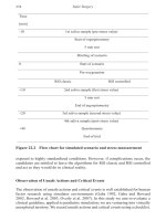
5.1 Diagnosis
e clinical suspicion of a progressive lesion in the
petrous apex is based on recognition of one or more of
the signs and symptoms related to adjacent anatomical
.
Core Messages
•
e petrous apex may be aected by cystic
and solid lesions. Cystic lesions are more
common and are benign. Solid lesions are less
common and may be benign or malignant.
• Clinical signs and symptoms of expanding le-
sions of the petrous apex include Eustachian
tube compression, third and sixth nerve de-
cits, and headache.
• Both magnetic resonance imaging and com-
puterized tomography are recommended in
the diagnosis and management of petrous
apex lesions.
• Surgical approaches to biopsy or stulize
petrous apex lesions include perilabyrinthine
cell tracts, sphenoid sinus, middle cranial
fossa, transcochlear.
Z
Petrous Apex Lesions
structures (Eustachian tube, cranial nerves III through
VIII, dura, and internal carotid artery).
Presently, thin-section (1–1.5 mm) CT scanning
and MRI are capable of identifying such lesions in the
petrous apex much earlier in their development, with
minimal risk. e evaluation of a suspected lesion in
the petrous apex should require CT scanning and MRI
[15, 16]. e nature of a lesion as solid or cystic is usu-
ally revealed by the enhancement on CT and signal
picture on T
1
and T
2
images with MRI. Arteriography
may be added to dene a vascular lesion or to locate
displacement of the internal carotid artery.
5.2 Management
5.2.1
Solid Tumors
e management of solid tumors requires identica-
tion of the histopathology prior to denitive treatment
[7]. Several factors determine the approach to biopsy
of solid petrous apex lesions. If the tumor has extended
into an area that is easily accessible without risk to lab-
yrinthine function such as the infralabyrinthine and
hypotympanic cell tracts or into the sphenoid sinus,
then these compartments should be accessed for sam-
pling the tumor (Fig. 5.2.). However, if the tumor is
contained within the petrous apex and labyrinth func-
tion is normal, then a middle cranial fossa extradural
approach skirting the temporal bone is the most direct
approach to the tumor while preserving seventh and
eighth cranial nerve functions.
Denitive management is guided by the histologic
nature of the lesion. A benign lesion may be excised
totally or subtotally, depending on the progressive
nature of the clinical symptoms and the patient’s age
and medical status. Postoperative surveillance of the
petrous apex tumor may be carried out with imaging
studies to determine possible recurrence and pro-
gression. A malignant tumor will require nonsurgical
management (radiation therapy, chemotherapy) since
en bloc resection of this portion of the temporal bone
Fig. 5.1 Diagram of the skull
base with anatomical structures
related to the petrous apex and
foramen lacerum
.
5
Chapter • Petrous Apex Lesions
is not feasible for cure, particularly if one considers
the morbidity associated with the surgery (Fig. 5.3).
However, low-grade malignancy such as eosinophilic
granuloma may be treated more eectively by exten-
sive subtotal removal using curettage, followed by ra-
diation therapy.
5.2.2 Cystic Lesions
Cystic lesions may be vascular or nonvascular, and
their management will vary from case to case. If im-
aging studies are not able to demonstrate the vascular
nature of a lesion with certainty, then arteriography
is required. e major vascular lesion of the petrous
apex is an internal carotid aneurysm (Figs. 5.4, 5.5).
If the lesion is causing minimal nonprogressive clini-
cal symptoms, then the aneurysm should be conserva-
tively followed clinically and neuroradiologically. If
the aneurysm becomes progressive and is responsible
for signicant neurologic symptoms, then a team ap-
proach (neurosurgery and otolaryngology) to man-
agement of the aneurysm is necessary. Progressive
preoperative occlusion followed by resection is one
method of management if the patient tolerates the oc-
clusive maneuver.
Occasionally, imaging studies are misleading. We
have seen two patients presenting with episodic ver-
tigo that revealed on MRI a high-signal-intensity mass
resembling a cholesterol cyst in the petrous apex (Fig.
5.6). However, CT scan did not reveal bone erosion,
but rather a compartment in the petrous apex was
present (Fig. 5.7). It has been suggested that this may
represent bone marrow. However, exploration of the
petrous apex in these cases with severe sensorineural
hearing loss in the involved ear revealed a thin-walled
space with brisk venous bleeding requiring rm pack-
ing control. It is possible that these lesions represent
venous anomalies (venous lakes). e sigmoid sinus
and the jugular bulb were anatomically separated from
this vascular compartment. e importance of this en-
tity is that if it is properly recognized with the posi-
Fig. 5.2 Axial CT demonstrating a solid lesion (schwannoma)
arising from the petrous apex (arrowhead) and presenting into
the sphenoid sinus, where it was biopsied through an ethmoid–
sphenoid approach. Presenting signs were Eustachian tube ob-
struction and sixth cranial nerve palsy in a 60-year-old female.
Observation with CT scanning was recommended
.
Fig. 5.3 Metastatic breast carcinoma in the bone marrow
of the petrous apex presented with a fth-nerve decit (arrow-
head)
.
. Management