Ear Surgery - part 6 docx
Bạn đang xem bản rút gọn của tài liệu. Xem và tải ngay bản đầy đủ của tài liệu tại đây (848.75 KB, 13 trang )
CO M P L I C ATI O N S TO AVO I D
1. Remove cholesteatoma membrane completely
to avoid recurrence.
2. Avoid exposing the subarachnoid space to
cholesteatoma prevents a chemical meningi-
tis.
Pearl
• Aer removal of cholesteatoma from the
mastoid, drill the bony surfaces lightly to
eliminate microscopic foci of squamous epi-
thelium.
Z
References
1. Chole RA (1984) Cellular and subcellular events of bone resorp-
tion in human and experimental cholesteatoma: the role of osteo-
clasts. Laryngoscope 94:76–95
2. Gacek RR (1975) Diagnosis and management of primary tumors
of the petrous apex. Ann Otol Rhinol Laryngol 84:1–20
3. Gacek RR (1980) Evaluation and management of primary petrous
apex cholesteatoma. Otolaryngol Head Neck Surg 88:519–523
4. Gacek RR (2005) Unpublished observation.
5. Heumann H (1989) Cholesteatoma in childhood, surgical treat
-
ment and results. In: Tos M, omsen J, Peiterson E (eds) Chole-
steatoma and mastoid surgery. Kugler & Ghedini, Amsterdam, pp
671–676
6. Levenson MJ, Michaels L, Parisier SC, Juarbe C (1988) Congenital
cholesteatoma in children: an embryological correlation. Laryn-
goscope 98:949–955
7. Morigama H, Huang CC, Abramson M, Kato M (1984) Bone re
-
sorption factors in chronic otitis media. Otolaryngol Head Neck
Surg 92:322–328
8. Piepergerdes MC, Kramer BM, Behnke EE (1980) Keratosis ob
-
turans and external auditory canal cholesteatoma. Laryngoscope
90:383–391
9. Portmann M (1982) Surgery of retraction pockets versus attic
cholesteatoma. In: Sade J (ed) Cholesteatoma and mastoid sur-
gery. Kugler & Ghedini, Amsterdam, pp 509–510
10. Sade J (1982) Treatment of retraction pockets and cholesteatoma.
In: Sade J, ed. Cholesteatoma and mastoid surgery. Kugler &
Ghedini, Amsterdam, pp 511–525
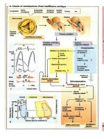
Fig. 6.10 Horizontal temporal
bone section demonstrates a canal
cholesteatoma (*) secondary to
stenosis of the cartilaginous exter-
nal auditory canal (arrowheads)
.
6
Chapter • Cholesteatoma
Obstructive lesions of the external auditory canal,
requiring surgical correction, are primarily of three
types.
7.1 Bony Lesions
Bony lesions of the ear canal (osteoma, exostosis) are
the most common of these obstructive lesions. Os-
teoma, usually solitary, has the normal structure of pe-
riosteal bone and is uncommonly large enough to cause
obstruction of the ear canal, leading to accumulation
of debris and/or cholesteatoma in the deep ear canal
[5, 7]. Exostosis on the other hand, are more common,
represent the formation of usually three locations of
laminated periosteal bone in the external auditory ca-
nal. e histologic make up of exostoses is shown in
Fig. 7.1. It is thought that since these occur in patients
who have the practice of swimming in very cold water
that the periosteal irritation from such a cold stimulus
promotes the laying down of periosteal bone matrix
in a repeated fashion leading to the gradual enlarge-
ment of bony lesions in the ear canal [21]. While of
no clinical signicance when they are small, as they
become large enough to cause recurrent entrapment of
cerumen and/or debris in the deep ear canal, repeated
external canal infection occurs (Fig. 7.2). Rarely, they
may cause complete obstruction of the lumen of the
bony ear canal and a conductive hearing loss. ese
are the primary indications for surgical removal.
Removal is performed through an endaural ap-
proach under general anesthesia, with preservation
Core Messages
• Obstructive lesions of the external auditory
canal require surgical management when
conductive hearing loss, retained epithe-
lial debris, and recurrent canal infection is
present.
• Surgical method requires adequate enlarge-
ment of the bony and cartilaginous segments
with re-epithelialization employing skin aps
or split thickness skin gras.
Z
External Auditory Canal Lesions
Fig. 7.1 The histological composition of external canal exos-
tosis reects multiple periosteal bone insults with the deposi-
tion of bone matrix (arrows)
. Fig. 7.2 Axial CT scan demonstrates near obstruction of the
external canal lumen by exostosis (arrowheads)
.
of as much ear canal skin both laterally and medially
to the location of the exostoses. e exostoses are re-
moved with a rotating burr, rst with a cutting burr,
and nally with a diamond burr when nearing the tym-
panic membrane. e diamond burr is used to hollow
out the rounded exostosis, leaving a shell-like cover.
ese thin bony portions of the exostoses can then be
removed with a curette and/or small diamond burrs.
It is important to avoid contact with the manubrium
or the lateral process of the malleus when drilling in
the deep ear canal to avoid transmitted energy to the
labyrinth causing sensorineural hearing loss [18]. Suf-
ciently large canal wall skin aps can be preserved to
allow for adequate coverage of the exposed canal bone.
If this is not possible, then the application of split-
thickness skin gras to bone, held in place with pack-
ing for at least 1 week to 10 days is eective. is surgi-
cal exercise is demonstrated in accompanying video.
7.2 Congenital Aural Atresia
Congenital aural atresia may aect the external au-
ditory canal by merely causing a narrow canal with
a small external meatus, a normal bony canal with
a small external meatus, or in its fullest expression,
complete absence of the bony and cartilaginous ca-
nal. is congenital lesion may occur unilaterally or
bilaterally [8, 9, 14, 20]. When it is bilateral, the in-
dications for surgical correction are clear-cut and are
usually carried out at the age of 5 or 6 years, when the
patient is more capable of tolerating the postoperative
care involved and the mastoid compartment has been
fully pneumatized. e usual criteria in a candidate for
this surgery is that they have a pneumatized middle
ear and mastoid compartment, that there is a normally
developed labyrinth with evidence of normal bone
conduction, and that parts of the ossicular chain, that
is the malleus and the incus are visible on CT scanning
[10] (Fig. 7.3).
Generally, two approaches have been used to cor-
rect the congenital atresia. One is a posterior approach
through the mastoid compartment, identifying the
central mastoid tract and then performing a canal
wall down mastoidectomy with the middle ear [20].
However, this surgical approach, while oering a wide
exposure of possible anomalous middle ear and facial
nerve structures, leaves a patient with a mastoid cavity
to care for with attendant water precautions and po-
tential for recurrent infection.
A preferred approach is the anterior one, following
the middle fossa dura medially to the epitympanic re-
cess of the middle ear (Fig. 7.4). An endaural so tissue
approach is used [10]. e head of the malleus and the
body of the incus are identied in the epitympanum,
and the new ear canal is created by drilling bone from
the epitympanum anteriorly and inferiorly. With this
approach, the facial nerve is not at any increased risk,
and a satisfactory bony ear canal can be created in an
orderly fashion. Split-thickness skin gras are used to
line the newly created ear canal, and temporalis fascia
is used as graing material for a new tympanic mem-
7
Fig. 7.3 Axial CT of external canal bony atresia (arrow), with
pneumatized middle ear and mastoid. O ossicles
. Fig. 7.4 Coronal CT demonstrates absence of the tympanic
bone (arrow)
.
Chapter • External Auditory Canal Lesions
brane. e ossiculoplasty is dependent on the presence
of usable ossicles in the middle ear [4]. If a malleus is
present along with the incus, then releasing the man-
ubrium of the malleus from the bony ear canal will
mobilize the ossicular chain and provide an eective
way of providing good hearing by way of a type II tym-
panoplasty. It is crucial that skin gras be applied to all
surfaces in the bony and cartilaginous canal as well as
the lateral surface of the tympanic membrane fascial
gra to prevent brous stenosis of the newly created
ear canal. e issue of reconstruction of the auricle is
dependent on the degree of aplasia or hypoplasia of
the auricle and of the willingness of the patient and
family to undergo the multiple procedures necessary
to recreate a cosmetically acceptable auricle [10].
7.3 Stenosing Chronic External Otitis
An obstructive lesion of the ear canal not usually rec-
ognized as a surgical condition is the brosing chronic
external otitis [17, 22]. A chronic inammatory proc-
ess in the ear canal skin may be responsible for not
only pain and discharge refractory to medical treat-
ment, but also for a conductive hearing loss. Recogni-
tion of canal stenosis as a result of recurrent or chronic
external otitis as well as hearing loss from thickening
of the tympanic membrane can be conrmed with CT.
e anatomical structures responsible for the reten-
tion of oending organisms are hair follicles and ceru-
minous glands located in the cartilaginous segment
of the ear canal. e denitive recommended treat-
ment is excision of the involved skin and so tissue
of not only the external cartilaginous canal, but also
of the bony canal and the lateral surface of the tym-
panic membrane. is procedure is shown in an ac-
companying video. Following removal of the brous
and epithelial components of the ear canal, the appli-
cation of split-thickness skin gras held in place with
a bolus-type dressing (rosebud dressing) is eective in
not only controlling the symptoms of external otitis,
but also in correction of the conductive hearing loss
caused by this ear canal lesion.
7.4 Necrotizing External Otitis
Necrotizing or malignant external otitis is a poten-
tially lethal form of osteitis of the ear canal, which
occurs in immunocompromised patients, particu-
larly elderly diabetics, by the organism pseudomonas
aeruginosa. is pathologic entity rst described by
Keleman and Meltzer [13] was more fully described
with eective management by Chandler [1, 3] in the
1960s. Although the progressive osteitis responsible
for this ear canal infection occurs in the oor of the
bony ear canal with the capability of extension to the
base of the skull, its lethal nature results from involve-
ment of the major vascular and neural structures in
this area [6, 16]. e primary treatment is by eective
antibiotics delivered intravenously as well as topically.
Gentamycin has been shown to be an eective topical
antibiotic in the area of involvement in the ear canal
while the preferred systemic antibiotic is ciprooxacin
[11, 12, 15, 19]. Gentamycin used systemically is held
in reserve if ciprooxacin is ineective because of the
Fig. 7.5 Coronal CT in a patient
with necrotizing external otitis and
facial paralysis demonstrates ero-
sion of the oor of the osseous ex-
ternal canal (arrow)
.
. Necrotizing External Otitis
potential ototoxic properties of gentamycin. e CT
image in Fig. 7.5 demonstrates bony destruction in the
oor of the external ear canal of a patient with malig-
nant external otitis and facial paralysis caused by in-
volvement of the descending fallopian canal near the
stylomastoid foramen (Fig. 7.6). When a cranial nerve
such as the facial nerve is involved by the process, sur-
gical curettage of diseased bone and removal of granu-
lation tissue is helpful for the resolution of this most
serious of external ear infections [2].
CO M P L I C ATI O N S TO AVO I D
1. In the removal of exostoses of the external
ear canal, avoid contact of the lateral process
of the malleus with the drill to prevent sen-
sorineural hearing loss.
2. Use split-thickness skin grafts to re-line the en-
larged bony ear canal following canalplasty to
prevent stenosis.
3. Surgery to correct congenital aural atresia
should follow the level of the middle cranial
floor to avoid facial nerve injury.
4. Avoid drill contact of the malleus fused to the
atresia plate to prevent sensorineural hearing
loss.
Pearl
• Canalplasty with split-thickness skin gra-
ing is useful in the treatment of ear canal le-
sions.
Z
References
1. Chandler JR (1968) Malignant external otitis. Laryngoscope
78:1257–1294
2. Chandler JR (1972) Pathogenesis and treatment of facial paraly
-
sis due to malignant external otitis. Ann Otol Rhinol Laryngol
81:648–658
3. Chandler JR (1977) Malignant external otitis: further considera
-
tions. Ann Otol Rhinol Laryngol 86:417–428
4. Crabtree JA (1968) Tympanoplastic techniques in congenital
atresia. Arch Otolaryngol 88:63–70
5. DiBartolomeo JR (1979) Exostoses of the external auditory canal.
Ann Otol Rhinol Laryngol 88(Suppl):1–20
6. Faden A (1975) Neurological sequelae of malignant external otitis.
Arch Neurol. 32:204–205
7. Graham MD (1979) Osteomas and exostoses of the external audi
-
tory canal: a clinical, histopathologic and scanning electron mi-
croscopic study. Ann Otol Rhinol Laryngol 88:556–572
8. House HP (1953) Management of congenital ear canal atresia.
Laryngoscope 63:916–946
9. Jafek BW, Nager GT, Strife J, Gayler RW (1975) Congenital au
-
ral atresia: an analysis of 311 cases. Trans Am Acad Ophthalmol
Otolaryngol 80:588–595
10. Jahrsdoerfer RA, Hall JW III (1986) Congenital malformations of
the ear. Am J Otol 7:267–269
11. Levy R, Shpitzer T, Shvero J, Pitlik SD (1990) Oral ciprooxacin as
treatment of malignant external otitis: a study of 17 cases. Laryn-
goscope 100:548–551
12. Mader JT, Love JT (1982) Malignant external otitis-cure with ad
-
junctive hyperbaric oxygen therapy. Arch Otolaryngol 108:38–40
13. Meltzer PE, Kelemen G (1959) Pyocyaneous osteomyelitis
of the temporal bone, mandible and zygoma. Laryngoscope
69:1300–1316
14. Meurman Y (1957) Congenital microtia and meatal atresia: obser-
vations and aspects of treatment. Arch OtoLaryngol 66:443–463
15. Meyer BR, Mendelson MH, Parisier SC, Hirschman SZ (1987)
Malignant external otitis—comparison of monotherapy vs. com-
bination therapy. Arch Otolaryngol Head Neck Surg 113:974–978
Fig. 7.6 A more posterior
view through the temporal bone
revealed erosion of bone around
the fallopian canal (arrows)
.
7
Chapter • External Auditory Canal Lesions
16. Nadol JB Jr (1980) Histopathology of Pseudom onas osteomyelitis
of the temporal bone starting as malignant external otitis. Am J
Otolaryngol 1:359–371
17. Nadol JB, Schuknecht HF (1993) Surgery of the ear and temporal
bone. Raven, New York
18. Paparella MM (1962) Acoustic trauma from the bone cutting bur.
Laryngoscope 72:116–26
19. Raines JM, Schindler RA (1980) e surgical management of re
-
calcitrant malignant external otitis. Laryngoscope 90:369–378
20. Schuknecht HF (1989) Congenital aural atresia. Laryngoscope
99:908–917
21. Schuknecht HF (1993) Pathology of the ear. In: Disorders of the
bone. Lea & Febiger, Philadelphia
22. Tos M, Balle V (1986) Post inammatory acquired atresia of
the external auditory canal: late results of surgery. Am J Otol
7:365–370
References
While cerebral spinal uid otorrhea (CSFO) second-
ary to head trauma and surgery is usually expectant
and obvious, spontaneous cerebral spinal uid otor-
rhea (SCSFO) is frequently overlooked because it may
be subtle and intermittent. Both types require a defect
in dura mater that normally represents a substantial
barrier to the spread of inammatory and neoplastic
disease from the middle ear and mastoid compart-
ments. Traumatic tears in the dura mater are respon-
sible for the former type, but the latter are caused by
congenital dural defects that may be divided into two
groups. In one type, a preformed bony pathway around
or through the bony labyrinth allows the higher sub-
arachnoid pressure to communicate with the middle
ear as a result of herniation of dura (meningocele) or
erosion through the labyrinthine windows because of
an absent or thin bony barrier to the middle ear [8, 13,
16, 18, 26]. is form of SCSFO usually presents early
in life, from the ages of 1 to 5 years.
e clinical presentation is usually meningitis af-
ter acute otitis media or as serous otitis media (SOM),
which is resistant to medical treatment. e presence
of CSF in the middle ear is oen rst recognized aer
myringotomy. ree such preformed pathways have
been described [8, 13, 16, 18, 26]: (1) enlarged petrosal
fallopian canal (Fig. 8.1); (2) patent tympanomenin-
geal (Hyrtl’s) ssure (Fig. 8.2); and (3) communica-
tion of the internal auditory canal with the vestibule
(Mondini dysplasia) (Fig. 8.3). e fallopian canal her-
niation of the subarachnoid space may be responsible
for SCSFO in the adult, while all three pathways have
been shown to cause SCSFO in the pediatric age group.
A contrast CT examination is an eective technique to
document a preformed pathway for CSF leak into the
temporal bone (TB).
Core Messages
• Two categories of spontaneous cerebral spi-
nal uid otorhhea: (1) pediatric: ages 1–5
years, (2) adult: over 50 years of age
• Pediatric preformed pathways are:
– Enlarged fallopian canal
– Patent tympanomeningeal (Hyrtl’s) s-
sure
– Mondini dysplasia with communication
to internal auditory canal
• e adult form is caused by enlarging arach-
noid granulations through the middle fossa
or posterior fossa surfaces of the temporal
bone.
• CT (1-mm cuts) of the temporal bone in
both axial and coronal planes is best to dem-
onstrate the bony defect and associated so
tissue mass.
• Surgical repair (middle fossa approach for
tegmen defects; mastoidectomy for posterior
fossa defects) with so tissue repair is recom-
mended.
Z
Spontaneous Cerebral Spinal Fluid Otorrhea
Fig. 8.1 Axial CT scan of enlarged fallopian canal in the epit-
ympanum (arrowhead) representing the potential for spontane-
ous cerebral spinal uid leak into the middle ear
.
e second type of congenital defect manifests it-
self clinically later in life (aer age 50 years) because
the congenital structures (arachnoid villi) carrying
CSF enlarge with increased age and physical activity
as a result of intermittent subarachnoid pressure [7, 9,
10, 12, 22]. is pulsatile pressure is capable of bone
erosion over the course of many years [10, 11]. If the
bone erosion occurs over a pneumatized part of the
skull such as the TB or paranasal sinuses, then CSF ot-
orrhea or rhinorrhea may develop [9, 10]. e clinical
presentation is usually unilateral SOM, which at rst
is recurrent but eventually is persistent [1, 20, 23, 24].
SCSFO in the adult age group may be frequently over-
looked when the CSF leak is slow and intermittent.
e reports of surgically repaired adult SCSFO
have described a tissue mass as glioma, meningomy-
elocele, or encephalocele [17, 23, 25] at the site of
leak, which was controlled with surgical excision and
repair. On the basis of TB review and histopathologic
examination of surgical specimens removed from pa-
tients with adult onset SCSFO [9], we have concluded
that the responsible congenital structures are arach-
noid granulations (AG), which, in development, are
aberrantly located over a pneumatized part of the skull
(TB, paranasal sinuses) rather than invaginated in the
intracranial venous system enclosed in dura (lateral,
sigmoid sinus, petrosal, and sagittal).
AGs are formed during development of the sub-
arachnoid space as the primary method of CSF resorp-
tion into the venous system [6, 19, 21, 27, 31]. ey
normally penetrate the dural wall of venous sinuses
to lie within the vessel lumen. Forming a sponge-like
arrangement of channels lined by arachnoid cell proc-
esses, AGs carry CSF driven by a higher pressure in
the subarachnoid space to the lower intraluminal ve-
nous pressure [15, 31]. Passage of CSF into the venous
lumen occurs through gaps between endothelial cells
covering the AG and by pinocytosis through this cell
layer [14, 30, 32].
It has been known for more than 70 years that a
variable number of AGs do not nd a venous termi-
nation in development, and aer penetrating dura
mater they come to lie against the bony surface of the
skull where they may produce pitholes over a period
of years [4, 11] (Fig. 8.4). Some AGs are surrounded
by ossifying mesenchyme and become separated from
Fig. 8.2 Axial CT of the tympanomeningeal (Hyrtl’s) ssure
(arrowhead) between the jugular bulb (J) and the basal turn of
the cochlea. ME middle ear
.
Fig. 8.3 Axial CT of Mondini
malformation of the temporal
bone in a 2-year-old boy with re-
current meningitis and CSF in the
middle ear. Arrow points to defect
between the internal auditory
canal and the vestibule
.
8
Chapter • Spontaneous Cerebral Spinal Fluid Otorrhea
Fig. 8.4 Drawing of the inside of the skull base shows the
location of aberrant arachnoid villi in the anterior, middle, and
posterior cranial fossa (stippled areas)
.
the dural defect by a narrow stalk that passes through
bone. e most common locations [4] for aberrant
AGs are lateral to the cribriform plate in the anterior
cranial fossa, and along the oor of the middle fossa
from the tegmen tympani to the lateral surface of the
sella turcica. Aberrant AGs may be infrequently lo-
cated in the posterior fossa plate of the TB between
the sigmoid sinus and bony labyrinth (Fig. 8.5) and
in the region of the jugular foramen. ere may be an
increased incidence of the AG on the right side of the
skull, which reects a right side predominance of the
venous system.
It is well known that AGs become larger and more
complex with time. At least part of the reason for this
change is the pulsation of CSF pressure that is in-
creased in the upright position and with physical ac-
tivity [14, 30]. e pressure from CSF pulsation over a
long time is capable of eroding bone. Erosion of bone
is not clinically signicant unless it is located near a
pneumatized part of the skull, such as the middle ear/
mastoid (Fig. 8.6) or the paranasal sinuses (ethmoid
and sphenoid) (Figs. 8.7, 8.8).
Fig. 8.5 Horizontal temporal
bone section shows the typical
location for an arachnoid villus
(arrowhead) in the posterior fossa
surface of the mastoid. PF poste-
rior fossa, PC posterior semicircular
canal
.
Fig. 8.8 a Herniation of arachnoid granulation (arrow) through the roof of ethmoid responsible for cerebrospinal uid rhinor-
rhea. b Metrizamide contrast CT of patient in a demonstrates continuity of the subarachnoid space with the arachnoid granulation
(arrow)
.
Fig. 8.6 This horizontal TB sec-
tion shows a large cystic arachnoid
granulation (C) that has eroded the
bone of the mastoid cortex and
trabeculae (arrowheads). PF poste-
rior fossa, A mastoid antrum
.
Fig. 8.7 Coronal CT through sphenoid sinus demonstrates
herniation of an arachnoid granulation (arrow) responsible for
cerebrospinal uid rhinorrhea
.
8
Chapter • Spontaneous Cerebral Spinal Fluid Otorrhea
CSF ow from such an AG may be slow and in-
termittent initially, presenting clinically as recurrent
SOM. e CSF may dissect submucosally and distend
mucosa, enhancing the appearance of a tissue mass
(Fig. 8.9). Initially, perforation of the mucosa may in-
termittently leak CSF into the middle ear. If the bone
erosion is extensive, reecting a large AG, then the
CSF leakage may be copious. Although the SOM is
usually unilateral, bilaterality is possible because ab-
errant AGs are frequently symmetrical. Because AGs
are far more numerous in the oor of the middle fossa
[4] and the bony tegmen plate is usually thin [3], most
reported cases of adult SCSFO are associated with an
AG located in the roof of the epitympanum or mastoid
compartment. Reports of a posterior fossa source for
SCSFO are uncommon [10].
e diagnosis of SCSFO in the adult depends on
a high index of suspicion. e late age at presentation
supports the contention that AG enlarges with time to
the point where they cause SCSFO in the adult. Every
adult older than 50 years with a negative history of
otologic disease who has refractory or recurrent SOM
should be evaluated for SCSFO [11].
Undetected AGs in the TB may be partly respon-
sible for an increased incidence of intracranial infec-
tion, particularly meningitis in patients older than age
60 years. A bimodal incidence of bacterial meningitis,
one with a peak between 1 and 10 years and a second
older than 50 years, has been demonstrated in sev-
eral studies [28, 29]. In the series (n = 164) reported
by Rasmussen et al. [29], more than half (n = 88) of
the patients were older than 50 years, with 32 older
than 70 years. e most common source of infection
in meningitis is the ear, with paranasal sinus infection
second [2, 5]. An unrecognized AG may be responsi-
ble for bacterial meningitis in the elderly patient. is
portal of entry in the TB has been documented his-
topathologically [10].
Aer elimination of neoplastic causes of unilateral
SOM, a search for CSFO should be made. A ow of
clear watery uid aer myringotomy is a strong indica-
tor of subarachnoid communication to the middle ear
cle. A small amount of aspirate from myringotomy
may be misleading, but should be considered as CSF if
it recurs. Analysis of the aspirate for beta-2-transferrin
will support a suspicion of CSFO. However, this test is
an added expense and may be falsely negative.
A time and cost-ecient approach is to proceed
with imaging of the TB or paranasal sinuses. CT (1-mm
slices) of the TB or paranasal sinuses in coronal and
axial planes is the most helpful study in locating the
lesion responsible for SCSFO. CT is superior to MRI
for the detection of bone erosion, which is the primary
mechanism by which AG reach the middle ear cle.
CT is also sensitive in demonstrating the presence of a
so tissue mass in the middle ear or mastoid. Because
AGs are most commonly located in the middle fossa,
examination of the tegmen tympani and mastoid with
coronal CT is most likely to reveal the source of CSF
communication. e presence of a so tissue mass
near a dehiscence in the bony tegmen is strong evi-
dence of AG CSFO (Fig. 8.10). If a dehiscence in bone
is present but no tissue mass can be demonstrated on
CT, then a repeat CT aer intrathecal administration
of contrast is useful to demonstrate an AG responsible
for CSFO. Symptomatic AG located in the posterior
fossa plate of the TB are uncommon (Figs. 8.11, 8.12).
A TB review [10] found AGs, many of them small and
asymptomatic, in 9% of TB. e cortical bone of the
posterior fossa surface of the TB is thick and does not
contain developmental dehiscences as in the tegmen.
Localized erosion of cortical and trabecular bone in
the posterior mastoid compartment indicates poste-
rior fossa AGs.
Early diagnosis of AG CSFO is important to prevent
the morbidity and mortality associated with bacterial
meningitis (Fig. 8.13). Surgical repair is recommended
regardless of age. e approach and method of repair
of middle fossa and posterior fossa AG diers. Middle
fossa craniotomy with extradural elevation of the tem-
poral lobe avoids the sound transmission system and
permits complete exposure and closure of the dural
defect. Furthermore, additional asymptomatic AG in
Fig. 8.9 Coronal CT demonstrates soft tissue herniation into
the mastoid antrum through a bony defect in the tegmen mas-
toidea (arrow)
.
Fig. 8.10 a Coronal CT shows a
soft tissue mass (arrow) adjacent
to a bony defect in the tegmen of
a 57-year-old male with recur-
rent serous otitis media. b MRI
in same patient demonstrates a
soft tissue mass (arrow) separate
from temporal lobe. ME middle ear
space. c Tissue mass removed via a
middle fossa craniotomy illustrates
the histological features of an
arachnoid granulation. Arrows
indicate tubules with CSF
.
8
Chapter • Spontaneous Cerebral Spinal Fluid Otorrhea
the middle fossa oor can be excised prophylactically.
If the dural defect is small (1–2 mm), then closure may
be accomplished with a suture ligature and coverage,
using temporalis fascia held in place by absorbable
packing, and the reexpanded temporal lobe. A larger
dural defect is covered with a thick temporal fascia
gra secured in place with absorbable packing.
e posterior fossa AG is usually solitary and best
managed through an intact canal wall mastoidectomy.
Aer thorough exenteration of mastoid air cells and
mucosa, the dural defect is widely exposed by removal
of the surrounding posterior fossa bony plate. e
mastoid defect is obliterated generously with a free
adipose tissue gra.
Many minor CSF leaks in the adult age group are
overlooked and treated as recurrent SOM. Observa-
tion of these patients, particularly where the SOM is
preceded by a negative otologic history, should include
analysis of middle ear aspirate for beta-2-transferrin.
If the aspirate is positive for this protein, then the ini-
tial examination is made with CT (1-mm slices) of the
TB (coronal and axial).
Fig. 8.11 MRI in a 64-year-old
male with CSF otorrhea after my-
ringotomy for conductive hearing
loss. Arrow points to collection of
CSF in a mastoid defect and air
cells. CE cerebellum, BS brainstem
.
Fig. 8.12 Axial CT shows the
bony defect (arrow) from a large
arachnoid granulation on the pos-
terior fossa surface of the temporal
bone. S sigmoid sinus
.