Ear Surgery - part 9 ppt
Bạn đang xem bản rút gọn của tài liệu. Xem và tải ngay bản đầy đủ của tài liệu tại đây (930.07 KB, 13 trang )
Fig. 11.5 The posterior fossa approach for hearing preserva-
tion in vestibular schwannoma removal requires exposure of
the internal auditory meatus (arrow). T tumor, F FN, CRN lower
cranial nerves
. Fig. 11.6 The canalicular portion of tumor (arrow) has been
dissected from the internal canal (*), with preservation of the
facial (F) and cochlear (C) nerves
.
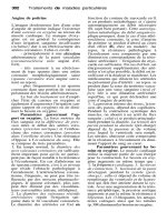
Fig. 11.7 Photomicrograph
of an intralabyrinthine cochlear
nerve schwannoma (T). The
endolymphatic hydrops (arrows) is
caused by tumor compression of
the ductus reuniens. R Reissner’s
membrane
.
. Internal Auditory Canal and Cerebellopontine Angle
IAC. In this location, the nerve is encountered before
tumor dissection is initiated. e results with hear-
ing preservation, however, are better than with other
approaches because the labyrinthine blood supply
is remotely located inferiorly in the IAC and can be
avoided.
11.2 Intralabyrinthine Vestibular/
Cochlear Schwannoma
e proliferation of Schwann cell neoplasms may be
limited to the bony labyrinth [1, 12, 22]. ese tumors
arise from the peripheral vestibular nerve branches,
aer leaving the cribrose portions of the otic capsule
and before supplying the vestibular sense organs. In
the cochlea, they arise from the dendrites of spiral
ganglion cells adjacent to the scala tympani (Fig. 11.7).
ese tumors are usually limited to the bony labyrinth
and are referred to as intralabyrinthine schwannomas.
e clinical presentation of the vestibular variety is
frequent recurrent vertigo, while the cochlear nerve
type is associated with sensorineural hearing loss, usu-
ally in the low frequencies. If an intracanalicular com-
ponent has been excluded by imaging studies, then
excision of the intralabyrinthine schwannoma may be
accomplished through the middle ear aer removal of
the promontory.
In the past, most cases of intralabyrinthine schwan-
noma have been recognized during labyrinthectomy
surgery for severe Ménière’s disease [12]. Now they
may be diagnosed preoperatively by MRI (Fig. 11.8).
e most common sensorineural hearing loss pattern
associated with the intralabyrinthine tumor is the as-
cending threshold pattern, seen with endolymphatic
hydrops [1], (Fig. 11.9). e most eective surgical ap-
proach to the detection and removal of these neural
tumors is by transcanal middle ear exposure of the ves-
tibule and cochlea aer removal of the promontory.
11.3 Benign Tumors of the Middle Ear
and Mastoid
Examples of these are glomus tumors, adenoma, low-
grade adenocarcinoma, and neurogenic tumors of
the middle ear. Clinical presentation is heralded by a
progressive conductive hearing loss, pulsatile tinnitus,
and a mass in the middle ear conrmed by neuroim-
aging of the TB. Computed tomography of the TB is
recommended to compliment MRI by evaluating bony
connes of the middle ear, especially the jugular fo-
ramen (JF).
Paraganglioma tumors that arise from glomus bod-
ies located along the course of the tympanic branch of
cranial nerve IX (Jacobsen’s) in the middle ear are clas-
sied as glomus tympanicum tumors. ose paragan-
glioma tumors that arise from glomus bodies located
in the adventia of the jugular bulb are classied as
glomus jugulare tumors. When these tumors are large
enough to be visible in the hypotympanum, the bony
margins of the jugular foramen have been eroded with
or without decits of the nerves passing through the
foramen.
Small glomus tympanicum tumors can be excised
though a tympanotomy approach (Fig. 11.10). Larger
Fig. 11.8 Gadolinium-en-
hanced MRI demonstrates an
intralabyrinthine cochlear schwan-
noma (arrow) in a patient with the
audiogram in Fig. 11.9
.
11
Chapter • Tumor Surgery
tumors lling the middle ear space require more expo-
sure provided by atticotomy and canaloplasty in order
to accomplish tumor removal with preservation of the
sound transmission system (20), (Fig. 11.11).
If CT indicates erosion of the bony limits of the
jugular foramen [21], then the presence of tumor
(glomus jugulare) arising in the JF with extension into
the middle ear must be assumed (Fig. 11.12). Addi-
tional neuroradiological studies are necessary to deter-
mine the size of tumor [11]. ese include MRI (Fig.
11.13) and arteriography (Fig. 11.14). Lateral skull
base approaches to the JF and middle ear are neces-
sary to control major vessels supplying the tumor, be-
Fig. 11.10 Axial CT scan demonstrates a small glomus tym-
panicum tumor (arrow)
.
Fig. 11.11 Coronal CT of a patient with larger glomus tym-
panicum lling the middle ear space (arrow )
.
Fig. 11.9 Low-frequency
sensorineural hearing associated
with intralabyrinthine cochlear
schwannoma
.
Fig. 11.12 Axial CT scan of skull base demonstrates erosion
of the jugular foramen (a rrows) in a patient with a glomus jugu-
lare tumor. F FN canal (mastoid)
.
. Benign Tumors of the Middle Ear and Mastoid
Fig. 11.17 Drawing of the
ndings at surgery in same pa-
tient. The tumor was completely
removed
.
Fig. 11.13 Gadolinium-enhanced coronal MRI of the glomus
jugulare tumor (arrow)
.
Fig. 11.14 Venous phase of arteriogram shows the intralumi-
nal extension of glomus jugulare tumor into the internal jugular
vein (arrow)
.
Fig. 11.15 Axial CT of an enlarged jugular foramen (arrows)
in a young woman with a red mass in the hypotympanum
.
Fig. 11.16 Arteriogram in same patient as in Fig. 11.15 shows
a spherical mass with mild vascular blush (arrows)
.
11
Chapter • Tumor Surgery
fore tumor resection. Transposition of the FN may or
may not be required for exposure of the JF and venous
structures [8]. Embolization of the tumor through the
external carotid system has not been eective in re-
ducing intraoperative bleeding, probably because of
signicant ow from tumor vessels arising from the
internal carotid artery. Preoperative assessment for
a catecholamine secreting paraganglioma should be
performed especially in the patient with a history of
elevated blood pressure.
Neurogenic (schwannomas) tumors arising from
nerves in the jugular foramen may closely mimic the
more vascular glomus jugulare tumor (Fig. 11.15).
Arteriography is the denitive study for this dier-
entiation [2]. e vascular supply in the neurogenic
tumor is far less prominent (Fig. 11.16) than it is in
the glomus (paraganglioma) tumor. Accordingly, the
surgical approach need not control the major vascu-
lar supply (ascending pharyngeal) in the neck when
dealing with neurogenic tumors in this location (Figs.
11.17, 11.18).
e histopathologic demonstration of neuro-
broma arising from the jugular foramen is shown in
Fig. 11.19. is 89-year-old female was diagnosed
with a glomus jugulare tumor causing decits of cra-
nial nerves VII, VIII, and X, and erosion of the jugular
foramen [3]. She received low-dose radiation therapy
recommended by Dr. Harvey Cushing and lived for
over 50 years with the tumor (shown in Fig. 11.19.)
Careful interpretation of CT, MRI, with arteriog-
raphy should be employed to eliminate false-positive
radiologic ndings in the skull base by imaging tech-
niques. Figure 11.20 is an MRI taken of a patient with
a 6-month history of pulsatile tinnitus in the right ear.
CT conrmed a large right jugular foramen with an in-
tact cortical rim (Fig. 11.21). Recommended vascular
studies failed to demonstrate neoplasm (Fig. 11.22).
11.4 Malignant Tumors of the TB
Malignant tumors of the outer ear (auricle and exter-
nal auditory meatus) are common (60%) and usually
of squamous cell, basal cell, and melanoma types [13].
ese are recognized early and resected completely
with generous margins, allowing for high curability.
Rarely regional node dissection is required unless sur-
rounding so tissue structures (i.e., parotid gland, au-
ricle) are involved.
Carcinoma (usually squamous cell) of the external
auditory canal is next in frequency (30% of malignant
ear neoplasms) and is causally related to chronic irrita-
tion (external otitis). e bony and cartilaginous canal
forms a compartment with the tympanic membrane as
Fig. 11.18 Histopathologically the tumor was classied as
schwannoma
.
Fig. 11.19 This vertical section through the TB of an 89-year-
old female with a jugular foramen schwannoma (T) that was
treated with radiation therapy over 50 years before her death.
The tumor arose from nerves of the jugular foramen (J) and
compressed the seventh and eighth nerves in the internal audi-
tory canal (arrow)
.
. Malignant Tumors of the TB
Fig. 11.22 Arteriogram conrms no neoplasm
in jugular foramen
.
Fig. 11.20 This gadolinium-
enhanced jugular foramen (arrow)
resembles a neoplasm
.
Fig. 11.21 Axial CT in same
patient in Fig. 11.20 shows intact
cortical rim of the jugular foramen
(arrow)
.
11
Chapter • Tumor Surgery
its medial boundary, and has sparse lymphatic drain-
age. ese anatomical features tend to restrict tumor
growth, allow for en bloc surgical resection, and lead
to very good curability (80%) (Fig. 11.23).
Resection of the external ear canal compartment
is referred to as lateral or partial TB resection. e
key to successful en bloc extirpation is identication
of the intratemporal course of the FN and completion
of appropriate bone cuts lateral to the fallopian canal
through the facial recess, tympanic bone, and epitym-
panum [5]. Vascularized muscle ap coverage of the
mastoid cavity is appropriate for postoperative radia-
tion therapy (Fig. 11.24). Occasionally tumor involve-
ment of the lateral half of the external ear canal may be
encompassed by transection of the bony canal lateral
to the tympanic membrane (Fig. 11.25).
Fig. 11.23 Specimen removed with partial TB resection dem-
onstrates carcinoma in deep external auditory canal (arrow).
M manubrium of malleus
. Fig. 11.24 Coronal CT of patient after partial TB resection
demonstrates muscle ap obliteration of the defect (arrow)
.
Fig. 11.25 Section through a
celloidin-embedded TB demon-
strates the medial resection plane
from subtotal (B) and lateral TB
resection. A plane for partial resec-
tion of the external ear canal. M
mastoid compartment, P PA, ICA
internal carotid artery, F FN, TM
temporomandibular joint, 8 eighth
nerve
.
. Malignant Tumors of the TB
Malignancy arising in or extending into the mid-
dle ear spreads through preformed bony pathways to
deeper portions of the TB, into vascular and neural
structures, and intracranially. Subtotal TB resection
carries risk to major vascular structures (internal ca-
rotid artery), brain injury, and intracranial infection
(Fig. 11.25). Cure rates of squamous cell carcinoma of
the middle ear by subtotal TB resection average 30%
[13, 14]. Similar cure rates have been reported with
radical mastoid–middle ear exenteration, followed
by radiation therapy. erefore, a clear case for the en
bloc approach to treatment of carcinoma in the middle
ear has not been made. e management of such cases
is best decided on case-by-case basis.
An exception in the treatment of malignancy in
the middle ear is the management of low-grade ad-
enocarcinoma or adenoma of the middle ear [4]. ese
neoplasms cause a conductive hearing loss and present
as a middle ear mass behind an intact tympanic mem-
brane. Complete piecemeal removal of these tumors
from the middle ear and its recesses is sucient for
cure with low morbidity (Figs. 11.26, 11.27).
11.5 Pseudoepithelial Hyperplasia
of External Ear Canal
e importance of recognizing pseudoepithelial hy-
perplasia (PH) is that, although it is a benign lesion,
it can, on clinical and histopathologic examination,
simulate an epithelial malignancy of the external audi-
tory canal (EAC) [6]. It is important to correlate the
clinical history and ndings with the histopathologic
presentation of lesions in the EAC. ese features are
important in dierentiating benign from malignant le-
sions of the EAC. Malignancy of the EAC usually has a
preceding history of chronic inammation and irrita-
tion of the ear canal (external otitis) or chronic otitis
media. A long history (years) of symptoms is usually
present before the development of malignancy. Clini-
cal symptoms usually consist of bloody discharge from
the ulcerated lesion of the EAC and pain in the ear
with or without radiation locally. Examination of the
ear usually reveals an ulcerated lesion in the EAC and/
or middle ear. On histologic examination, malignan-
cies are usually of the squamous cell type (SCC). Basal
cell carcinoma, adenocystic carcinoma, and melanoma
are less frequent lesions of the ear canal. On radiologic
examination, malignancy of the EAC may be associ-
ated with evidence of destruction of the bony ear ca-
nal initially and with neural decits (i.e., facial) in ad-
vanced lesions.
Benign lesions of the EAC, on the other hand, are
not usually associated with a bloody discharge from
the ear canal or otalgia. ese supercial lesions are
usually covered with intact epithelium, although ul-
ceration may be present. However, such ulceration
oen resolves with conservative measures employing
antibiotic and steroidal eardrops. e discharge from
the ear canal is usually of a much shorter duration
than found with malignancy. PH represents a reaction
of the epithelium of the ear canal to chronic irritation
and may clinically and histopathologically simulate
SCC (Fig. 11.28).
Since malignancy involving the EAC represents a
grave prognosis that justies aggressive surgical and
nonsurgical (radiation therapy) treatment, it is essen-
tial that histologic conrmation of epithelial malig-
Fig. 11.26 Low-grade malignancy of the middle ear (arrow),
with no evidence of bone erosion on CT scan
.
Fig. 11.27 Histopathologically the tumor was classied as
carcinoid tumor
.
11
Chapter • Tumor Surgery
nancy be ensured before such treatment be initiated.
e distinction between PH and SCC may be dicult
to make with certainty. e surgeon should provide
the pathologist with a favorable opportunity to make
this distinction by supplying a suciently large tissue
sample that includes the transition from normal to ab-
normal squamous epithelium. Generally, this means
total or subtotal excision of the granular lesion with
some surrounding epithelium. In addition, the clini-
cal response to a course of conservative treatment de-
signed to eliminate the irritative stimulus may help to
support the diagnosis of PH.
CO M P L I C AT I O N S TO AV O I D
1. FN monitoring is essential in vestibular schwan-
noma surgery to avoid FN injury.
2. Soft tissue obliteration of the dural defect fol-
lowing translabyrinthine removal of vestibular
schwannoma will prevent cerebrospinal fluid
leak.
3. When the facial nerve is resected in large ves-
tibular schwannoma, facial–hypoglossal nerve
anastomosis will prevent significant facial dis-
figurement.
4. Blood loss can be minimized during glomus
tumor surgery by the use of minipacks to com-
press the tumor.
5. Ligation of the internal jugular vein and the
sigmoid sinus will greatly reduce blood loss in
glomus jugulare surgery.
Pearl
• Microscopic diagnosis of squamous cell car-
cinoma of the external ear canal should be
carefully assessed and consistent with the
clinical presentation.
References
1. DeLozier H, Gacek R, Dana S (1979) Intralabyrinthine schwan-
noma. Ann Otol Rhinol Laryngol 88:187–191
2. Gacek RR (1976) Schwannoma of the jugular foramen. Ann Otol
Rhinol Laryngol 85:215–224
3. Gacek RR (1983) Pathology of jugular foramen neurobroma.
Ann Otol Rhinol Laryngol 92:128–133
4. Gacek RR (1992) Management of malignancy in the temporal
bone. In: Nadol JB, Schuknecht HF (eds) Surgery of the ear and
temporal bone. Raven, New York
5. Gacek RR, Goodman M (1977) Management of malignancy of the
temporal bone. Laryngoscope 87:1622–1634
6. Gacek M, Gacek R, Gantz B, McKenna M, Goodman M (1998)
Pseudoepithelial hyperplasia versus squamous cell carcinoma of
the external canal. Laryngoscope 108:620–623
7. Glasscock ME III (1968) Acoustic neuroma: recent advances in
the diagnosis and treatment. Rev Laryngol Otol Rhinol 89:28–42
8. Glasscock ME, Kveton JF (1987) erapy of glomus tumors of the
ear and skull base. In: awley S, Panje W, Batsakis J, Lindberg
R (eds) Comprehensive management of head and neck tumors.
Saunders, Philadelphia, pp 222–246
9. House WF (1961) Surgical exposure of the internal auditory canal
and its contents through the middle cranial fossa. Laryngoscope
71:1363–1385
10. House WF (1968) Monograph II acoustic neuroma. Arch Otolaryn-
gol 88:576–715
11. Jackson CG, Glasscock ME, Nissen AJ, Schwaber MK (1982)
Glomus tumor surgery: the approach, results, and problems.
Otolaryngology Clin North Am 15:897–916
12. Karlan MS, Basek M, Potter GB (1972) Intracochlear neurilem
-
oma. Arch Otolaryngol 96:573–575
13. Lewis JS (1960) Cancer of the ear: a report of 150 cases. Laryngo
-
scope 70:551–579
14. Lewis JS (1983) Surgical management of tumors of the middle ear
and mastoid. J Laryngol Otol. 97:299–311
15. Nadol JB Jr, Levine R, Ojemann RG, Martuza RL. Montgomery
WW, Klevins de Sandolval P (1987) Preservation of hearing in
surgical removal of acoustic neuromas of the internal auditory ca-
nal and cerebellopontine angle. Laryngoscope 97:1287–1294
16. Nager GT (1985) Acoustic neuromas. Acta Otolaryngol (Stockh)
99:245–261
Z
Fig. 11.28 Histopathologically, pseudoepithelial hyperplasia
can resemble squamous cell carcinoma. Arrow points to areas of
squamous cell breakthrough into subepithelial tissue layers
.
References
17. Ojemann RG, Montgomery WW, Weiss AD (1972) Evalua-
tion and surgical treatment of acoustic neuroma. N Engl J Med
287:895–899
18. Schuknecht HF (1977) Pathology of vestibular schwannoma
(acoustic neurinoma) In: Silverstein H, Norrell H (eds) Neurologi-
cal surgery of the ear. Aesculapius, Birmingham, Ala., pp 193–197
19. Skinner H (1929) Origin of acoustic nerve tumors. Br J Surg
16:440
20. Spector GJ, Maisel RH, Ogura JH (1973) Glomus tumors in
the middle ear. I. An analysis of 46 patients. Laryngoscope
83:1652–1672
21. Spector GJ, Compagno J, Perez CA, Maisel RH, Ogura JH
(1975) Glomus jugulare tumors: eects of radiotherapy. Cancer
35:1316–1321
22. Wanamaker H (1972) Acoustic neuroma: primary arising in the
vestibule. Laryngoscope 82:1040–1044
11
Chapter • Tumor Surgery
Cochlear implantation (CI) has been a relatively new
addition to the realm of otologic surgery over the past
20–25 years [1, 4, 9]. Its intent is to produce meaning-
ful electrical stimulation of the auditory nerve in those
individuals where degeneration of the sense organ has
progressed to the point where the external stimulation
(amplication) provided by hearing aids is no longer
eective.
e details of the evaluation process used to iden-
tify candidates for this form of auditory rehabilitation
are not covered in this section. e criteria for the
postlingually deafened individual are the clearest. Bi-
lateral profound sensorineural hearing loss that is no
longer aidable represents the indication for CI consid-
eration. e longer the time from reaching this level of
SNHL to the evaluation for implantation, the greater
the chance for auditory nerve degeneration and fewer
neurons available for stimulation. Protocols for this
evaluation process vary among institutions. Com-
monly they include careful audiometric assessment of
the hearing loss, psychologic evaluation of the patients
and their expectations, radiologic (CT) evaluation of
the middle ear, mastoid, and inner ears, and some es-
timation of auditory nerve reserve [5]. A well-trained
team of audiologists, speech pathologists, and social
workers are vital to the success of the cochlear implan-
tation project. Implantation in the congenitally deaf
child is more controversial but the centers performing
CI in this group report encouraging results.
Although extracochlear implantation has been used
early in the development of this concept, intracochlear
implantation is the preferred method of stimulation
used. Extracochlear implantation avoids the trauma of
intracochlear insertion and allows for stimulation sites
in the upper turns, but it has a major disadvantage in
a greater distance of electrodes from auditory neurons
requiring elevated thresholds for stimulation.
Although several variations of the surgical ap-
proach for intra cochlear implantation have been de-
scribed, there are anatomical and neuropathological
issues that are crucial to this method of auditory reha-
bilitation. e scala tympani is chosen for CI because
it is the larger of the two perilymphatic compartments
in the cochlea and allows the stimulating bipolar elec-
trodes of the prosthesis to be in close proximity to
Cochlear Implant Surgery
Core Messages
• Implantation of a multiple-channel electrode
prosthesis has proven to be a successful ap-
proach to the profoundly deafened patient,
acquired or congenital.
• e results in the post lingual deafened pa-
tient are superior to those in the patients
with congenital forms of profound hearing
loss.
• Insertion of the prosthesis close to spiral gan-
glion cells in Rosenthal’s canal (scala tympani
is the desired location).
• e intracochlear prosthesis may be intro-
duced via a transmastoid approach through
the facial recess or a transcanal approach
through the posterior epitympanic space.
Z
Fig. 12.1 Photomicrograph of cross section through a co-
chlear turn shows the proximity of the scala tympani (ST) to
spiral ganglion cells (SG) in Rosenthal’s canal. SV scala vestibuli,
OC organ of Corti
.
spiral ganglion cells and dendrites (Fig. 12.1). Fur-
thermore, the ionic composition of perilymph (high
Na
+
, low K
+
) is favorable for neural impulse genera-
tion. is requires low stimulation thresholds, with lit-
tle risk of spread to adjacent neurons. e dendrites
of spiral ganglion cells travel within the osseous spiral
lamina as bundles of myelinated nerve bers to dis-
crete areas of the basilar membrane (Fig. 12.2). Since
these nerve bers supply inner hair cells in these dis-
crete locations, frequency localization is maintained.
ese anatomical features allow for selective activation
of remaining auditory neurons.
e lesser curvature and diameter of the cochlear
basal turn permits insertion of present-day prostheses
up to a distance of 20–21 mm from the RWM. is
permits stimulation of two thirds of spiral ganglion
cells including some at the speech frequencies. e
minimum number of surviving neurons necessary for
successful stimulation is not known. However, some
histopathological studies have indicated that a third
(approximately 10,000) of the normal complement of
cochlear neurons is necessary for successful reactiva-
tion [6, 7]. When technological changes in the pros-
thesis permit further insertion of the scala tympani
into the middle and apical turns, it may be possible to
stimulate a greater number of ganglion cells.
Although insertion of the prosthesis through a co-
chleostomy in the round window niche was originally
selected for CI, many surgeons have favored a cochle-
ostomy site further up the basal turn (away from the
round window niche). is location avoids the cur-
vature of the hook portion of the basal turn, allowing
for a straightforward insertion into the upper basal
turn. Such a higher cochleostomy location permits a
fuller insertion of the prosthesis, reaching the location
for speech frequencies and bypassing little-used and
oen-degenerated neurons in the hook region of the
basal turn (Fig. 12.3).
e histopathological correlate of profound sen-
sorineural hearing loss is based on the behavior of the
auditory nerve to the sensory hair cells in the organ
of Corti. When the hair cells (inner) in the organ of
Corti degenerate because of peripheral pathology
(ototoxicity, infection, trauma, heredity, immunol-
ogy) a secondary degeneration of type I spiral gan-
glion cells, (90% of auditory nerve) follows in time [6].
is degeneration process is dependent on the loss of
peripheral trophic factors, which may have a variable
loss aer hair cell loss. At least a part of this trophic
factor loss may depend on the integrity of supporting
elements [8] in the organ of Corti (Fig. 12.4).
erefore, CI should be planned as soon as possible
aer the hearing impairment has reached a profound
level. is reduces the risk of further secondary neu-
Fig. 12.2 The myelinated den-
drites (arrows) travel in bundles
within the osseous spiral lamina to
innervate inner hair cells located
in discrete segments of the organ
of Corti. P heads of pillar cells in
organ of Corti, SG spiral ganglion
.
12
Chapter • Cochlear Implant Surgery
ronal loss by providing electrical stimulation. On the
other hand, primary neuronal degeneration (neoplas-
tic, surgical transection) with preservation of labyrin-
thine blood supply preserves the structural integrity of
the organ of Corti (Fig. 12.5). is form of profound
sensorineural hearing loss is not amendable to CI.
12.1 Surgery for Cochlear Implantation
e transmastoid approach utilizes a canal wall up
mastoidectomy to expose the facial recess (FR) ap-
proach to the round window niche (RWN). Exposure
of RWN is dependent on size of FR. If the FR cell is
Fig. 12.3 The solid line shows
the location of the RWM facing
the basal end of the scala tympani
in the human cochlea. There is
degeneration of myelinated
dendrites in the hook portion of
the basal turn (B). Note the sharper
curvature of the middle (M) and
apical (A) turns, which prevent
insertion of present day prosthesis.
.
Fig. 12.4 Cochlea of cat sub-
jected to excessive acoustic trau-
ma shows degeneration of over
50% of type I spiral ganglion cells
(I) secondary to the loss of sensory
and supporting cells in the organ
of Corti (arrow). S Rosenthal’s canal
.
. Surgery of Cochlear Implantation