ANATOMY, PHYSIOLOGY, AND DISORDERS OF THE AUDITORY SYSTEM - PART 1 pps
Bạn đang xem bản rút gọn của tài liệu. Xem và tải ngay bản đầy đủ của tài liệu tại đây (3.34 MB, 33 trang )
HEARING:
ANATOMY, PHYSIOLOGY,
AND DISORDERS OF THE
AUDITORY SYSTEM
Second Edition
This page intentionally left blank
HEARING:
ANATOMY,
PHYSIOLOGY, AND
DISORDERS OF THE
AUDITORY SYSTEM
Second Edition
A. R. Møller
School of Behavioral and Brain Sciences
University of Texas at Dallas
Texas
AMSTERDAM • BOSTON • HEIDELBERG • LONDON
NEW YORK • OXFORD • PARIS • SAN DIEGO
SAN FRANCISCO • SINGAPORE • SYDNEY • TOKYO
Academic Press is an imprint of Elsevier
Academic Press is an imprint of Elsevier
30 Corporate Drive, Suite 400, Burlington, MA 01803, USA
525 B Street, Suite 1900, San Diego, California 92101-4495, USA
84 Theobald’s Road, London WC1X 8RR, UK
This book is printed on acid-free paper.
Copyright © 2006, 2000 Elsevier Inc. All rights reserved.
No part of this publication may be reproduced or transmitted in any form or by
any means, electronic or mechanical, including photocopy, recording, or any
information storage and retrieval system, without permission in writing from
the publisher.
Permissions may be sought directly from Elsevier’s Science & Technology Rights
Department in Oxford, UK: phone: (+44) 1865 843830, fax: (+44) 1865 853333,
E-mail: You may also complete your request on-line via
the Elsevier homepage (), by selecting “Support & Contact”
then “Copyright and Permission” and then “Obtaining Permissions.”
Front cover design concept by Milda Dorsett.
Library of Congress Cataloging-in-Publication Data
Møller, Aage R.
Hearing : anatomy, physiology, and disorders of the auditory system/A.R.
Moller, 2nd ed.
p. cm.
Includes bibliographical references and index.
ISBN-13: 978-0-12-372519-6 (casebound : alk. paper)
ISBN-10: 0-12-372519-4 (casebound : alk. paper)
1. Hearing Physiological aspects. 2. Hearing disorders Pathophysiology.
3. Ear Anatomy. I. Title.
RF290.M58 2006
617.8 dc22 2006014244
British Library Cataloguing-in-Publication Data
A catalogue record for this book is available from the British Library.
ISBN 13: 978-0-12-372519-6
ISBN 10: 0-12-372519-4
For information on all Academic Press publications
visit our Web site at www.books.elsevier.com
Printed in the United States of America
060708091011987654321
Preface ix
Acknowledgements xi
Introduction xiii
SECTION
I
THE EAR
CHAPTER 1
Anatomy of the Ear
1. Abstract 3
2. Introduction 3
3. Outer Ear 3
3.1. Ear Canal 5
4. Middle Ear 6
4.1. Tympanic Membrane 6
4.2. Ossicles 8
4.3. Middle-ear Muscles 8
4.4. Eustachian Tube 8
4.5. Middle-ear Cavities 9
5. Cochlea 10
5.1. Organ of Corti 10
5.2. Basilar Membrane 13
5.3. Innervation of Hair Cells 13
5.4. Fluid Systems of the Cochlea 15
5.5. Blood Supply to the Cochlea 16
CHAPTER 2
Sound Conduction to the Cochlea
1. Abstract 19
2. Introduction 19
3. Head, Outer Ear and Ear Canal 20
3.1. Ear Canal 20
3.2. Head 20
3.3. Physical Basis for Directional Hearing 21
4. Middle Ear 22
4.1. Middle Ear as an Impedance
Transformer 24
4.2. Transfer Function of the Human
Middle Ear 27
4.3. Impulse Response of the Human
Middle Ear 29
4.4. Linearity of the Middle Ear 29
4.5. Acoustic Impedance of the Ear 29
4.6. Contributions of Individual Parts of the
Middle Ear to Its Impedance 32
C
HAPTER 3
Physiology of the Cochlea
1. Abstract 41
2. Introduction 41
3. Frequency Selectivity of the Basilar
Membrane 42
3.1. Traveling Wave Motion 43
3.2. Basilar Membrane Frequency Tuning
Is Non-linear 44
3.3. Frequency Tuning of the Basilar
Membrane 45
3.4. Role of the Outer Hair Cells in Basilar
Membrane Motion 46
3.5. Epochs of Research in Cochlear
Mechanics 47
4. Sensory Transduction in the Cochlea 48
4.1. Excitation of Hair Cells 48
4.2. Which Phase of a Sound Excites Hair Cells
(Rarefaction or Condensation)? 48
4.3. Molecular Basis for Sensory
Transduction 50
4.4. Endocochlear Potential 52
4.5. Cochlea as a Generator of Sound 53
4.6. Efferent Control of Hair Cells 55
4.7. Autonomic Control of the Cochlea 55
5. Autoregulation of Blood Flow to the Cochlea 56
Contents
v
CHAPTER 4
Sound Evoked Electrical Potentials
in the Cochlea
1. Abstract 57
2. Introduction 57
3. Electrical Potentials in the Cochlea 57
3.1. Cochlear Microphonics 58
3.2. Summating Potential 59
3.3. Action Potential 59
3.4. Electrocochleographic Potentials 64
Section I References 68
SECTION
II
THE AUDITORY NERVOUS SYSTEM
CHAPTER 5
Anatomy of the Auditory Nervous System
1. Abstract 75
2. Introduction 76
3. Classical Ascending Auditory Pathways 76
3.1. Auditory Nerve 76
3.2. Cochlear Nucleus 80
3.3. Superior Olivary Complex 81
3.4. Lateral Lemniscus and Its Nuclei 81
3.5. Inferior Colliculus 81
3.6. Medial Geniculate Body 81
3.7. Auditory Cerebral Cortex 82
3.8. Differences between the Classical Auditory
Pathways in Humans and in Animals 84
4. Non-classical Ascending Auditory Pathways 85
5. Parallel Processing and Stream Segregation 87
5.1. Parallel Processing 88
5.2 Stream Segregation 88
5.3. Connections to Non-auditory Parts of the
Brain 89
6. Descending Pathways 89
CHAPTER 6
Physiology of the Auditory Nervous
System
1. Abstract 93
2. Introduction 94
3. Representation of Frequency in the Auditory
Nervous System 95
3.1 Hypotheses about Discrimination
of Frequency 95
3.2. Frequency Selectivity in the Auditory
Nervous System 97
3.3. Cochlear Non-linearity Is Reflected in
Frequency Selectivity of Auditory Nerve
Fibers 99
3.4. Frequency Tuning in Nuclei of the
Ascending Auditory Pathways 102
3.5. Tonotopic Organization in the Nuclei of the
Ascending Auditory Pathways 104
3.6. Extraction of Information from Place
Coding of Frequency 106
4. Coding of Temporal Features 106
4.1. Coding of Periodic Sounds 107
4.2. Extraction of Information from the
Temporal Pattern of Neural Discharges 111
5. Is Temporal or Place Code the Basis for
Discrimination of Frequency? 112
5.1. Temporal Hypothesis for Frequency
Discrimination of Complex Sounds 112
5.2. Place Hypothesis for Frequency
Discrimination of Complex Sounds 113
5.3. Preservation of the Temporal Code of
Frequency 115
5.4. Preservation of the Place Code 117
5.5. Robustness of the Temporal Code 117
5.6. Robustness of the Place Code of
Frequency 117
5.7. Coding of Speech Sounds 117
5.8. A Duplex Hypothesis of Frequency
Discrimination 118
5.9. Cochlear Spectral Filtering May Be
Important in other Ways than Frequency
Discrimination 118
5.10. Speech Discrimination on Spectral
Information Only 118
5.11. Conclusion 119
6. Coding of Complex Sounds 119
6.1. Response to Tone Bursts 120
6.2. Coding of Small Changes in
Amplitude 121
6.3. Response to Tones with Changing
Frequency 128
6.4. Selectivity to Other Temporal Patterns
of Sounds 138
6.5. Coding of Sound Intensity 140
6.6. Conclusion 140
7. Directional Hearing 142
7.1. Physical Basis for Directional Hearing 142
7.2. Neurophysiologic Basis for Sound
Localization 143
7.3. Localization in the Vertical Plane 146
7.4. Representation of Auditory Space
(Maps) 146
8. Efferent System 149
9. Non-classical Pathways 149
10. Effect of Anesthesia 150
vi Contents
CHAPTER 7
Evoked Potentials from the Nervous
System
1. Abstract 151
2. Introduction 152
3. Near-field Potentials from the Auditory
Nervous System 152
3.1. Recordings from the Auditory Nerve 152
3.2. Recordings from the Cochlear
Nucleus 160
3.3. Recordings from More Central Parts of the
Ascending Auditory Pathways 163
4. Far-field Auditory Evoked Potentials 163
4.1. Auditory Brainstem Responses 165
4.2. Middle Latency Responses 175
4.3. Far-field Frequency Following Responses
in Humans 176
4.4. Myogenic Auditory Evoked Potentials 177
CHAPTER 8
Acoustic Middle-ear Reflex
1. Abstract 181
2. Introduction 181
3. Neural Pathways of the Acoustic Middle-ear
Reflex 182
4. Physiology 183
4.1. Responses to Stimulation with Tones 184
4.2. Functional Importance of the Acoustic
Middle-ear Reflex 187
4.3. Non-acoustic Ways to Elicit Contraction of the
Middle-ear Muscles 190
4.4. Stapedius Contraction May Be Elicited before
Vocalization 190
5. Clinical Use of the Acoustic Middle-ear Reflex 190
Section II References 192
SECTION
III
DISORDERS OF THE AUDITORY
SYSTEM AND THEIR
PATHOPHYSIOLOGY
CHAPTER 9
Hearing Impairment
1. Abstract 205
2. Introduction 206
3. Pathologies of the Sound Conducting
Apparatus 206
3.1. Ear Canal 207
3.2. Middle Ear 207
3.3. Impairment of Sound Conduction in the
Cochlea 213
3.4. Accuracy of Measurements of Conductive
Hearing Loss 213
3.5. Implications of Impairment of Conduction
of Sound to the Cochlea 214
4. Pathologies of the Cochlea 215
4.1. General Audiometric Signs of Cochlear
Pathologies 215
4.2. Age-related Hearing Loss (Presbycusis) 216
4.3. Noise Induced Hearing Loss 219
4.4. Implications of Hearing Loss on Central
Auditory Processing 226
4.5. Modification of Noise Induced Hearing
Loss 226
4.6. Hearing Loss Caused by Ototoxic Agents
(Drugs) 227
4.7. Diseases that Affect the Function of the
Cochlea 229
4.8. Congenital Hearing Impairment 233
4.9. Infectious Diseases 234
4.10. Perilymphatic Fistulae 234
4.11. Changes in Blood Flow in the Cochlea 234
4.12. Injuries to the Cochlea from Trauma 234
4.13. Sudden Hearing Loss 234
5. Implications of Hearing Loss on Central Auditory
Processing 235
5.1 Neural Components of Hearing Loss 236
5.2. Role of Expression of Neural Plasticity 237
6. Pathologies from Damage to the Auditory
System 239
6.1. Auditory Nerve 239
6.2. Other Space-occupying Lesions 243
7. Pathologies of the Central Auditory Nervous
System 243
7.1. Disorders of the Brainstem Auditory
Pathways 244
7.2. Auditory Cortices 244
7.3. Efferent System 246
7.4. Pathologies that Can Affect Binaural
Hearing 246
7.5. Viral Infections 246
7.6. Ototoxic Drugs 247
7.7. Sudden Hearing Loss 247
8. Role of Neural Plasticity in Disorders of the
Central Auditory Nervous System 247
8.1. What Is Neural Plasticity? 247
8.2. What Can Initiate Expression of Neural
Plasticity? 250
Contents vii
CHAPTER 10
Hyperactive Disorders of the Auditory
System
1. Abstract 253
2. Introduction 253
3. Subjective Tinnitus 254
3.1. Assessment of Tinnitus 254
3.2. Disorders in which Tinnitus Is
Frequent 255
3.3. Causes of Subjective Tinnitus and Other
Hyperactive Symptoms 255
3.4. Role of Expression of Neural Plasticity
in Tinnitus 259
4. Abnormal Perception of Sounds 260
4.1. Hyperacusis 261
4.2. Phonophobia 262
4.3. Misophonia 262
4.4. Recruitment of Loudness 262
5. Treatment of Subjective Tinnitus 263
5.1. Medical Treatment 264
5.2. Electrical Stimulation 264
5.3. Surgical Treatment 265
5.4. Desensitization 266
6. Treatment of Hyperacusis 266
CHAPTER 11
Cochlear and Brainstem Implants
1. Introduction 267
2. Cochlear Implants 268
2.1. Development of Cochlear Implants 268
2.2. Function and Design of Cochlear
Implants 268
2.3. Physiological Basis for Cochlear Implants 273
2.4. Coding of Sound Intensity 275
2.5. Functions that Are Not Covered by Modern
Cochlear Implants 275
2.6. Success of Cochlear Implants 276
2.7. Selection Criteria for Cochlear Implant
Candidates 277
3. Cochlear Nucleus Implants 277
3.1. Function and Design of Auditory
Brainstem Implants 277
3.2. Physiological Basis for Auditory Brainstem
Implants 277
3.3. Success of Auditory Brainstem Implants 278
3.4. Patient Selection for Auditory Brainstem
Implants 279
4. Role of Neural Plasticity 279
Section III References 280
APPENDIX A
Definitions in Anatomy 289
APPENDIX B
Hearing Conservation Programs 291
1. Introduction 291
2. Purpose and Design of Hearing Conservation
Programs 292
2.1. Basis for Hearing Conservation Programs 292
3. Establishment of Noise Standards 295
3.1. Noise Level and Exposure Time 296
3.2. Effect of Age-related Hearing Loss 296
3.3. What Degree of Hearing Loss is
Acceptable? 296
4. Measurement of Noise 297
4.1. Sound Level Meters 297
4.2. Noise Dosimeters 298
5. Personal Protection 299
5.1. Earplugs and Earmuffs 299
5.2. Active Noise Cancellation 300
5.3. Other Means of Reducing the Risk of Noise
Induced Hearing Loss 300
6. Non-occupational Noise Exposure 300
7. Effect of Noise on Bodily Functions 300
7.1. Effect of Ultrasound and Infrasound 300
Appendices References 301
List of Abbreviations 303
Index 305
viii Contents
Preface
ix
This book is intended for otologists, audiologists,
neurologists and researchers in the field of hearing. The
book will also be of interest to psychologists and psy-
chiatrists who treat patients with tinnitus and other
hyperactive auditory disorders. The book provides the
basis for a broad understanding of the anatomy and
function of the ear and the auditory nervous system,
and it discusses the cause and treatment of hearing
disorders. Most books on hearing focus either on the
anatomy and function of the ear, the auditory nervous
system or on peripheral or central hearing disorders.
This book covers both anatomy and physiology of the
ear and the nervous system. The book also provides a
comprehensive coverage of disorders of the auditory
system emphasizing the interaction between patholo-
gies of the middle ear and the cochlea and the function
of the nervous system and vice versa. Hyperactive dis-
orders of the auditory nervous system and the role of
expression of neural plasticity in causing auditory
symptoms are also topics of the book. An extensive list
of references makes it possible for the reader to find
original work on the different subjects.
Understanding of the anatomy and the function of
the auditory system together with knowledge about
the pathophysiology of the auditory system are essen-
tial for all clinicians who are involved in diagnosis and
treatment of disorders of the auditory system. The
book prepares the clinician and the clinical researcher
for the challenges of the modern clinical auditory disci-
pline. The book also provides basic information about
the auditory system in a form that is suitable for the sci-
entist who does basic research on the auditory system.
The book thus aims at cross-fertilization between clini-
cians, clinical researchers and basic scientists. It is my
hope that such knowledge can guide basic auditory
research into clinically relevant questions.
The book is the third edition of books on the auditory
system, the first, Auditory Physiology, published in 1983
by Academic Press, and the second, Hearing: Its
Physiology and Pathophysiology, published in 2000, also
by Academic Press.
The book has 11 chapters that are organized in three
sections. Chapters from earlier editions have been
re-organized and most parts have been re-written and
new information has been added. A separate chapter
is devoted to an extended coverage of hyperactive
disorders, most importantly tinnitus, the cause and
treatment of which is discussed in detail. A new chap-
ter describes cochlear and brainstem implants and
hearing conservation programs are discussed in an
appendix.
The four chapters of Section I cover anatomy and
physiology of the middle ear and the cochlea, includ-
ing a chapter on the electrical potentials that are gen-
erated by the cochlea. Section II has two chapters that
cover anatomy and physiology of the nervous system.
Both the classical and the less known non-classical
(extralemniscal) auditory pathways are covered exten-
sively. The latter is involved in some forms of tinnitus
and may be activated in other disorders also. A third
chapter is devoted to evoked potentials from the nerv-
ous system. The neural generators of the ABR are dis-
cussed in detail. The anatomy and physiology of the
acoustic middle-ear reflex is covered in a fourth chapter
in this section.
The final section (Section III) discusses disorders of
the auditory system. Two chapters regard hearing
impairment and hyperactive disorders, focusing on tin-
nitus, its etiology, and treatment. These two chapters
stress the role of expression of neural plasticity. A third
chapter in this section concerns cochlear implants and
auditory brainstem implants. The basic design and
function of the processors in these modern auditory
prostheses are described and the physiologic basis for
the function of these prostheses is discussed. An
appendix discusses hearing conservation programs.
This page intentionally left blank
Acknowledgements
xi
I want to thank Hilda Dorsett for help with the new
artwork and for revising some of the illustrations from
the first edition of the book and Karen Riddle for tran-
scribing many of the revisions of the manuscript. I also
want to thank Johannes Menzel, Senior Publishing
Editor, Elsevier Science, and Heather Furrow and John
Donahue, Project Managers, Elsevier, Burlington, MA
for their excellent work on the book.
I would not have been able to write this book with-
out the support from the School of Behavioral and
Brain Sciences at the University of Texas at Dallas.
Last but not least I want to thank my dear wife,
Margareta B. Møller, MD, DMedSci., for her support
during writing of this book and for her valuable
comments on earlier versions of the manuscripts for
this book.
Dallas, November, 2005
Aage R. Møller
This page intentionally left blank
It is now recognized that disorders of one part of the
auditory system often affect the function of other parts
of the auditory system. This is especially apparent
with regard to hyperactive disorders such as tinnitus
and hyperacusis, but even noise induced hearing loss
and presbycusis are not isolated cochlear phenomena,
for the auditory nervous system is involved in these
disorders. Expression of neural plasticity and a com-
plex series of events seem to be necessary in order that
such pathologies become manifest. This means that it
is no longer valid to divide disorders of the auditory
system in to peripheral and central disorders. This
book therefore takes an integrated approach to disor-
ders of the auditory system.
While most disorders of the auditory system have
detectable morphologic abnormalities, hyperactive
disorders lack such detectable morphologic changes,
and even other objective signs are often absent.
Symptoms such as tinnitus, hyperacusis, and phono-
phobia even involve physiological abnormalities in
other parts of the central nervous system than the clas-
sical auditory pathways. A part of the auditory nervous
system, known as the non-classical, or extralemniscal,
auditory pathways, seems to be involved in some of
these hyperactive disorders, and that may also cause
abnormal activation of structures of the limbic system,
which can explain why patients with tinnitus often
present with symptoms of affective disorders such as
fear and depression. The role of the non-classical audi-
tory nervous system may have much wider importance
than previously known. This book provides a thorough
description of the anatomy and physiology of this part
of the auditory nervous system and it discusses how
their function can change and cause different symp-
toms. The book also covers less common disorders such
as bilirubinemia and cortical lesions and it discusses
vestibular Schwannoma and their diagnosis.
Because of the complexity of many disorders of the
auditory system the clinician must have a thorough
understanding of the basic functions of the entire audi-
tory system and the interactions between the periph-
eral and the central portions of the auditory system
that may occur in various hearing disorders.
Cochlear implants now provide an effective way to
treat severe hearing loss. The implementation of
cochlear and brainstem implants requires a thorough
knowledge not only about the function of such devices
but also an understanding of the way sounds are nor-
mally coded and processed in the nervous system is a
prerequisite for understanding how such prostheses
can provide useful hearing. The more recent addition
to auditory prostheses, namely auditory brainstem
(cochlear nucleus) implants, present an even greater
challenge for the clinician and there are ample possi-
bilities to do important research in this area. Aseparate
chapter in the book deals with cochlear and brainstem
(cochlear nucleus) implants and the physiological
basis for their success is discussed.
Cochlear implants and auditory brainstem implants
do not provide the same coding of sounds in the nerv-
ous system as provided by the normal ear and expres-
sion of neural plasticity is essential for the success of
such prostheses. Thus, optimal implementation of
such prostheses requires understanding of basic audi-
tory physiology.
The advent of these new aspects in treatment of dis-
orders of the auditory system should not detract atten-
tions from classical problems such as hearing loss from
middle ear and cochlear pathologies. Also in these areas
of hearing new knowledge has contributed to better
understanding of pathologies of the auditory system.
The surprising research results that show that exposure
to sound can reduce presbycusis and that noise induced
hearing loss is affected by pre-exposure to sound are
examples of signs of a greater complexity of disorders
of the auditory system than previously assumed. The
results indicate that the auditory nervous system is
involved in disorders that earlier were assumed to be
Introduction
xiii
caused solely by morphological changes in the cochlea.
This means that altered function of the nervous system
caused by altered input contributes to the symptoms
and signs of such disorders. This book provides insight
into the physiologic basis for the involvement of the
auditory nervous system in disorders that earlier were
assumed to only involve the ear. The role of expression
of neural plasticity in creating the symptoms and signs
of these disorders is discussed.
Understanding how electrical potentials are gener-
ated in the auditory nervous system is a prerequisite
for correct interpretation of clinical tests that make use
of such recordings. The book describes the various
electrical potentials that are generated in the auditory
nervous system, what anatomical structures generate
the different components of such far-field potentials as
the ABR and the MLR, and how these potentials are
affected by different types of pathologies.
Prevention of hearing loss is important and audio-
logists and otolaryngologists play important roles in
reducing the risk of noise induced hearing loss. The
basis for that is discussed in several chapters in the
book. The practical and legal aspects of hearing con-
servation are covered in an appendix.
xiv Introduction
The ear as a sensory organ is far more complex than other sensory organs. The sensory
cells are located in the cochlea but the cochlea not only serves to convert sound into
a code of neural impulses in the auditory nerve but it also performs the first analysis
of sounds that prepare sounds for further analysis in the auditory nervous system.
This analysis consists primarily of separating sounds into bands of frequencies before
they are coded in the discharge pattern of individual auditory nerve fibers. The separa-
tion of sounds is accomplished by the properties of the basilar membrane and the
sensory cells that are located along its length. The cochlea is more frequency selective
for weak sounds than louder sounds, which facilitates detection of weak sounds. The
cochlea also compresses the amplitudes of sounds, which makes it possible to code
sounds within the very large range of sound intensities that is covered by normal hearing.
Without such amplitude compression the ear could not detect and analyze sounds in
the intensity range of normal hearing.
The cochlea is fluid filled and that means that sounds must be converted into vibra-
tions of fluid in order to activate the sensory cells. Direct transfer of sound to a fluid is
ineffective. The middle ear facilitates the transfer of sound to the cochlea by acting as
a transformer that matches the impedance of the air to that of the cochlea. The middle
ear is the only part of the entire auditory system where medical or surgical interven-
tions can remedy hearing loss from disease processes or trauma.
During the past decade or so, our understanding of the function of the cochlea has
changed in a fundamental way and its function now appears far more complex than
perceived earlier. Earlier it was believed that the basilar membrane was a linear system
where properties determined at one sound intensity were directly applicable to all
sound intensities. More recently, it has become evident that the frequency selectivity of
SECTION
I
THE EAR
Chapter 1 Anatomy of the Ear
Chapter 2 Sound Conduction to the Cochlea
Chapter 3 Physiology of the Cochlea
Chapter 4 Sound Evoked Electrical Potentials in the Cochlea
the basilar membrane depends on the sound intensity. Earlier it was believed that the
function of hair cells was limited to transducing the vibration of the basilar membrane
into a neural code. The discovery that hair cells also can change their length in response
to sound and thus interact with the vibration of the basilar membrane in addition to
being transducers radically changed our perception of the function of the cochlea. The
best description of the function of outer hair cells is that they act as “motors” that coun-
teract the frictional losses of energy in the cochlea. This particular function of outer hair
cells increases the sensitivity of the ear by approximately 50dB. The discovery of the
active role of outer hair cells explains how the loss of outer hair cells causes hearing
loss. The interaction between the hair cells and the basilar membrane vibration makes
the cochlea more complex than other sensory organs. Extensive research during many
years has resulted in more knowledge being accumulated about the function of the
cochlea than of any other sensory organ.
3
HEARING: ANATOMY, PHYSIOLOGY, Copyright © 2006 by Academic Press, Inc.
AND DISORDERS OF THE AUDITORY SYSTEM Second Edition All rights of reproduction in any form reserved.
1. ABSTRACT
1. The ear consists of the outer ear, the middle ear
and the inner ear.
2. The outer ear consists of the pinna and the ear canal.
3. The skin of the ear canal is innervated by four
cranial nerves: the trigeminal; the facial; the
glossopharyngeal; and the vagus nerves.
4. The middle ear consists of the tympanic membrane
and three ossicles: malleus; incus; and stapes.
5. Two muscles are attached to the ossicles: the tensor
tympani to the manubrium of malleus; and the
stapedius to the stapes. The tensor tympani muscle
is innervated by the trigeminal nerve and the
stapedius muscle is innervated by the facial nerve.
6. The cochlea in humans has a little more than
2
1
/
2
turns.
7. The cochlea has three fluid-filled compartments: the
scala tympani; scala media; and the scala vestibuli.
The basilar membrane separates the scala media
from the scala tympani and Reissner’s membrane
separates the scala vestibuli from the scala media.
8. The ionic composition of the fluid in scala tympani
and scala vestibuli (perilymph) is similar to that
of extracellular fluid (high contents of sodium,
low contents of potassium), while the fluid in
scala media (endolymph) is similar to intracellular
fluid (high contents of potassium, low contents
of sodium).
9. The fluid space of scala tympani and scala vestibuli
communicates with the cerebrospinal fluid space
through the cochlear aqueduct. The fluid space in
scala media communicates with the endolymphatic
sac through the endolymphatic canal.
10. Hair cells are organized along the basilar
membrane in one row of inner hair cells and
3–5 rows of outer hair cells.
11. The hair cells of the cochlea differ from vestibular
hair cells in that they lack a kinocilium.
12. Each inner hair cell is innervated by many
(type I) auditory nerve fibers, while each
(type II) nerve fiber innervates many outer
hair cells.
13. Efferent nerve fibers terminate directly onto outer
hair cells while other efferent fibers terminate on
the dendrites of the type I fibers that innervate
the inner hair cells.
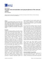
2. INTRODUCTION
The ear (Fig. 1.1) consists of three parts: the outer
ear; the middle ear; and the inner ear. The inner ear
consists of two parts: the vestibular apparatus for bal-
ance; and the cochlea for hearing. The outer ear and
the middle ear conduct sound to the cochlea, which
separates sounds with regard to frequency before they
are transduced by the hair cells into a neural code in
the fibers of the auditory nerve.
3. OUTER EAR
The different parts of the external ear, “the auricle,”
have specific names (Fig. 1.2). The groove called the
CHAPTER
1
Anatomy of the Ear
4 Section I The Ear
FIGURE 1.1 (A) Localization of the ear in the head (after Melloni, 1957). (B) Cross-section of the human ear
(reprinted from Brodel, 1946).
concha is acoustically the most important. The outer
ear enlarges in older individuals, especially in men.
3.1. Ear Canal
The ear canal has a length of approximately 2.5 cm
and a diameter of approximately 0.6 cm. It has the
shape of a lazy S. The most medial part is a nearly
circular opening in the skull bone, and the outer part
is cartilage. The outer cartilaginous portion of the
ear canal is also nearly circular in young individuals
but with age the cartilaginous part often changes
shape and attains an oval shape. In addition to chang-
ing its shape with age, the lumen of the ear canal often
becomes smaller with age, and in avid swimmers, it
may become very narrow.
The ear canal is covered by skin that secrets ceru-
men (wax) and it has hairs on its surface. There are no
sweat glands in the ear canal. Since the skin is not
rubbed naturally, as exposed skin on other parts of
the body, it must self clean dead cells and cerumen.
Two types of cells contribute to secretion of cerumen,
namely sebaceous cells located close to the hair folli-
cles and ceruminous glands. The sebaceous glands
cannot secrete actively but form their secretion by pas-
sive breakdown of cells. Two kinds of cerumen exist,
dry and wet.
Chapter 1 Anatomy of the Ear 5
FIGURE 1.2 Schematic drawing of the human external ear showing
components that are of importance for sound conduction: pinna flange
(helix, antihelix, and lobule), concha (cymba and cavum), and ear canal
(reprinted from Shaw, E. A. C. 1974. The external ear. In: Keidel, W. D.
and Neff, W. D. (eds) Handbook of sensory physiology V(1). New York:
Springer-Verlag, pp. 455–490, with permission from Springer).
BOX 1.1
CERUMEN
The dry type is found mostly in people in the Orient
and in Mongolians while the wet cerumen is found mostly
in Caucasians, Africans and Hispanic people [60, 143].
The kind of cerumen is genetically related and chromo-
some 16 has been identified as carrying the cerumen
locus.
Accumulation of cerumen in the ear canal to an extent
that it becomes occluded is a common cause of hearing
impairment. Cerumen may also cover the tympanic
membrane, which causes hearing loss. Individuals who
attempt to clean their ear canals by cotton swaps often
push cerumen deeper into the ear canal. The cerumen is
supposed to become dry and leave the ear canal. The
secreted cerumen has a slight anti-bacterial and anti-
fungal property and it may act as an insect repellant.
The outer layer of the skin (epidermis) in the ear canal,
together with that of the tympanic membrane migrates
outwards. The migration helps heal small injuries and
move scars outwards as well as transporting cerumen out
of the ear canal. It has been suggested that failure in this
migration of the epidermis may cause several kinds of
pathology such as development of cholesteatoma and it
may play a role in causing inflammation of the ear canal.
The skin of the ear canal has an unusual nerve supply.
Its sensory receptors (including bare axons) are inner-
vated by four different cranial nerves (CN), namely the
sensory portion of the mandibular division of the trigem-
inal nerve (CN V), the facial nerve (CN VII) the glos-
sopharyngeal nerve (CN IX) and the auricular branch of
the vagal nerve (CN X), which supplies the posterior wall
of the ear canal and the tympanic membrane. This nerve
branch is a part of Arnold’s nerve, which also receives
contributions from the glossopharyngeal nerve. The
innervation of the ear canal by the glossopharyngeal
nerve explains why many people cough when the skin of
the inner part of the ear canal is touched. The innervation
by the glossopharyngeal and the vagal nerve explain why
mechanical stimulation of the ear canal can affect the
heart and blood circulation and cause sensitive individu-
als to faint when the ear canal is cleaned for wax.
6 Section I The Ear
4. MIDDLE EAR
The middle ear consists of the tympanic membrane
that terminates the ear canal (Fig. 1.3) and the three
small bones (ossicles), the malleus, the incus and the
stapes (Fig. 1.3 and Fig. 1.4). Two small muscles, the
tensor tympani muscle and the stapedius muscle, are
also located in the middle ear. The manubrium of
malleus is imbedded in the tympanic membrane and
the head of the malleus is connected to the incus that
in turn connects to the stapes, the footplate of which is
located in the oval window of the cochlea. The chorda
tympani is a branch of the facial nerve (the nervous
intermedius) that travels across the middle ear cavity
(Fig 1.4). It carries taste fibers and probably also pain
fibers. The Eustachian tube connects the middle ear
cavity to the pharynx.
4.1. Tympanic Membrane
The tympanic membrane (Fig. 1.5) is a slightly oval,
thin membrane that terminates the ear canal. It is
cone-shaped, with an altitude of 2 mm with the apex
pointed inward. Seen from the ear canal, the mem-
brane is slightly concave and is suspended by a bony
ring. Normally it is under some degree of tension. Its
surface area is approximately 85 mm
2
. The main part
of the tympanic membrane, the pars tensa with an area
of approximately 55 mm
2
(Fig. 1.5), is composed of
radial and circular fibers overlaying each other. These
fibers are comprised of collagen and they provide a
lightweight stiff membrane that is ideal for converting
sound into vibration of the malleus. A smaller part of
the tympanic membrane, the pars flaccida, located
above the manubrium of malleus, is thicker than the
pars tensa and its fibers are not arranged as orderly as
FIGURE 1.3 (A) Cross-section showing the middle ear (reprinted from Brodel, 1946, with permission from
Elsevier).
Chapter 1 Anatomy of the Ear 7
FIGURE 1.3 (Continued) (B) Schematic drawing of the human middle ear seen from inside the head (from
Møller, 1972, with permission from Elsevier).
FIGURE 1.4 The ossicular chain as it is normally placed within the middle-ear cavity (adapted from Tos,
1995, with permission from Thieme Medical Publishers).
the collagen fibers of the pars tensa. The tympanic
membrane is covered by a layer of epidermal cells,
continuous with the skin in the ear canal. This outer
layer of the tympanic membrane migrates from its
center outwards and this moves small injuries and
scars and transports small foreign bodies out into the
ear canal. Small holes in the tympanic membrane
usually heal spontaneously.
4.2. Ossicles
The middle-ear bones are suspended by several liga-
ments (Figs 1.3 and 1.4). The manubrium of the malleus
is embedded in the tympanic membrane with the tip of
the manubrium located at the apex of the tympanic
membrane (Fig. 1.5). The head of the malleus is sus-
pended in the epitympanum. The short process of the
incus rests in the fossa incudo of the malleus, and it is
held in place by the posterior incudal ligament. The long
process, also called the lenticular process, of the incus
forms one side of the incudo-stapedial joint. The head of
the malleus and the incus are fused together in a double
saddle joint and the joint between these two bones is
regarded to be rigid. The joint between the incus and
the stapes is rigid for movement of the stapes towards
the cochlea (piston like movements), but the joint is
flexible for movements of the stapes that are induced
by contraction of the stapedius muscle. The stapes is
suspended in the oval window of the cochlea by two
ligaments and one ligament is stiffer than the other.
4.3. Middle-ear Muscles
Two small muscles are located in the middle ear. One,
the tensor tympani muscle, is attached to the manubrium
of the malleus and the other, the stapedius muscle, is
attached to the stapes (Figs 1.3 and 1.4). The tensor
tympani muscle extends between the malleus and the
wall of the middle-ear cavity near the entrance to the
Eustachian tube. When contracting, it pulls the manu-
brium of the malleus inward, displacing the tympanic
membrane inwards and stretching the membrane. The
stapedius muscle is the smallest striate muscle of the
body. It is attached to the head of the stapes and most
of the muscle is located in a bony canal. It pulls the
stapes in a direction that is perpendicular to its piston-
like motion, tilting the stapes so that it rotates around
its posterior ligament. The tensor tympani muscle is
innervated by the trigeminal nerve (CN V) and the
stapedius muscle by the facial nerve (CN VII).
4.4. Eustachian Tube
The Eustachian tube consists of a bony part (the
protympanum) that is located close to the middle ear
cavity, and a cartilaginous part that forms a closed
slit where it terminates in the nasopharynx (Fig. 1.6).
8 Section I The Ear
FIGURE 1.5 The tympanic membrane and the position of the
malleus and incus (reprinted from Anson and Donaldson, 1973, with
permission from Elsevier).
FIGURE 1.6 (A) Cross-section of the human middle ear to show
the Eustachian tube.
The optimal function of the middle ear depends on
keeping the air pressure in the middle-ear cavity close
to the ambient pressure. That is accomplished by
briefly opening the Eustachian tube. In the adult, the
Eustachian tube is 3.5–3.9 cm long and it follows an
inferior (caudal) – medially – anterior (ventral) direc-
tion in the head, tilting downwards (caudally) by
approximately 45 degrees to the horizontal plane (Fig.
1.6B). The Eustachian tube is shorter in young children
and it is directed nearly horizontally.
The cartilaginous part of the Eustachian tube forms
a valve that closes the middle ear off from pressure
fluctuations in the pharynx such as occurs during
breathing and it decreases transmission of a person’s
voice to the middle-ear cavity. The mucosa inside the
Eustachian tube (which really is not a tube except for
the bony part) is rich in cells that produce mucus and
it has cilia that propel mucus from the middle ear to
the nasopharynx. The slit shaped cartilaginous part
of the Eustachian tube allows transport of material
from the middle-ear cavity to the nasopharynx but not
the other way.
The most common way the Eustachian tube opens
is by contraction of a muscle, the tensor veli palatini
muscle. The tensor veli palatini muscle is located in
the pharynx and innervated by the motor portion of
the fifth cranial nerve. This muscle contracts naturally
when swallowing and yawning, and some individuals
have learned to contract their tensor veli palatine
muscle voluntarily. The Eustachian tube can also be
opened by positive air pressure in the middle ear
cavity but not by negative pressure, which in fact may
close it harder.
4.5. Middle-ear Cavities
The middle-ear cavities consist of the tympanum (the
main cavity) that lies between the tympanic membrane
and the wall of the inner ear (the promontorium),
a smaller part (the epitympanum) that is located above
the tympanum, and a system of mastoid air cells. The
head of the malleus is located in the epitympanum
(Fig. 1.3). The middle-ear cavity and the Eustachian
tube are covered with mucosa. The total volume of the
middle-ear cavities is often given to be approximately
2 cm
3
, but the size of the middle-ear cavities varies
Chapter 1 Anatomy of the Ear 9
FIGURE 1.6 (Continued) (B) Orientation of the Eustachian tube
in the adult. The tensor veli palatini is shown (both reprinted from
Hughes, 1985, with permission from Thieme Medical Publishers).
FIGURE 1.7 (A) Schematic drawing of the ear showing the cochlea as a straight tube (reprinted from
Møller, 1983, with permission from Elsevier).
(Continued)
considerably from person to person and if the volume
of the mastoid air cells is included, the total volume
can be as large as 10 cm
3
.
5. COCHLEA
The cochlea is a snail-shaped bony structure that
contains the sensory organ of hearing. The cochlea in
humans has a little more than 2 1/2 turns. Uncoiled
the cochlea has a length of 3.1–3.3 cm. The height of
the cochlea is approximately 0.5 cm in humans and
similar in small animals such as the chinchilla. The
cochlea, together with the vestibular organ, is totally
enclosed in the temporal bone, which is one of the
hardest bones in the entire body. Together the cochlea
and the vestibular organs are often referred to as the
labyrinth. The bony structures are known as the bony
labyrinth and the content is the membranous
labyrinth. The cochlea has three fluid-filled canals: the
scala vestibuli; the scala tympani; and the scala media
(Fig. 1.8). The scala media, located in the middle of the
cochlea, is separated from the scala vestibuli by
Reissner’s membrane and from the scala tympani
by the basilar membrane. The ionic composition of
the fluid in the scala media is similar to that of intra-
cellular fluid, thus rich in potassium and low in
sodium, while the fluid in the scala vestibuli and scala
tympani is similar to that of extracellular fluid such as
the cerebrospinal fluid, thus rich in sodium and poor
in potassium.
The scala media narrows towards the apex of the
cochlea ending just short of the apical termination
of the bony labyrinth. An opening near the apical
termination of the bony labyrinth, called the helicotrema,
allows communication between the scala vestibuli and
scala tympani. In humans, the area of this aperture is
approximately 0.05 mm
2
. The basilar membrane sepa-
rates sounds according to their frequency (spectrum)
and the organ of Corti, located along the basilar
membrane, contains the sensory cells (hair cells) that
transform the vibration of the basilar membrane into
a neural code.
While the gross anatomy of the cochlea has been
known for many years, recent studies of its morphol-
ogy and function have produced what seems to be an
endless series of surprising and intriguing results. In
fact, the sensory transduction in the cochlea has
attracted more research effort than any other part of
the auditory system and the function of the auditory
receptor organ is better known than that of any other
sensory system.
5.1. Organ of Corti
The organ of Corti contains many different kinds of
cells. The sensory cells, the hair cells, so called because
of the hair-like bundles that are located on their top,
are arranged in rows along the basilar membrane
(Fig. 1.9). The hair cells have bundles of stereocilia on
their top but the hair cells in the mammalian cochlea
have no kinocilia (Fig. 1.10). The hair cells are of two
10 Section I The Ear
FIGURE 1.7
(Continued) (B) Schematic drawing of the human ear. (C) Cross-section of the cochlea ((B) and
(C) reprinted from Møller, A.R. 1975. Noise as a health hazard. Ambio 4: 6–13, with permission from The Royal
Swedish Academy of Sciences).