ANATOMY, PHYSIOLOGY, AND DISORDERS OF THE AUDITORY SYSTEM - PART 8 docx
Bạn đang xem bản rút gọn của tài liệu. Xem và tải ngay bản đầy đủ của tài liệu tại đây (1.96 MB, 33 trang )
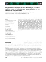
The results show that the high frequency hearing loss
increases with age (Fig. 9.10). The data in Fig. 9.10
show the averages of eight published studies compris-
ing data from more than 7,600 men (Fig. 9.10A) and
almost 6,000 women (Fig 9.10B) (310). Such studies
rarely define which criteria were used for inclusion in
the studies and it is therefore possible that the results
may reflect hearing loss that is caused by factors other
than age. In large population studies such as those
compiled by Spoor [310], many individuals have been
exposed to noise, which results in greater hearing loss
at 4 kHz than other frequencies (see p. 219).
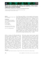
A cross-sectional and longitudinal population study
of hearing loss and speech discrimination scores in an
unselected population of individuals aged 70 (Fig. 9.11)
(228) showed that both these groups of individuals had
high speech discrimination scores (Fig. 9.12), some-
what lower in men than women. Exposure to noise
affected hearing in men more than in women and that
appears as a slightly greater hearing loss for high fre-
quencies. The reason for this gender difference may be
that many men had noise induced hearing loss, but
there may be other reasons related to hormonal influ-
ence on the progression of age-related changes in the
cochlea and possibly differences in the age-related
change in neural processing of sounds. M.B. Møller
[228] also provided the distributions of hearing loss
among the individuals of the study (Fig. 9.11) and
these data show that the hearing loss in the men and
women studied is far from being normally (Gaussian)
distributed. For low frequencies, the distributions are
skewed with a long tail towards larger hearing loss
while the distribution of hearing loss for higher fre-
quencies is more symmetrical although it is far from
being a normal distribution. The mean value and stan-
dard deviation are therefore not adequate descriptions
of the hearing loss as a function of age. Despite that,
mean and median values of hearing loss are com-
monly the only data provided in population studies of
hearing [310].
Age related hearing loss (presbycusis) is associated
with morphological changes in the cochlea in the form
of loss of outer hair cells. As for other causes of cochlear
impairments (noise exposure and ototoxic drugs), the
loss of outer hair cells is more pronounced in the basal
portion of the cochlea, thus affecting the cochlear
amplifier for high frequency sounds more than for low
frequency sounds. Loss of outer hair cells is the most
obvious change, and it has received more attention than
other changes, but there are also changes in the auditory
nerve, and the variations in fiber diameter of the axons
in the auditory nerve increases with age (Fig. 5.3).
Evidence of age-related changes in the function
of the auditory nervous system such as changes in syn-
thesis of inhibitory neurotransmitter such as gamma
butyric acid (GABA) have been presented [44].
Chapter 9 Hearing Impairment 217
FIGURE 9.10 (A) Average hearing loss in different age groups of men. Results from eight different pub-
lished studies based on a total of 7,617 ears. (B) Average hearing loss in different age groups of women.
Results from eight different published studies based on 5,990 ears (reprinted from Spoor, 1967).
Expression of neural plasticity from reduced high fre-
quency input from the cochlea may cause functional
changes in the nervous system [190]. There is also evi-
dence of changes in the function of the corpus callosum,
affecting binaural hearing, and perhaps impairing the
ability to fuse sound from the two ears [49, 137].
Some unexpected results of animal experiments have
shown that the progression of age related hearing loss
can be slowed by sound stimulation [328] (see p. 237).
That the progression of sensorineural hearing loss
can be slowed has only been shown in a few studies
because of the obvious difficulties in performing
218 Section III Disorders of the Auditory System and Their Pathophysiology
FIGURE 9.11 Distribution of hearing loss at different frequencies from a cross-sectional population study
of hearing in people of age 70; men and women. Solid lines represent left ear and dashed lines represent right
ears (data from Møller, 1981, with permission from Elsevier).
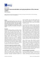
FIGURE 9.12 Distribution of speech discrimination scores from a cross-sectional population study of
hearing in people of age 70. The speech discrimination scores were obtained using phonetically balanced
word lists presented at 30 dB SL or at the most comfortable level. Solid lines represent left ears and dashed
lines represent right ears (data from Møller, 1981, with permission from Elsevier).
controlled studies. This type of hearing loss is similar
to presbycusis and is primarily a result of degenera-
tion of cochlear hair cells.
The mechanisms for that reduction in hearing loss
are unknown but several possibilities have been sug-
gested [328], such as neural activity in the cochlear
efferents that could affect outer hair cells, effects on
neurotrophin action, effects on some unknown factors
that are elicited by stimulation (excitotoxicity), regula-
tion of certain genes, and possibly an effect of intracel-
lular calcium concentration. It is important to point
out that it is the progression of hearing loss that is
affected (slowed) but the degenerative process does
not seem to be reversed by such sound exposure. The
fact that the progression of this kind of hearing loss
can be reduced means that appropriate sound stimula-
tion can actually affect cochlear degeneration. In the
past it has been the negative aspects of exposure to
sound that have been studied, and it is only recently
that it has been shown in a few studies that there are
also positive aspects of sound exposure. Thus as more
knowledge about age related changes accumulate, it
appears that presbycusis is more complex than just
normal age related changes of cochlear hair cells.
4.3. Noise Induced Hearing Loss
Noise induced hearing loss (NIHL) is normally
associated with noise exposure in industry and thus
thought of as a product of modern civilization. It is
mainly thought of as being caused by injury to cochlear
hair cells but as our knowledge about disorders of the
auditory system increases it has become evident that
the effect of noise exposure is complex. It has been
mainly the loss of hearing sensitivity that has been
studied but NIHL has many other effects on hearing.
Tinnitus may accompany any of the different forms
of cochlear hearing deficits but it is more common in
NIHL and in fact most incidences of tinnitus are asso-
ciated with NIHL (see Chapter 10).
The effect on the cochlea in NIHL has been studied
extensively and it was for a long time believed that the
morphological changes in the cochlea could explain
the changes in hearing. However, it has more recently
become evident that the effect of exposure to traumatic
noise also causes both morphological and functional
changes in the auditory nervous system. Expression of
neural plasticity plays an important role in creating
the symptoms from the auditory nervous system.
Exposure to a moderately loud noise causes hearing
loss that decreases gradually after the end of the noise
exposure. The hearing threshold may return to its normal
value after minutes, hours or days depending on the
intensity and duration of the noise exposure and the
individual person’s susceptibility to noise exposure.
Exposure to noise above a certain intensity and duration
results in hearing loss that does not fully recover to its
pre-exposure level. This remaining hearing loss is known
as permanent threshold shift (PTS). Hearing loss that
resolves is known as temporary threshold shift (TTS).
Hearing loss caused by noise exposure affects high
frequencies more than low frequencies. The audiogram
of a person with noise induced hearing typically has
a dip at 4 kHz (Fig. 9.13) and the hearing threshold at
8 kHz is better than it is at 4 kHz, at least for moderate
Chapter 9 Hearing Impairment 219
BOX 9.4
EFFECT OF AAE ON AGE-RELATED HEARING LOSS
Experiments by Willott and co-workers [328, 349] in
strains of mice that have early deterioration of hearing
have shown that low level sound stimulation (augmented
acoustic environment [AAE]) can reduce or slow the age
related hearing loss in these animals. The mouse that
these investigators used, DBA/2J, had progressive hear-
ing loss from early adolescence.
FIGURE 9.13 Typical audiogram for an individual who has
suffered noise induced hearing loss (data from Lidén, 1985).
degrees of noise induced hearing loss. This distinguishes
noise induced hearing loss from age related hearing
loss (presbycusis), which results in threshold elevation
that increases with the frequency (Fig. 9.10). The 4 kHz
dip is more or less pronounced depending on the noise
exposure and it is most pronounced in individuals
who have been exposed to impulsive noise, thus noise
with a broad spectrum.
The amount of acquired hearing loss depends not
only on the intensity of the noise and the duration of
exposure but also on the character of the noise (fre-
quency spectrum and time pattern). The hearing loss
from noise exposure is thus distinctly related to the
physical characteristics of the noise exposure but great
individual variations exist. The combination of noise
level and duration of exposure is known as the immis-
sion level and it is used as a measure of the effective-
ness of noise in causing PTS. However, the PTS caused
by exposure to noise with the same immission level
shows large individual variations (Fig. 9.14) [33].
Exposure to pure tones or sounds with a narrow
spectrum causes the greatest hearing loss at about one
half octave above the frequency of the highest energy
of the sound. The reason for this half octave shift is
most likely the shift of the maximal vibration of the
basilar membrane towards the base of the cochlea with
increasing sound intensity (see Chapter 3). Exposure
to loud noise is expected to cause the most damage to
hair cells at the location on the basilar membrane where
the noise gives rise to the largest vibration amplitude.
That means that the most damage is done at a location
that is tuned to the frequency of the maximal energy of
the noise at the intensity of the noise. The location of
maximal vibration amplitude is not the same for high
intensity sounds as for sounds at the threshold used to
measure the hearing loss. This is because the frequency
to which a certain location along the basilar membrane
is tuned shifts along the basilar membrane with
increasing stimulus intensity.
The audiograms obtained in individuals who have
been exposed to many different kinds of noise have
similar shape, but the 4 kHz dip is probably most pro-
nounced for exposure to impulsive noise. Studies have
shown evidence that the enhancement of sound from
the resonance of the ear canal [246] is the cause of the
selective damage. Ear-canal resonance amplifies sounds
in the region of 3 kHz (cf. Chapter 2). That the greatest
hearing loss from exposure to sound with their highest
energy around 3 kHz occurs near 4 kHz can be explained
by the half octave shift discussed above. The point on
the basilar membrane that was tuned to 3 kHz at a high
sound intensity (e.g., 90 dB) will be tuned to a higher
frequency when tested near the threshold. This is why
the largest threshold shift from exposure to 3 kHz sound
occurs at a higher frequency, approximately 4 kHz.
Individual variation is a characteristic feature of
all forms of hearing impairment including NIHL, but
the reason for this individual variation in acquired
hearing loss from similar noise exposure is not well
understood. It is characteristic of NIHL that the same
noise exposure causes different degrees of hearing
impairment in different individuals (see Fig. 9.14). This
individual variation in susceptibility to noise induced
hearing loss has many sources. Genetic variations
are one [61], and age and health status are also impor-
tant factors that affect injury to hair cells from noise
exposure. Drugs of various kinds most likely also
increase susceptibility to noise induced hearing loss.
Hearing loss of conductive type also affects the risk of
NIHL [237]. Absence or impairment of the acoustic
middle ear reflex results in increased hearing loss from
noise exposure [356]. Ingestion of alcohol and other
drugs that impair the function of the acoustic middle
ear reflex (cf. Chapter 8) may also affect susceptibility
to NIHL.
Numerous hypotheses have been presented but
published experimental evidence is rare. Besides vari-
ability in the exposure conditions, genetic differences,
age, gender, pigmentation, differences in the sound
conducting apparatus, blood supply and innervation
of the cochlea have all been suggested as causes of the
variability in NIHL to the same noise exposure. The
hypothesis that age is a factor in the observed varia-
tions in susceptibility to NIHL has been supported by
studies in mice [120]. Other factors that affect NIHL
include a history of sound exposure, as discussed below.
220 Section III Disorders of the Auditory System and Their Pathophysiology
FIGURE 9.14 Hearing loss at 4 kHz as a function of noise expo-
sure. Each dot represents the elevation in hearing threshold at 4 kHz
for one ear. The solid line is the mean value. The horizontal axis rep-
resents both the sound level and the time of exposure (known as the
noise immission level which is equal to the noise level (in dB) + 10
times the logarithm of the duration of exposure) (modified from
Burns and Robinson, 1970, with permission from Her Majesty’s
Stationery Office).
Much of the individual variations in NIHL in
humans can be explained by genetic differences,
environmental factors, and inaccuracies in determina-
tion of the level and the duration of the noise to which
they were exposed. The noise level and environmental
facts can be controlled in animal experiments in the
laboratory. Animals can be exposed to noise in the
laboratory in a much more accurate way than humans.
When normal guinea pigs are exposed to noise the
acquired hearing loss varies considerably (Fig. 9.15)
[186].
The fact that different animals are affected to different
degrees from the same insults is an indication of indi-
vidually different genetic makeup. This assumption
Chapter 9 Hearing Impairment 221
BOX 9.5
FREQUENCY OF GREATEST NIHL DEPENDS ON EAR CANAL LENGTH
Studies of the correlation between the resonance fre-
quency of the ear canal and the frequency of the greatest
hearing loss in people with noise induced hearing loss
[246] have shown that the mean resonance frequency of the
ear canal in the group of people studied was 2.814 kHz and
the maximal hearing loss occurred at 4.481 kHz. Assuming
that the maximal energy of broad band noise occurred at
the resonance frequency of the ear canal (2.814 kHz) and
that the greatest hearing loss occurs at a frequency that is
1.5 times the frequency of the maximal energy of the noise
exposure, then the maximal hearing loss would be
expected to occur at 4.221 kHz. The mean frequency of
maximal hearing loss was 4.481 kHz, thus very close to the
expected value. This study also showed a high correlation
between ear-canal resonance frequency and the frequency
of the maximal hearing loss in individuals.
Earlier studies [38], showed that extending the ear
canal by a tube that caused the resonance frequency to
decrease caused a similar decrease in the frequency of the
maximal TTS in volunteers who were exposed to broad
band noise. The greatest hearing loss (TTS) occurred at
frequencies about one half octave higher than the fre-
quency of maximal sound energy.
These studies thus support the hypothesis that the
typical 4 kHz dip in the audiograms of individuals who
have suffered noise induced hearing loss is a result of the
resonance of the ear canal. (It has been pointed out [270]
that the maximal transfer of sound power to the cochlea
does not necessarily occur at the frequency of the ear
canal resonance but depends on other factors that
are frequency dependent, such as the transformation ratio
of the middle ear.)
FIGURE 9.15 NIHL in animals with various degrees of genetic variations. (A) Data obtained in male
guinea pigs (400–500 g); the exposure was a 2–4 kHz octave band of noise at 109 dB SPL for 4 h with a 1-week
survival. The mean peak PTS was 35.1 dB at 7.6 kHz (SD of 21.33 dB) (reprinted from Maison, S.F. and
Liberman, M.C. 2000. Predicting vulnerability to acoustic injury with a non-invasive assay of olivocochlear
reflex strength. J Neurosci 20: 4701–4707, with permission from the Society for Neuroscience; Courtesy
Charles Liberman. Copyright © 2000 Society for Neuroscience). (B) Inbred mice, males (23–29 g) exposed to
octave band noise (8–16 kHz) at 100 dB for 2 h with a 1-week survival. The mean peak PTS was 38 dB at
17.5 kHz (SD of 4.06 dB) (reprinted from Yoshida and Liberman, 2000, with permission from Elsevier).
is supported by the finding that the variation is less
when inbred animals are used in such experiments
(Fig. 9.15) [353].
That genetics is important for acquiring NIHL is
supported by the results of other animal experiments
that have shown that animals with genetically related
hypertension acquire more hearing loss than normoten-
sive animal from the same noise exposure [26, 28].
The amount of hearing loss acquired by genetically
identical animals from noise exposure under controlled
laboratory conditions shows individual variation (Fig.
9.15). These variations in NIHL in genetically identical
animals can be explained by difference in epigenetics
1
[140] or “noise in gene expressions” [260]. The variations
that occur in the susceptibility to noise exposure
between animals that are regarded to be genetically
identical can be purely stochastic in nature or caused
by differences in the internal states of a population of
cells. Ongoing mutations are another source of varia-
tions that can manifest as differences in the physical
characteristic of genetically identical organisms.
Naturally, environmental factors can also affect the
development of an animal.
These factors (epigentics and “noise” in gene
expression) and perhaps other yet unknown ones, can
explain the variations in the effect of insults such as
noise exposure but it also explains why, for example,
only one of two identical (homozogotic) twins acquires
an inherited disease, despite both twins having exactly
the same genetic set-up.
Other factors than genetics and epigentics may affect
the susceptibility to noise exposure, such as hearing
loss due to middle-ear pathologies. Middle-ear patho-
logy acts as an ear protector and actually decreases the
person’s hearing loss from exposure to noise [237]. The
conductive hearing loss does not affect hearing to any
great extent at high frequencies but the protective
effect from the low frequency conductive hearing loss
against noise induced hearing loss is substantial. The
result is that the acquired NIHL can be considerably
less in the ear with conductive hearing loss than in the
ear without conductive loss (Fig. 9.16).
NIHL has many similarities with presbycusis. It
mainly affects outer hair cells and speech discrimina-
tion is little affected when the hearing loss is moderate
and limited to frequencies around 4 kHz. It is mainly
outer hair cells in the basal portion of the cochlea that
are injured or totally destroyed, thus causing impair-
ment of the cochlear amplifier. It is not known why
hair cells located in the base of the cochlea are more
susceptible to insults from noise exposure (and from
ototoxic agents and aging, see pp. 216, 227) compared
to hair cells in other parts of the cochlea. Pure tones or
noise that has a narrow spectrum cause lesions within
a restricted region of the basilar membrane.
Little damage to the stereocilia can be detected by
light microscopic examination after noise exposure
that produces 40–60 dB hearing loss [178]. In moderate
degrees of cochlear hearing loss, inner hair cells are
intact when examined by the light microscope. High
resolution light microscopy (using Nomansky optics)
and scanning electron microscopy (SEM) have shown
that noise exposure causes a disarray of stereocilia on
both inner and outer hair cells (Fig. 9.18) [178]. High-
resolution light microscopy has revealed that the stere-
ocilia of inner hair cells are altered to almost the same
extent as were the stereocilia of outer hair cells after
exposure to moderate levels of noise.
It has been shown that noise exposure causes dis-
connection between stereocilia of outer hair cells and
the tectorial membrane. It should be noted that this is
different from other types of insults to the cochlea such
222 Section III Disorders of the Auditory System and Their Pathophysiology
BOX 9.6
HYPERTENSIVE RATS ACQUIRE GREATER NIHL THAN
NORMOTENSIVE ANIMALS
Experiments in rats [26, 28] have shown that sponta-
neous hypertensive rats acquire more PTS from noise
exposure than normotensive rats. However, hypertension
caused by impairing blood supply to the kidney does not
show such increased PTS [27]. Thus, hypertension in itself
is probably not the cause of the higher susceptibility to
noise induced hearing loss. The increase in susceptibility
to noise induced hearing loss seen in the spontaneous
hypertensive rats is probably related to factors that occur
together with the predisposition for hypertension.
1
Epigentics: This term is used to describe activation and de-
activation of genes. It is defined as the study of heritable changes
in gene function that occur without a change in the DNA sequence.
This mainly occurs in the uterus but can also occur after birth. It has
become increasingly evident that epigenetic mechanisms such as
DNA methylation, histone acetylation, and RNA interference, and
their effects in gene activation and inactivation, are important
factors in phenotype transmission and development [107].
as from ototoxic antibiotics, which affect the integrity
of the cell bodies of hair cells.
Only exposure to extreme loud noise causes other
structural damages besides the damage to hair cells.
Thus exposure to sounds with levels in excess of 125
dB SPL seems to be necessary to cause mechanical
damage to the cochlea of the guinea pig [308]. The
level of noise exposure that causes structural damage
varies between species and it may thus be different in
humans from the values obtained in the guinea pig.
Hearing loss caused by injury to outer hair cells
does not affect sensory transduction but rather the
mechanical properties of the basilar membrane. Recall
from Chapter 3 that the outer hair cells function as
“motors” that increase the sensitivity and the frequency
selectivity of the ear and that it is the inner hair cells
that transduce the motion of the basilar membrane
and control the discharge pattern of auditory nerve
fibers. Also, recall that the amplification caused by
outer hair cells is most effective for sounds of low
intensity and that it has little effect for sounds that are
more than 50-60 dB above (normal) hearing threshold.
This explains why hearing loss caused by impairment
of the function of outer hair cells rarely exceeds 50 dB.
It is also the reason why tests that employ high inten-
sity sounds such as ABR and the acoustic middle ear
reflex are largely normal in patients with hearing loss
caused by malfunction of outer hair cells.
The most prominent physiological signs of noise
induced hearing loss as revealed in animal studies are
deterioration of the tuning of single auditory nerve
fibers, loss of sensitivity at the fiber’s CF and a down-
wards shift in frequency of the CF (Fig. 9.19) [51]. The
widening of basilar membrane tuning after noise
exposure is typical for loss of function of the active role
of outer hair cells, that is to increase the sensitivity and
frequency selectivity of the ear (cf. Chapter 3). The
widening of the tuning of the basilar membrane
broadens the “slices” of the spectrum of broad band
sounds from which the cochlea provides information
to the (temporal) analyzer in the central nervous system.
This broadening may cause interference between dif-
ferent spectral components (impair “synchrony capture”,
see p. 109) and it may increase masking. The impair-
ment of the cochlear amplifier from injury of the outer
hair cells also impairs the amplitude compression that
is prominent in the normal cochlea and that may be
the reason why recruitment of loudness accompanies
NIHL. The sensitivity of a single auditory nerve fiber
for frequencies below a fiber’s CF (in the tail region
of the tuning curves) increases after noise exposure
[179] and that may also contribute to the symptoms
of NIHL.
While published reports of morphological changes
of the cochlea as a result of noise exposure are abundant,
few studies that concern the cause of these changes have
been published. It is poorly understood how noise
exposure causes the observed damage to the hair cells.
It has been suggested that impairment of blood supply,
or simple exhaustion of the metabolism could be the
cause of the hair cell injury and destruction. These
hypotheses have received little experimental support.
Oxygen free radicals have been implicated in caus-
ing injury to hair cells from noise exposure, aging and
ototoxic antibiotics [94, 252]. It has been shown that
the level of glutathione, an enzyme that defends cells
against the toxic effects of reactive oxygen species,
decreases with age and depend on the physiologic
state of a person and on environmental challenges. It
has been shown that oxygen free radical scavengers
can reduce the effect of noise exposure on hearing. The
best effect was obtained when a free radical scavenger
Chapter 9 Hearing Impairment 223
FIGURE 9.16 Audiograms of a welder exposed to shipyard noise
for 30 years and who had conductive hearing loss in one ear (top
audiogram). The bottom audiogram is from the ear without conduc-
tive hearing loss (data from Nilsson and Borg, 1983, with permission
from Taylor & Francis).
BOX 9.7
HAIR CELL LOSS, HEARING LOSS AND STEREOCILIA DAMAGE
Light microscopic studies of cochlear hair cells in ani-
mals that have been exposed to a moderately loud noise
that causes hearing loss show loss of some hair cells,
mainly outer hair cells (Fig. 9.17). Exposure to more
intense sounds for longer periods causes more extensive
damage and inner hair cells may be affected. An incre-
ment of only 5 dB in the intensity of the sound to which
the animals were exposed caused a considerable increase
in the injury of hair cells and in the PTS (Fig 9.17) [75].
Cell counts using surface preparation of the cochlea (cyto-
cochleograms) reveal damage mainly to outer hair cells
in the first row in an animal where the loss of sensitivity
was moderate (30–40 dB) while high resolution light
microscopy reveal abnormalities in stereocilia in both outer
and inner hair cells (Fig. 9.17) [75]. An animal exposed to
the same noise but studied at different times after noise
exposure (right hand graphs in Fig. 9.18) showed much
greater hearing loss and more extensive hair cell damage,
including missing inner hair cells. There is a clear correla-
tion between loss of hair cells and threshold shift at the
characteristic frequency (CF) but there is considerable
individual variation in the extent of the damage even
in animals that are genetically similar and treated in
similar ways.
FIGURE 9.17 Relationship between hearing loss and loss of hair cells in cats exposed to 2 kHz tones for
1 h and three different intensities (reprinted from Dolan et al., 1975, with permission from Blackwell
Publishing Ltd).
Chapter 9 Hearing Impairment 225
BOX 9.7 ( cont’d)
FIGURE 9.18 Results of recordings from single auditory nerve fibers and morphologic examination of the
cochleae of two cats after exposure to 2 h of noise, 2 octaves wide, centered at 3 kHz and with an intensity of
115 dB SPL. The cats were examined, 620 (left panel) and 63 days (right panel) after noise the exposure. Upper
graphs: sample tuning curves, centered at approximately 3.6 kHz of single auditory nerve fibers and thresh-
old at CF. Middle graphs: cytocochleograms of the cochleae showing loss of hair cells. Bottom graphs: stere-
ocilia damage in the first row of outer hair cells and inner hair cells as revealed by high resolution (Nomarsky)
light microscopy with 100X objectives (reprinted from Liberman, 1987, with permission from Elsevier).
was administered before the noise exposure but some
effect was also achieved when it was administered
after the noise exposure [252]. Oxygen free radicals are
associated with activity of mitochondria, and the prop-
erties of mitochondria are inherited from mothers.
The finding that the cochlea can recover from noise
induced hearing loss shows that hair cells can cease to
function, or have a reduced function, without perma-
nent injury occurring. That also explains the recovery of
threshold shift after noise exposure of moderate degree
(temporary threshold shift [TTS]). Only when the insult
has reached a certain level does the recovery become
incomplete and the result is permanent injury (PTS).
4.4. Implications of Hearing Loss on
Central Auditory Processing
While NIHL is usually assumed to be caused only
by the loss or injuries of outer hair cells it has been
shown that NIHL is also associated with specific mor-
phologic changes in the central nervous system [148,
205]. In addition to that, neural plasticity may result in
functional changes in the nervous system because of
the deprivation of input to specific groups of neurons
that is caused by the injury to the cochlea [101]. This
may alter the balance between inhibition and excita-
tion, and that may cause hyperactivity (see Chapter 11).
Animal studies of evoked potentials recorded
from the cerebral cortex showed enhancement of the
responses after exposure to noise that caused hearing
loss [318]. The authors concluded that their results
indicate that the enhancement of the amplitude of the
evoked potentials that are recorded from the auditory
cortex is caused by changes in the processing of infor-
mation in the central auditory nervous system. These
changes are caused by expression of neural plasticity.
Even exposure to sounds that do not cause hearing
loss can cause changes in frequency tuning of neurons
in the cerebral cortex of animals consisting of greater
frequency selectivity and greater sensitivity to quiet
sounds [88].
4.5. Modification of Noise Induced
Hearing Loss
It has generally been assumed that exposure to loud
sounds (noise) caused hearing loss only because it
affected hair cells, either by mechanical stress or by
changing the chemical composition inside or outside
the hair cells. The finding that prior noise exposure can
226 Section III Disorders of the Auditory System and Their Pathophysiology
BOX 9.8
NOISE EXPOSURE CAUSES CHANGES IN THE COCHLEAR NUCLEUS
Animal experiments have shown morphological changes
occur in the cochlear nucleus after noise exposure [204,
205]. Recordings made from the inferior colliculus shows
signs of hyperactivity after noise exposure [320]. Several
studies have shown that exposure to traumatizing noise
alter frequency tuning of neurons in the auditory cortex.
FIGURE 9.19 Deterioration in tuning and sensitivity of auditory
nerve fibers as a result of exposure to pure tones. The data were
pooled from many nerve fibers and the frequency scale is normali-
zed. The arrows show the frequency of the exposure tones and the
different curves represent different exposure times (reprinted from
Cody and Johnstone, 1980, with permission from Elsevier).
affect the hearing loss from subsequent exposure to
loud noise [42, 198, 302] brought a new and unexpected
angle to the relations between the physical noise expo-
sure and the acquired hearing loss. It became evident
that the physiological mechanisms involved in noise
induced hearing loss are more complex than earlier
believed [171]. The finding that noise induced hearing
loss is affected by prior stimulation and by simultane-
ous stimulation of the opposite ear may explain some
of the individual variation in susceptibility to noise
induced hearing loss.
It was shown by Miller et al. [198] in animal experi-
ments that the TTS caused by noise exposure decreased
gradually during repeated exposures. That was taken
to indicate that the ear’s susceptibility to noise expo-
sure is affected by previous exposure. This “toughening”
of the ear with regard to TTS from noise exposure
has been extensively studied in a variety of animals
and in humans by several investigators [42, 302] and it
has been confirmed that it is also possible to reduce the
effect of noise exposure on PTS by pre-exposure to
noise. The exposure pattern of such “conditioning” is
important for achieving this effect. Several studies
have suggested this toughening of the cochlea against
noise-induced injury is related to induced changes in
the hair cells by the “conditioning” noise exposure.
The mechanism for such toughening is not com-
pletely understood but evidence has been presented
from animal (guinea pig) experiments that both the
medial and the lateral olivocochlear (efferent) system
is involved [10, 171]. There is evidence that activity in
the olivocochlear fibers can adjust the intracellular
potential in the outer hair cells and thereby protect the
hair cells from damage from noise exposure. Other
possibilities that have been suggested involve intracel-
lular pathways that can provide protection from noise-
induced cellular damage in the cochlea. It has been
suggested that pathways that regulate and react to
levels of reactive oxygen species in the cochlea, stress
pathways for the heat shock proteins, and neurotrophic
factors may be involved [171]. However, none of these
possibilities have been confirmed.
The concept of augmented acoustic environment has
been pursued in other animal experiments [238], which
showed that such “enriched acoustic environment,”
affects the tuning of neurons in the auditory cortex.
Perhaps more surprising, it can alter the changes in the
tuning that occur as a result of subsequent exposure to
noise at levels that cause permanent damage to the ear.
Such “enriched acoustic environment” also affect the
development of changes in the cerebral cortex after
exposure to traumatizing noise. Animal experiments
have shown that NIHL can be reduced when the ani-
mals were placed in an enriched acoustic environment
after the noise exposure, thus, animals that were exposed
to sounds at moderate levels had less hearing loss com-
pared with similarly exposed animal that were placed
for the same time in a quiet environment [238]. These
authors also showed that the cortical re-organization
that normally occurs after noise exposure was reduced
in the animals that were exposed to such enriched
acoustic environment after noise exposure.
These findings are important for two reasons:
1) Exposure to such enriched acoustic environment
immediately after exposure to the traumatizing noise
prevented the plastic tonotopic map changes in pri-
mary auditory cortex that normally occur after expo-
sure to traumatizing noise; and 2) the hearing loss
from the noise exposure was less than in animals that
were placed in a quiet environment after the noise
exposure. These studies also support the hypothesis
that the nervous system is involved in noise induced
hearing loss (see also tinnitus, p. 254).
4.6. Hearing Loss Caused by Ototoxic
Agents (Drugs)
Many commonly used medications can cause hear-
ing loss. Antibiotics of the aminoglycoside type can
cause permanent hearing loss [94, 291]. Streptomycin
(dihydrostreptomycin) was the first of this family of
antibiotics found to cause hearing loss, but now com-
monly used antibiotics of the same family such as gen-
tamycin, kanamycin, amikacin and tobramycin have
also been found to be ototoxic but to a varying degree.
Erythromycin and polypeptide antibiotics such as
vancomycin have produce hearing loss but it is mostly
reversible once the drugs are terminated. Commonly
used agents in cancer therapy (chemotherapy) such as
cisplatin and carboplatin are also ototoxic.
Aspirin (acetylsalicylic acid) can produce tinnitus
and transient hearing loss but only at high dosages and
the hearing loss normally resolves when the drug is
terminated or the dosage reduced [234]. Administration
of 5–10 g per day of acetylsalicylic acid (aspirin) can
cause hearing loss and it can abolish the spontaneous
otoacoustic emission [45]. Certain diuretic drugs such
as furosemide and ethacrynic acid can produce tran-
sient hearing loss and tinnitus but they rarely cause per-
manent hearing loss. The same is the case for quinine.
Hearing loss caused by these substances may be
reversible when the drug treatment is terminated or
it may be permanent.
These substances are used to treat diseases of differ-
ent kinds and therefore they are almost always used in
people with various kinds of illnesses that may increase
the ototoxic effect. The experimental results upon which
recommendations on the safe limits of such drugs are
Chapter 9 Hearing Impairment 227
based were obtained in healthy individuals and these
recommendations may not be applicable to humans with
diseases for which these substances are administrated.
The ototoxic effect of these substances varies widely
among individuals and it is different in different
animal species. There is evidence that older individu-
als have a higher susceptibility and individuals with
diseases of various kinds may also be more susceptible.
Whether or not aminoglycoside antibiotics such as
gentamycin and Kanamycin may cause hearing loss
and vestibular disturbance depends on the way they
are administered. Antibiotics that are ototoxic are often
given in fixed dosages to treat life-threatening infec-
tions in individuals who are generally weakened and
often have impaired kidney function. Many of these
drugs are excreted through the kidneys and if kidney
function is impaired, they are excreted more slowly
than normal. The blood levels of the drug will there-
fore increase and become higher than anticipated if
the excretion is slower than normal. The dosages used
are designed to maintain a certain plasma level with
normal excretion rates but in individuals with impaired
kidney function such dosages may cause a pile up of
the drug because it is excreted at a slower rate than it is
administered. Since many of the ototoxic drugs are also
nephrotoxic, a vicious circle may result from impaired
kidney function that becomes aggravated by higher
blood levels of an ototoxic drug. Monitoring of plasma
levels of ototoxic antibiotics can reduce the risk of
hearing loss considerably.
Antibiotics usually enter the cochlear fluid space
from systemic administration but these substances may
also enter the cochlear fluid space when administered
in the middle-ear space, such as to treat infections. It is
interesting that an ototoxic antibiotic, neomycin, that
is not allowed for systemic administration because of
its ototoxicity is approved for local administration
including in the ear. Evidence has been presented
that inflamed mucosa of the middle ear acts as a bar-
rier for neomycin and prevents it from entering the
cochlea [305]. It is also possible that toxic substances
generated by bacterial activity in the middle ear fluid
can enter the cochlear fluid space through the mem-
branes of the round and oval windows and cause
injuries to hair cells [185, 305].
Ototoxic antibiotics may cause hearing loss by
changing important biochemical processes leading to
metabolic exhaustion of hair cells and that can event-
ually lead to cell death. It is generally assumed that
oxygen free radicals are involved in causing injuries to
the cochlea by ototoxic substances [94, 191]. Attempts
have been made to prevent the ototoxic effect of drugs
by administration of substances developed to protect
against the effect of radioactivity but such drugs also
reduce the ototoxic effect of these antibiotics [247, 274]
and so far practical application of this method has not
been demonstrated.
Most ototoxic drugs induce hearing loss by injuring
outer hair cells and thus impairing the function of the
cochlear amplifier, in a similar way as occurs in pres-
bycusis and in NIHL. Inner hair cells are usually unaf-
fected. However, the effect of toxic substances such as
salicylate is different from that of noise in that it affects
the cell bodies of the outer hair cells, while noise also
causes a decoupling between the outer hair cell stere-
ocilia and the tectorial membrane. Hearing loss caused
by ototoxic drugs seldom exceeds 50–60 dB and it usu-
ally begins at high frequencies and extends gradually
towards lower frequencies as it progresses. Most drugs
cause the greatest damage to hair cells in the basal
region of the cochlea and the greatest hearing loss thus
occurs at high frequencies. High frequency audiome-
try (i.e., determination of the pure tone threshold at
frequencies above 8 kHz) may therefore detect a begin-
ning hearing loss before it reaches frequencies that
affect speech discrimination.
While the effect on the cochlear hair cells from
ototoxic substances have been studied extensively,
little is known about the subsequent effect on the func-
tion of the central nervous system. Impairment of the
228 Section III Disorders of the Auditory System and Their Pathophysiology
BOX 9.9
CARBOPLATIN AFFECTS INNER HAIR CELLS
While most ototoxic substances mainly affect outer
hair cells, carboplatin causes injury mainly to inner hair
cells in one animal species, the chinchilla, leaving outer
hair cells intact. In the guinea pig, carboplatin injures
outer hair cells, mainly in the basal region of the cochlea,
thus similar to other ototoxic substances. This means that
the nature of the resulting hearing loss caused by carbo-
platin is different from that of other ototoxic substances
because it affects neural transduction in the cochlea
rather than the mechanical properties of the basilar mem-
brane. Its effect in humans is unknown [70].
function of outer hair cells affects tuning in auditory
nerve fibers. As we have discussed earlier, impairment
of cochlear function also affect the function of the
auditory nervous system.
Studies in animals have shown that administration
of ototoxic substances and metabolic insults to the
cochlea can affect cochlear frequency tuning [90] (see
Chapter 6). Tuning of single auditory nerve fibers in
animals that were treated with Furosemide shows sim-
ilar changes to those caused by anoxia. Treatment with
Kanamycin also results in deterioration of tuning of
auditory nerve fibers in a similar way to that caused by
metabolic insult to the cochlea (see Chapter 6, Fig. 6.7).
4.7. Diseases that Affect the Function
of the Cochlea
Several diseases may affect the function of the
cochlea. The most common disease that causes hearing
impairment is Ménière’s disease. Hearing impairment
may also result from hereditary causes. Infectious dis-
eases such as meningitis and certain viral infections
can also cause destruction to cochlear hair cells, caus-
ing hearing impairment.
Ménière’s disease is a progressive disorder that is
defined by a triad of symptoms, namely vertigo with
nausea, fluctuating hearing loss and tinnitus [199]. It is
one of a few kinds of sensorineural hearing loss that
affects hearing initially at low frequencies. The incidence
of Ménière’s disease is different in different geographic
locations. A study made in Rochester, Minnesota, showed
an incidence of 15.3 per 100,000 people with a small
preponderance for women (16.3 vs. 9.3 for men) [352].
A study in Italy [47] showed an incidence of 8.2, and a
study in Sweden [311] arrived at an incidence of 46 per
100,000 people. A part of this variation is probably
caused by differences in the definition of the disease.
In the early stages of the disease the patient experi-
ences acute attacks of vertigo, often preceded by brief
aural fullness in the affected ear, hearing loss and tin-
nitus that may last from several hours to 24 h. Longer
lasting symptoms of vertigo are caused by other disor-
ders of the inner ear. Typically, hearing loss in the early
stages of Ménière’s disease affects only low frequen-
cies and it fluctuates and increases during an acute
attack (Fig. 9.20). The hearing returns to normal after
each attack in the beginning of the disease but as the
disease progresses, residual hearing loss from each
attack accumulates and the hearing loss spreads to
higher frequencies. After years of disease, some patients
may experience ‘drop attacks,” i.e., sudden severe ver-
tigo that occurs without warning, and which causes
the patient to fall to the ground. Over time, hearing
loss progresses and extends to higher frequencies; but
it rarely exceeds 50 dB. Speech discrimination is little
affected in the early stages of the disease but may
become affected in the advanced, late stage of the dis-
ease. The end stage of the disease, reached 10–15 years
after its debut, is flat hearing loss of approximately 50 dB
and speech discrimination scores of approximately
50%. The symptoms are initially unilateral but many
patients experience bilateral symptoms after 10–15 years.
Ménière’s disease can be diagnosed by the patient’s
history and standard audiological tests. Recently it has
been hypothesized that the cochlear summating
potential (SP) is abnormal in patients with Ménière’s
disease and recording of the SP is in common use for
diagnosis of Ménière’s disease and for monitoring
treatment. The SP is the sum of the cochlear distortion
products (Chapter 3) and its amplitude depends on
several factors, one of which is endolymphatic pres-
sure (or volume) and this is why it has been suggested
as a way of detecting endolymphatic hydrops. The SP
has been reported to be high in patients with Ménière’s
disease. SP varies considerably between individuals
without signs of Ménière’s disease and it may also vary
from time to time in the same individual. The large
variability of the SP in individuals without cochlear
hydrops hampers the use of SP in diagnosis of disor-
ders with hydrops including Ménière’s disease. Some
investigators have therefore expressed doubt about
the significance of such findings and refer to the large
individual variation in the SP. The value of recordings
of SP as a diagnostic tool to diagnose Ménière’s dis-
ease has therefore been set in question by some inves-
tigators [41, 83] whereas others [283] find that the SP
anomaly and the ratio between the action potential
(AP) and the SP are important signs of the disease.
Chapter 9 Hearing Impairment 229
FIGURE 9.20 Typical audiogram from a person with Ménière’s
disease showing hearing loss during an attack (I) and between
attacks (II) (data from Møller, M. B., 1994, with permission form
Lippincott).
BOX 9.10
SP/AP RATIO IS A MEASURE OF COCHLEAR HYDROPS
The ratio between the amplitude of the SP and the AP
components are used as indication of cochlear hydrops.
In response to clicks, the SP appears before the AP and the
SP occurs at the same time as the cochlear microphonics
(CM) making it difficult to distinguish the SP from the
CM components of the electrocochleogram (ECoG). Some
investigators [284] have made the SP appear more clearly
by using clicks of high repetition rate as stimuli (Fig. 9.21),
which reduces the amplitude of the AP component of the
ECoG without affecting the SP. Subtracting the response
to high stimulus rate from a response to low stimulus
rate eliminates the SP component and thus shows a clean
AP waveform. However, tone-bursts would be a more
adequate stimulus than clicks for recording the SP. In the
ECoG elicited by tone bursts, the SP appears as a plateau
that occurs during and after the AP and it is thus easy to
measure the amplitude of both AP and SP (Fig. 9.22).
Note that the polarity of the SP is different in response to
tones of different frequency.
It has been the ratio between the SP and the AP that
has been used as an indicator of endolymphatic hydrops
when Sass et al. [283] found a mean SP/AP ration of 0.26
in normal individuals with a standard deviation of 0.11.
In patients with Ménière’s disease, the mean SP/AP was
0.46 with a standard deviation of 0.15. The SP was signifi-
cantly larger in Ménière’s patients at 1 and 2 kHz but not
at 4 and 8 kHz. Campbell et al. [41] used 6 kHz tones as
stimuli and that may be the reason these investigators
found little difference between the SP/AP ratio in
patients with Ménière’s disease compared with patients
who did not have Ménière’s disease. The sensitivity of
transtympanic ECoG using the SP/AP ratio of the
response to 1 kHz tone bursts was 82% and the specificity
was reported to be 95% [283]. Thus the choice of stimuli
affects the sensitivity if the SP/AP ratio as an indicator of
endolymphatic hydrops, and that may be one of the rea-
sons why different investigators have arrived at different
values of sensitivity of this test.
FIGURE 9.21 ECoG response obtained at two different click rates
(A: 9 pps and B: 90 pps) in a patient with Ménière’s disease, and the
difference between the two responses (A-B) (reprinted from Sass et al.,
1998, with permission from Blackwell Publishing Ltd).
FIGURE 9.22 ECoG in response to tone bursts of different fre-
quencies obtained in a patient with Ménière’s disease (reprinted
from Sass et al., 1998, with permission from Blackwell Publishing Ltd).
Chapter 9 Hearing Impairment 231
Ménière’s disease is probably not one disorder but
rather a group of different disorders. Some variations
of Ménière’s disease have predominantly cochlear
signs and some investigators have called these dis-
eases cochlear Ménière’s disease. Thus, while the clas-
sical definition of Ménière’s disease is a triad of
symptoms (fluctuating hearing loss, tinnitus, and ver-
tigo), over time physicians have accepted patients
with variations to that classical pattern and labeled
these disorders Ménière’s disease as well.
The fact that Ménière’s disease has a distinct name
while many disorders that have symptoms from the
vestibular system have less distinct names or no names
at all makes it attractive to use the name Ménière’s dis-
ease for disorders that resembles Ménière’s disease.
Recordings of the SP during operations for Ménière’s
disease may be of value because it involves compari-
son of the SP over a short time in the same individual
and thus it is not subjected to the effect of individual
variations. The SP is affected by vestibular nerve sec-
tion [164] probably because of severance of the olivo-
cochlear bundle that occurs in these operations.
Neural activity in the olivocochlear bundle influences
the function of hair cells, which contribute to the SP
and that may explain the effect of severance of the
olivocochlear bundle on the SP.
It is believed that the symptoms of Ménière’s
disease are caused by pressure (or rather volume)
imbalance in the fluid compartments of the inner ear
(endolymphatic hydrops). The hearing loss in Ménière’s
disease can be explained by a distension of the basilar
membrane causing the largest distension where its
stiffness is least, i.e., in the apical portion (Chapter 3).
Permanent damage to hair cells does not seem to
occur, at least not in the early stage of the disease. The
fluctuations in hearing are assumed to be caused by
varying degrees of endolymphatic hydrops in the
cochlea. That is supported by studies by Kimura [150]
who showed that blocking the endolymphatic duct in
guinea pigs mimicked the signs of attacks of Ménière’s
disease.
Little is known about the effects of elevated peri-
lymphatic pressure on the function of the ear and it
is a matter of diverse opinion whether moderately
abnormal pressure in the perilymphatic space causes
any signs of pathology. It is, however, generally recog-
nized that an increase in the volume of the endolym-
phatic space is associated with similar disturbances of
hearing and balance as seen in Ménière’s disease but it
is not known whether the abnormal volume of inner-
ear fluid compartments is a cause of the disorder or
a result of the pathology of Ménière’s disease.
Elevated endolymphatic volume causes Reissner’s
membrane to bulge and the basilar membrane to bow.
That is assumed to give rise to the low frequency hear-
ing loss and perhaps tinnitus that are two of the triad
of symptoms that defines Ménière’s disease, at least in
its early stage. If the volume of the endolymphatic
space increases beyond a certain value Reissner’s mem-
brane may rupture resulting in fluids of widely differ-
ent ionic composition mixing, which would have a
dramatic effect on the function of the cochlea and the
vestibular system [74]. It has been suggested that such
an event increasing the concentration of potassium in
the perilymphatic space was the cause of the most vio-
lent symptoms [74]. However, it is not known how the
imbalance in pressure (or volume) comes about and
why it only occurs at certain times.
That the pathophysiology of Ménière’s disease is
complex is evident from the finding that application of
air puffs to the middle-ear cavity reduces the symp-
toms and normalizes electrophysiologic signs (SP) [68].
The applied air pressure affects the fluid pressure in
the inner ear and thus presumably stimulates receptors
in the labyrinth. The effect of that on the symptoms
indicates that expression of neural plasticity probably
is involved in generating the symptoms of Ménière’s
disease [213].
A few reports on single cases have found indications
that the symptoms of Ménière’s disease were related to
vascular compression of the auditory-vestibular nerve
roots [191]. However, these examples could have been
misdiagnosed, disabling positional vertigo (DPV),
which can be successfully treated by moving a blood
vessel off the root of the vestibular nerve [231].
The pressure, or rather the volume, in the different
fluid compartments of the cochlea is normally kept
within narrow limits by mechanisms that are poorly
understood [278]. It is known that imbalance of the
volume in the endolymphatic and perilymphatic
spaces causes malfunction of the cochlea and results
in symptoms from both the auditory system and the
vestibular system. Thus, proper balance in the pres-
sure or rather the volume of the fluid in these compart-
ments is essential to achieve optimal functioning of
the cochlea.
It is not known what mechanisms keep the
endolymphatic volume within its normal range but it
seems reasonable to assume that pressure sensitive
areas of membranes that limit the endolymphatic space
may act as the sensors. Since the pressure in the peri-
lymphatic space is closely coupled to that of the
intracranial pressure (ICP) it seems unlikely that the
pressure in the perilymphatic space can be regulated
locally in the cochlea, at least in individuals in whom
the cochlear aqueduct is patent. The role of the
endolymphatic sac in pressure regulation in the inner
ear is incompletely known but it is the target for some
232 Section III Disorders of the Auditory System and Their Pathophysiology
BOX 9.11
NON-INVASIVE MEASUREMENT OF COCHLEAR FLUID PRESSURE
Measurement of pressure (or volume) in the cochlea
has been done in animals for research purposes for many
years, but it is only recently that it has become possible to
measure intralabyrinthine pressure non-invasively. A
method for measuring intralabyrinthine pressure that
makes use of the effect of contractions of the middle ear
muscles on the displacement of the tympanic membrane
has been described [99, 187]. This method is based on the
assumption that increased pressure in the perilymphatic
space pushes the stapes footplate out of the oval window
and that the displacement of the incus by contraction of
the stapedius muscle will depend on how much the
stapes is pushed out of the oval window. Displacement of
the incus causes the tympanic membrane to displace and
that can be measured as a small change in the air pressure
in the sealed ear canal (Fig. 9.23) [336]. Normally, contrac-
tion of the stapedius muscle causes only a very small shift
of the position of the incus as shown above for animals
such as the cat or the rabbit (Chapter 2, Fig. 2.23). This is
because contraction of the stapedius muscle normally
causes the stapes to move perpendicular to the surface of
the flat portion of the incudo-stapedial joint and that does
not cause any movement of the incus and thus no dis-
placement of the tympanic membrane. If the pressure in
the perilymphatic space is abnormally elevated, the stapes
tilts because the elasticity in the two ligaments of the
stapes footplate is different. Contraction of the stapedius
muscle then does not displace the stapes exactly perpen-
dicular to the surface of the incudo-stapedial joint, but it
will cause the incus to displace, and the tympanic mem-
brane will move, and that results in a small change in
the air pressure in the sealed ear canal. This test is used
clinically to measure intralabyrinthine pressure non-
invasively. The outcome of the test depends on fine
details of the anatomy of the stapes and its suspension in
the oval window, the incudo-stapedial joint and the ori-
entation of its plane surface. This causes considerable
individual variation in the displacement of the tympanic
membrane from contraction of the stapedius muscle. The
method is therefore best suited for measuring changes
that occur over time in the same individual.
Measurement of the displacement of the tympanic
membrane has also been proposed as a (non-invasive)
method for measurement of intracranial pressure (ICP) or
rather as an indicator of elevated ICP [89, 187]. The valid-
ity of this method for measuring ICP assumes that the
perilymphatic space communicates with the intracranial
space and that depends on the patentcy of the cochlear
aqueduct (see Chapter 1).
Measurements of the change in the air pressure in the
sealed ear canal from contraction of the stapedius muscle
are technically difficult and the air pressure in the ear
canal may change from other reasons such as pulsation of
the blood. These unrelated changes act as background
noise that interferes with measurements of the displace-
ment of the tympanic membrane from contraction of the
stapedius muscle. These difficulties may be overcome by
using laser interferometry to measure the displacement of
the tympanic membrane (mentioned in Chapter 2).
Since the method described above for detecting ele-
vated intracochlear pressure or ICP relies on contraction
of the stapedius muscle, the method is limited to individ-
uals who have an acoustic middle ear reflex. Hearing loss
of conductive type, lesions to the auditory nerve, presence
of hypnotic drugs such as barbiturate, alcohol, anesthet-
ics etc. are all factors that can affect or abolish the acoustic
middle-ear reflex.
FIGURE 9.23 Illustration of how intracochlear (and intracra-
nial) pressure can be measured by recording changes in the air
pressure in the sealed ear canal (as a measure of the displace-
ment of the tympanic membrane) during contraction of the
stapedius muscle (reprinted from Wable et al., 1996, with per-
mission from Springer-Verlag).
of the different treatments used in disorders that are
believed to be caused by inner-ear hydrops for which
Ménière’s disease is one. The endolymphatic sac plays
an important role in correcting imbalance of volume. The
endolymphatic sac responds to endolymph volume
disturbance and responds in opposition to volume
increases and decreases. The endolymphatic sac can
thereby correct volume disturbances caused by imbal-
ance of the ion transport system in the labyrinth [278].
Treatment of Ménière’s disease is mainly directed
towards the vestibular symptoms. Vestibular nerve
section was an early treatment used to relieve the ver-
tigo in patients with Ménière’s disease [95] and it is
still often done [5, 299]. Other treatments aim at releas-
ing the endolymphatic pressure and surgically estab-
lishing an artificial drainage of the endolymphatic sac
(endolymphatic shunt [251, 292]). Modern treatments
of Ménière’s disease now include the use of infusion of
Streptomycin or gentamycin (ototoxic antibiotics), into
the middle-ear cavity [43] to destroy parts of the sen-
sory epithelium. A method has been described to infuse
gentamycin into the cochlea through a catheter that is
passed through the tympanic membrane and the end
of which is placed over the round window [298].
Medical treatment is successful in controlling the
vestibular symptoms in 80% of patients with Ménière’s
disease but has little effect on hearing impairment and
tinnitus. Acute treatment has consisted of intravenous
Droperidol, atropine sulfate or diazepam (Valium).
For long term treatment Valium, 2 mg, or alprazolam
(Xanax), 0.25 mg B.I.D. (twice a day). Medrol dose-pak
(a corticosteroid) has also been found useful for
immune-mediated symptoms. Of the 20% of patients
who do not respond satisfactorily to medical treatment,
vestibular nerve section is effective in 90% of such
patients [5].
The observation that changing the ambient pressure
(using a pressure chamber) could influence the hearing
threshold in patients with Ménière’s disease [66] led to
the development of a method for treatment of patients
with Ménière’s disease [67] that consists of applying
pulses of air pressure to the inner ear through a device
place in the sealed ear canal. Ventilation (PE) tubes in
the tympanic membrane are a prerequisite for the use
of this method. That such stimulation of the vestibular
system has beneficial effect indicates as that expression
of neural plasticity is involved in the development of
the symptoms of the disease.
Thus, many different treatments are in use to treat
Ménière’s disease, but it is also a question of how
much these treatments affect the normal course of the
disease. The disease seems to be unpredictable in its
short course but more predictable in the long term,
supporting the assumption that it is a complex disorder
that is affected by many factors, probably including the
autonomic nervous system and psychological factors.
These assumptions are supported by the finding that
some of the vestibular symptoms can be controlled by
psychological counseling and controlled lifestyle with
restricted diet [92]. Treatment of the vestibular symp-
toms of Ménière’s disease has been summarized as
avoiding caffeine, alcohol, tobacco and stress (“CATS”)
[5] and provides re-assurance to the patient.
4.8. Congenital Hearing Impairment
Congenital hearing disorders most often affect
cochlear hair cells and result in hearing loss of a cochlear
type. The hearing loss is usually bilateral and high fre-
quencies are affected more often than low frequencies,
but the audiograms may have widely different shapes.
In some cases, the largest hearing loss in the mid-
frequency range (“cookie bite” audiograms) (Fig. 9.24).
The cause of most congenital hearing impairments is
unknown, but conditions during pregnancy such as
rubella or cytomegalovirus (CMV) infections can increase
the risk of congenital hearing impairment. It has been
shown that the gap junction protein connexin 26 is
involved in many cases of congenital deafness [174].
Congenital hearing impairment may progress after
birth and it may reach various degrees of severity.
Hearing loss may accompany genetically related dis-
orders. Rare congenital malformations include ear canal
atresia and atresia of the internal auditory meatus
[110]. Malformations of the internal auditory meatus
are often accompanied by malformations of the inner
ear. It is important to diagnose these malformations so
Chapter 9 Hearing Impairment 233
FIGURE 9.24 Typical "cookie bite" audiogram from a person
with hereditary cochlear hearing loss (data from Lidén, 1985).
that the children with such causes of hearing loss do
not receive cochlear implants. They should instead
have auditory brainstem implants (ABI), where the
auditory nerve is bypassed by directing the stimula-
tion to the cochlear nucleus (see p. 277) [53].
Since the most common congenital hearing prob-
lems affect outer hair cells, newborns are now screened
using recording of otoacoustic emission. Such screen-
ing will not find those with internal auditory meatus
malformations, however.
Atresia of the ear canal can be detected by visual
inspection whereas it is only recently that internal audi-
tory meatus atresia has been recognized and diagnosed.
Genetic factors account for at least half of all cases of
profound congenital deafness [236]. Hearing loss occurs
in malformation such as Mondini syndrome, Cogan’s
disease, Usher, Turners, Waardenburg and Pagett’s
disease.
4.9. Infectious Diseases
Infections diseases affect both the middle ear (see
p. 207) and the cochlea. Bacterial meningitis was one of
the most common causes of childhood hearing impair-
ment before immunization came in common use.
Several bacteria can cause meningitis and the hearing
loss is a result of inflammation of the labyrinth that
destroys hair cells and replaces the membranous
labyrinth with fibrous tissue. The hearing loss is usu-
ally bilateral and permanent. Sometimes the cochlea
fills with bone after meningitis, which makes it difficult
to use cochlear implants to provide hearing. CMV infec-
tions can also cause congenital hearing impairment.
4.10. Perilymphatic Fistulae
Perilymphatic fistulae are small perforations that
develop around the cochlear windows. They are most
likely a result of slight weakening of the membranes
that seal the cochlea fluid (perilymph). Such fistulae
cause the perilymph of the cochlea to leak and the result
is hearing loss and vestibular symptoms. Perilymphatic
fistulae can appear spontaneously but they more often
occur as a result of increased venous pressure from
accidents, scuba diving, rapid descend by airplanes,
extreme strain, etc., large or abrupt changes in middle
ear pressure, as in barotraumas. Perilymphatic fistulae
may present with similar symptoms and signs as
Ménière’s disease. The hearing loss is purely cochlear
with normal or near normal acoustic middle-ear reflexes,
and normal ABR.
Many children with hearing loss have hearing at
birth but lose hearing at the time they begin to move
around. It is possible that such hearing loss, often
called hereditary hearing loss, may be caused by peri-
lymphatic fistulae that appear when the children begin
to stand upright and thus experience fluctuations in
the pressure of the inner-ear fluid. Perhaps the weak-
ness cannot sustain normal fluctuations in the pres-
sure of the inner-ear fluid that cause the hearing loss or
deafness. It is possible to repair such leaks surgically
although it is similar to repairing a leaking boat from
the inside and therefore not always successful, at least
not the first time.
Diagnosis of perilymphatic fistulae is a challenge
and several tests have been designed to detect such
leakage of cochlear fluid. Observation of vestibular
responses (nystagmus) to a sudden change in air pres-
sure in the ear canal is used to detect perilymphatic
fistulae. The patient’s eye movements are studied
using either direct observation of eye movements or
by using electrical recordings of eye movements
(electronystagmography). ECoG recordings in connec-
tion with changes in posture have in animal experi-
ments shown some promising results as indicators of
perilymphatic fistulae. The changes in the ratio of the
amplitudes of the summating potential and action
potential elicited by click sounds or tone bursts
(SP/AP) have been used as indicator of the presence of
a fistula [40]. These methods are not precise indicators
of fistulae and it is most important to take the patient’s
history into consideration.
4.11. Changes in Blood Flow in the
Cochlea
Normal function of the cochlea depends on correct
blood supply. The labyrinthine artery (see p. 16) is an
end-artery and the inner ear has no collateral blood
supply. Variation in perfusion may therefore give rise
to abnormal function of the cochlea [235]. Thromboses
or bleedings of the labyrinthine artery or surgical
injury to the artery results in deafness on that ear.
4.12. Injuries to the Cochlea from Trauma
Injuries to the cochlea may be caused by trauma
and skull fractures sometimes cause fractures of the
cochlear bone causing total deafness. Auditory brain-
stem implants are now used to restore hearing in
patients with bilateral traumatic cochlear injuries [54].
4.13. Sudden Hearing Loss
Sudden hearing loss (sudden deafness) [124, 263,
300] is characterized by sudden unexplained onset.
The hearing loss is often total, fortunately almost
always only in one ear. It can occur without any other
234 Section III Disorders of the Auditory System and Their Pathophysiology
symptoms, but often the patient has the feeling of a
plugged ear or observes a “pop” in the ear before the
hearing loss occurs. Tinnitus and imbalance or vertigo
may accompany the loss of hearing at its onset. This is
why the disorder is known as “idiopathic sudden sen-
sorineural hearing loss” (SSNHL). It has been esti-
mated that there are approximately 4,000 new cases of
SSNHL in the USA every year [124].
SSNHL can occur during disorders such as myel-
ogenous leukemia and other disorders where plasma
viscosity is altered. It is often regarded to be caused by
pathology of the cochlea although the exact anatomical
location of the pathology is unknown. Perilymphatic
fistulas may be one cause of sudden hearing loss and
it has been suggested that SSNHL might also be a
result of viral infections or interruption of the blood
supply to the cochlea because it often results in total
deafness in the affected ear. However, none of these
causes have been proven and even suggestive evi-
dence is rarely obtained.
There may in fact be several pathologies that can
cause symptoms of sudden hearing loss, and the
anatomical location of the pathology may not be the
same in all patients with these symptoms. The symp-
toms are so characteristic that the disorders have often
been treated as a single entity.
Many treatments have been tried [262, 263] but
few have shown better results than no treatment. In
approximately one-third of the patients hearing returns
to near normal, one third will improve, and one third
will remain deaf in the ear for life, with or without
treatment. Treatment with antiviral agents and
steroids [263] is common and studies have shown that
such treatment is slightly better than no treatment in
causing restoration of (some) hearing provided that it
is done shortly after the hearing loss occurs [355].
Steroids injected directly into middle-ear cavity through
the tympanic membrane have been reported to be more
effective than systemic (intravenous) administration.
Hyperbaric oxygen treatment [168] has also been tried.
In general, treatment results are difficult to interpret
because sudden hearing loss is a heterogeneous group
of disorders, probably with different pathology. Adding
to these difficulties is the fact that different investiga-
tors have selected their patients according to different
criteria and treated the patients at different times after
onset of the symptoms.
Recovery from SSNHL depends on many factors
such as the patient’s age and other symptoms that
accompany the loss of hearing, such as vertigo, and it
depends on the shape of the audiogram [167].
5. IMPLICATIONS OF HEARING
LOSS ON CENTRAL AUDITORY
PROCESSING
Morphological abnormalities of hair cells are often
associated with changes in function of the auditory
nervous system. The signs of such changes in function
are deteriorated temporal resolution, (increased gap
detection thresholds), and impaired speech discrimi-
nation [190]. The symptoms of presbycusis and other
disorders that involve abnormalities of cochlear hair
cells thus represent a combination of impaired func-
tion of the auditory periphery with altered function of
the central auditory system. With a few exceptions, it
has been difficult to detect morphological changes in
the nervous system that could explain these functional
changes. It has therefore been assumed that the changes
in function may be caused by changes in synaptic effi-
cacy and altered balance between inhibition and exci-
tation. Expression of neural plasticity is most likely
involved in causing these changes in function [213].
Chapter 9 Hearing Impairment 235
BOX 9.12
TEMPORAL BONES SHOW NO DETECTABLE
ABNORMALITIES IN SSNHL
A study of temporal bones in 17 ears of individuals
who were known to have had SSNHL [192] showed that
there were no detectable histological abnormalities in
two ears where hearing had recovered. Of the remaining
15 ears, 13 ears had loss of hair cells and supporting cells
in the organ of Corti. One ear had loss of the tectorial
membrane, supporting cells and stria vascularis. One ear
had loss of auditory nerve fibers. Only one ear had signs
of possible vascular cause of SSNHL. One ear of the
17 temporal bones that was acquired acutely during
idiopathic SSNHL did not demonstrate any leukocytic
invasion, hypervascularity, or hemorrhage within the
labyrinth, as might be expected with a viral cochleitis.
The authors of this study concluded that the most likely
cause of SSNHL may be a pathologic activation of cellu-
lar stress pathways within the cochlea.
Studies in animals have shown that administration
of ototoxic substances and metabolic insults to the
cochlea can affect cochlear frequency tuning. Evans
[90] in 1975 demonstrated that anoxia changes the fre-
quency selectivity of single auditory nerve fibers. The
changes in the tuning of auditory nerve fibers caused
by anoxia consist of decreased sensitivity at a fiber’s
CF, broadening of the tuning and a shift of the CF
towards lower frequencies (Fig. 6.7). These changes of
the tuning of auditory nerve fibers have later been
interpreted to be caused by impairment of the active
function of outer hair cells (see Chapter 3). Tuning of
single auditory nerve fibers in animals that were treated
with Furosemide shows similar changes as those caused
by anoxia. Treatment with Kanamycin also results in
deterioration of tuning of auditory nerve fibers in a
similar way as caused by metabolic insult to the cochlea
(see Chapter 6, Fig. 6.7). Nerve fibers that did not respond
to sound stimulation at all did respond to electrical
stimulation of the cochlea, indicating that the nerve
fibers were still excitable, thus showing the possibility
of using electrical stimulation in cochlear prostheses in
individuals who are deaf due to loss of hair cells.
It is not possible to record from single auditory
nerve fibers in humans but estimates of the cochlear
tuning in humans can be obtained by recording of the
ECoG from the ear in connection with masking (two
tone masking [59]). This method was used to study the
effect of injuries to cochlear hair cells in humans and in
animals (see Fig. 4.12) [114]. The results confirmed that
cochlear tuning becomes broader when hair cells are
injured by administration of Kanamycin. Comparison
between the results obtained using electrophysiologic
methods and psychoacoustic methods show good
agreement, and the obtained tuning curves are similar
to those obtained in recordings from single auditory
nerve fibers. Interestingly, simultaneous masking and
forward masking gave different results in individuals
with hearing loss, while in individuals with normal
hearing the results of the two tests were similar.
Injuries to outer hair cells or other insults to the
cochlea change the representation of sounds in the
discharge pattern of auditory nerve fibers, thus chang-
ing the input to the central auditory nervous system.
Changes in tuning of auditory nerve fibers from injuries
to cochlear hair cells is the most apparent change but
the altered balance between inhibitory and excitatory
response areas of auditory nerve fibers also occurs
because of insults to the cochlea, and these changes
may have complex implications regarding the process-
ing of sounds in the auditory nervous system.
5.1. Neural Components of Hearing Loss
Ototoxicity is normally associated with injury to
the cochlea but some drugs affect neural processing
of sounds. Thus the drugs salicylate and quinine that
affect the cochlea (see Chapter 14) also change the
function of the auditory nervous system. Neural dis-
charges in neurons in the secondary auditory cortex
(AII) that receive their input from the non-classical
auditory system (see Chapter 5) are affected by admi-
nistration of both salicylate and quinine [84]. The spon-
taneous activity of neurons in the AII area increased
while administration of these drugs caused a decrease
in the spontaneous activity of neurons in the primary
auditory cortex (AI) and the anterior auditory field
(AAF) that are parts of the classical auditory system
(see Chapter 5). These drugs are known to cause tinni-
tus and these findings are therefore significant in
understanding the pathophysiology of tinnitus and
hyperacusis, which will be discussed in Chapter 10.
236 Section III Disorders of the Auditory System and Their Pathophysiology
BOX 9.13
COCHLEAR TUNING DETERMINED USING MASKING
The use of masking to determine the tuning of the
cochlea is based on the assumption that a weak tone acti-
vates only a few auditory nerve fibers. To obtain a tuning
curve, the electrophysiologic response (AP, CAP from the
auditory nerve or the ABR) to a weak tone (a few decibels
above threshold) is recorded while a masking tone is
applied. The intensity of the masking tone is adjusted so
that the test tone evokes a reduced response (e.g., two-thirds
of the response without a test tone). The test tone and the
masker are presented as short tone bursts, and the masker
is usually applied immediately before the test tone
(forward masking), but it can also be applied at the same
time as the test tone (simultaneous masking). This proce-
dure can be used in animals [59] as well as in humans
[114]. A similar procedure can be used to obtain psychoa-
coustic tuning curves in humans [357].
5.2. Role of Expression of Neural Plasticity
The auditory nervous system possesses great abili-
ties to change its function and there is increasing evi-
dence that neural plasticity may be involved in such
hyperactive hearing disorders as tinnitus and hypera-
cusis (Chapter 10). More recent studies indicate that
neural plasticity may be involved in many more forms
of hearing impairment than earlier believed. A variety
of studies have shown that different brain functions
such as processing of sensory information, pain and
even motor functions can change more or less perma-
nently as a result of altered input or lack of input. This
means that the brain is plastic to a much greater extent
than previously believed.
Earlier, it had been assumed that plastic changes
could only occur early in life during ontogenetic
development but it has become apparent that plastic
changes can indeed occur in the adult nervous system,
although to a lesser degree. The changes that occur are
mainly a result of change in synaptic efficacy but out-
growth of new connections or degeneration of existing
connections has also been demonstrated. The changes
develop over time as a result of abnormal input such
as overstimulation or deprivation of input but novel
stimulation can also cause such changes. These changes
may reverse spontaneously or become permanent after
the processes that initially caused them have been
eliminated.
The changes that occur over time in the function of
the cochlea may result in deprivation of input to neu-
rons that are tuned to high frequencies because of loss
of hair cells in the basal portion of the cochlea.
Deprivation of input has been shown in many stud-
ies to promote expression of neural plasticity. Such
expression may or may not include detectable morpho-
logical changes. The fact that mutant deaf mice have
been shown to have fewer synapses and different
synaptic organization in their auditory cortex show
that deprivation can be associated with morphological
abnormalities [243].
It is likely that slowly decreasing hearing such as
occurs in presbycusis may have similar effects in reor-
ganizing the central auditory system as NIHL and
hearing loss from administration of ototoxic drugs.
Recent studies have provided evidence that other
forms of hearing loss that have traditionally been asso-
ciated with injuries to cochlear hair cells, such as pres-
bycusis and drug induced hearing loss have neural
components that are similar to the reorganization of
the central nervous system discussed in connection
with NIHL [281].
Animal studies have shown that deprivation of
auditory input that results from hearing loss can affect
auditory processing through expression of neural plas-
ticity (see Chapter 10). The sound environment can
alter maps in the cerebral auditory cortex [88, 146].
Evidence has been presented that exposure to some
kinds of organized sounds such as music may be ben-
eficial for the development of mental skills in the
young individual and this has promoted the use of
amplification in children with hearing loss and the use
of cochlear implants in young children with severe
hearing loss. A study in rats has shown that exposure
in uteri to music improves the rats’ ability to complete
a maze test in a shorter time with fewer errors than
animals exposed to white noise or silence [264].
Individuals with newly acquired cochlear injury
have lower discrimination scores than individuals
with similar hearing impairment of a congenital type
when tested with distorted (low redundancy) speech
[157]. The reason that people with congenital hearing
loss have higher discrimination scores is probably an
expression of neural plasticity, indicating that the audi-
tory system can adapt to a poorly functioning cochlea.
Input to the central nervous system from the cochlea
(via the auditory nerve) is not only excitatory but also
inhibitory.
The changes in function of the central nervous
system are assumed to be the result of changes in
synaptic efficacy, change in the balance between inhi-
bition and excitation, and degeneration of nerve fibers
and nerve cells. Decrease in inhibition or increase in
excitation may cause hypersensitivity and hyperactivity.
Opening of dormant synapses
2
can result in rerouting
of information. Such changes are known to cause cer-
tain types of pain [213] but only relatively recently has
neural plasticity been implicated in disorders of the
auditory system. Changes in the function of the cochlea
can, however, also cause changes in the auditory nerv-
ous system that have morphologic correlates such as
degeneration of axons. [148, 204, 205].
There is evidence that the gain of the central audi-
tory pathways can be up- or down regulated to com-
pensate for the amount of neural activity from the
cochlea [281]. This is particularly pronounced for nerve
cells that are tuned to high frequencies. Animal exper-
iments have shown evidence of reduced inhibition
after injury to cochlear hair cells (though noise expo-
sure) [101] which over time may cause changes in the
function of higher auditory centers such as the IC [320]
(see Chapter 10). There are other causes for decrease in
inhibition in the auditory nervous system. It has been
shown that GABA in the central nucleus of the IC
Chapter 9 Hearing Impairment 237
2
The term “dormant synapses” was coined in 1977 by Wall
[339] as an explanation of certain forms of pain.
decreases with age, creating a deficit of an important
inhibitory neurotransmitter [44]. This may cause an
age-related shift in the balance between inhibition and
excitation in the central nervous system. All these fac-
tors may contribute to the hearing impairment that is
normally called presbycusis. These complex changes
in the ear and auditory nervous system are also assumed
to be involved in the development of tinnitus, which
often accompanies presbycusis (see Chapter 10)
The old traditional view that the nervous system
was hard wired has been replaced with a concept of
dynamic connectivity and the ability to change func-
tion by changes in synaptic efficacy. Such changes
can be brought about by novel stimulation of sensory
systems, deprivation of stimulation, and by injuries
of various kinds. The success in the use of prosthetic
devices, such as cochlear implants and brainstem
implants, has supported this view of flexibility of the
function of the auditory system. That injury and loss of
cochlear hair cells can cause profound changes in the
structure and function of the central auditory system
supports this hypothesis. Reorganization of frequency
maps in the midbrain [113] and auditory cortex [146]
and re-routing of information such as to non-classical
auditory pathways [223] are other expressions of the
ability of the nervous system to change its function.
Such changes in function of nerve cells in the adult
central auditory system are similar to the process of
learning.
Many studies have shown that changes in the func-
tion of the auditory nervous system can be induced
by deprivation of input [101], or overstimulation [119,
350], or by input that is abnormal in one way or
another. Overstimulation may induce changes in the
central auditory nervous system [148]. The changes
reported in these early studies consist of increased
sensitivity [100], altered temporal integration [101],
broadening of tuning in cochlear nucleus units [119],
or changes in the temporal pattern of responses from
IC neurons [12, 341].
It has been known for many years that exposure to
loud noise causes hearing loss. Until recently it has been
assumed that such NIHL was the result of injury to
cochlear hair cells. While a large part of NIHL can
indeed be explained by injury to the cochlea [112] it is
evident that exposure to loud noise can alter the func-
tion of parts of the auditory nervous system [148, 281].
Further, it has been shown that the amount of NIHL
is affected by prior exposure to sound [42, 198, 302]
and that this is likely to be caused by involvement of
the central nervous system. It is thus evident that the
hearing impairment from noise exposure is caused not
only by alteration of the function of the cochlea. These
changes in the auditory nervous system are only partly
related to detectable morphological changes [205].
Babigian et al. [12] showed in 1975 that there is
a central component to auditory fatigue and that the
response from the inferior colliculus decreased more
than the response from the ear and the auditory nerve
during a period of temporary threshold shift caused
by prior sound stimulation. Syka and co-workers [249]
have shown that evoked potentials recorded from
different places of the auditory nervous system are
altered differently after exposure to loud noise [319].
The responses to sound stimulation were altered in
different ways at three locations along the neural axis.
Thus while the reduction in the amplitude of the
evoked potentials was similar when recorded from the
auditory nerve and the IC, the response from the cortex
increased at a steeper rate as a function of sound inten-
sity after noise exposure than before. Other investiga-
tors [279] found that noise exposure resulted in altered
stimulus response functions of neurons in the IC.
Mainly, the response increased at a steeper rate with
increasing stimulus intensity after noise exposure, but
these changes depended on the frequency that was
tested and the spectrum of the noise to which the ani-
mals were exposed.
The amplitude compression in the cochlea might be
impaired from noise exposure (see Chapter 3) but this
should affect evoked potentials recorded from periph-
eral and central portions of the auditory nervous
system equally. The paradoxical change in the evoked
potentials recorded from the auditory cortex is likely
to be a result of changes in synaptic efficacy some-
where in the ascending auditory pathways, brought
about by expression of neural plasticity. Deprivation
of input, perhaps due to neurons responding to high
frequencies, or the changes in synaptic efficacy, could
have been a result of the overstimulation during the
noise exposure.
The changes in function in the central nervous
system from noise exposure that can be demonstrated
by electrophysiologic methods could be a result of
change in synaptic efficacy or a change in the balance
between inhibition and excitation. Some investigators
have proposed that a disinhibition may occur in the
auditory cortex after exposure to loud noise [317].
Morphologic studies by Morest and co-workers [204,
205, also 148] have shown that injuries to cochlear hair
cells cause degeneration of not only auditory nerve
fibers but also cells in the cochlear nucleus and that
transneural degeneration of axonal endings occurs in
the superior olivary complex and are thus signs of
morphological changes.
Whatever the cause of these plastic changes are, the
increased neural activity in the cortex may explain why
some people with NIHL have an abnormal perception
of the loudness of sounds and experience normal
sounds to be unpleasantly loud and even perceive loud
238 Section III Disorders of the Auditory System and Their Pathophysiology
sounds as being unpleasant or painful (hyperacusis).
The abnormal function of the auditory cortex that
these changes reflect may also explain why some
people with NIHL have lower than expected speech
discrimination.
6. PATHOLOGIES FROM
DAMAGE TO THE AUDITORY
SYSTEM
Hearing impairments from disorders of the central
nervous system are more difficult to assess than disor-
ders affecting the conductive apparatus and the cochlea.
Hearing impairments from disorders of the auditory
nerve differs from hearing loss caused by cochlear
impairments in the way that they affect the patient and
in how such disorders alter the outcome of audiomet-
ric tests.
Symptoms from the auditory system from patholo-
gies of central portions of the auditory nervous system
manifest themselves with even more complex symp-
toms and signs than those caused by injury to the
auditory nerve. Hearing impairments from disorders
of the central nervous system are more difficult to
assess than disorders affecting the conductive apparatus
and the cochlea. The terms “psychogenic dysacusis,”
“functional deafness,” or “non-organic deafness” have
been used for disorders of the nervous system the
(organic) cause of which could not be demonstrated by
availably tests. That, however, does not mean that
these disorders do not have an organic pathology and
they are different from malingering where the patient
knows that he/she can hear but pretends not to hear.
If no pathology can be found with the methods of
testing that are available, it does not mean that
patients’ complaints are false – it simply means that
we are unable to find their cause with present knowl-
edge and technology.
Diagnosis of lesions of the auditory nervous system
requires more sophisticated audiological tests than
diagnosis of lesions of the sound conducting appara-
tus and the cochlea. The patients’ own description of
his/her hearing loss is important for proper diagnosis
of disorders of the auditory nervous system. Detailed
knowledge about the anatomy and the function of the
auditory nervous system is necessary in order to make
an accurate diagnosis of central auditory disorders.
6.1. Auditory Nerve
Lesions to the auditory nerve are the most common
cause of disorders of the auditory nervous system.
Lesions to the auditory nerve may also affect the vestibu-
lar portion of the eighth cranial nerve and hearing
deficits may thus be accompanied by symptoms from
the vestibular (balance) system.
The most common disease process that affects the
auditory nerve is vestibular Schwannoma, which is
almost always associated with hearing impairment
(and tinnitus, see p. 255). Other space occupying lesions
in the cerebello-pontine angle are rare but any such
lesion may cause symptoms and signs from the audi-
tory nerve. Irritation or compression of the eighth cra-
nial nerve from blood vessels may also cause symptoms
such as tinnitus, hearing loss and vertigo. Surgical
injury to the auditory nerve from operations in the
cerebello pontine angle may cause hearing loss or deaf-
ness (together with tinnitus). Viral infections that affect
the auditory nerve may cause hearing impairment.
It is assumed that normal speech discrimination
depends on a high degree of temporal coherence.
Normally, the conduction velocity of different audi-
tory nerve fibers varies very little (see Chapter 5),
ensuring a high degree of temporal coherence of nerve
impulses that reach the cochlear nucleus. Mild injury
to the auditory nerve makes nerve fibers conduct slower
than normal and more severe injury can interrupt
neural conduction in auditory nerve fibers. The reduced
speech discrimination that is typical for injuries to the
auditory nerve is assumed to be caused by impaired
temporal coherence of nerve impulses that reach the
cochlear nucleus. This occurs because the reduction in
conduction velocity of auditory nerve fibers that occurs
after injury is different for different nerve fibers.
The auditory nerve may be affected by disease
processes such as vestibular Schwannoma and other
space occupying lesions of the cerebello pontine angle.
Viral infections may also affect neural conduction in
the auditory nerve and close contact with a blood
vessel can cause subtle changes in the function of the
auditory nerve. The auditory nerve can be injured in
operations in the cerebello pontine angle and head
trauma may involve injuries to the auditory nerve.
Close contact between the intracranial portion of the
auditory nerve and a blood vessel (vascular compres-
sion) can cause symptoms from the auditory systems
(mainly tinnitus, see p. 255).
Vestibular Schwannoma (earlier known as acoustic
tumors) are benign tumors that grow (mainly) from
the transition between peripheral (Schwann cell) myelin
and central (oligodendrocyte) myelin (the Obersteiner-
Redlich [OR] zone). Vestibular Schwannoma usually
grow from the superior vestibular nerve (a portion of
the eighth cranial nerve).
The earliest symptoms of vestibular Schwannoma
are tinnitus and hearing loss in one ear, with a larger
reduction in speech discrimination scores than occurs
with a similar hearing loss of cochlear origin. It may be
surprising that vestibular symptoms are not common.
Chapter 9 Hearing Impairment 239
The reason is that slowly decreasing vestibular func-
tion gives little or no symptoms because the other
side’s inner ear and other neural systems take over the
function of the impaired vestibular system. This may
be different when a tumor grows at a fast rate, espe-
cially if a person is in his or her sixties or older. This
is because the loss of vestibular function cannot be
compensated for by the remaining vestibular system in
elderly individuals as well as it can in younger indi-
viduals. An elderly person who loses vestibular func-
tion on one side at that age typically has a persistent
off-balance without vertigo.
The facial nerve travels in the internal auditory
meatus together with the eighth cranial nerve but
vestibular Schwannoma seldom cause noticeable signs
from the facial nerve and impairment of facial function
usually does not occur before the tumor is treated
surgically. Injury to the facial nerve may occur as a
result of surgical manipulations in connection with
removal of a tumor but the risk of such damage can be
reduced by the use of intraoperative neurophysiologic
monitoring [212]. While surgical removal of these tumors
is the most common treatment, gamma radiation
therapy (“Gamma Knife”) is also used to treat such
tumors.
The presence of a vestibular Schwannoma must be
ruled out in individuals who have asymmetric hearing
loss. While the most common early sign of vestibular
Schwannoma is tinnitus, only a few individuals with
tinnitus have a vestibular Schwannoma and many
people have asymmetric hearing loss without having
a tumor. Recording of ABR is an effective test for
vestibular Schwannoma because the tumor affects
neural conduction in the auditory nerve. Recently it
has been regarded to be more appropriate to use MRI
scanning but the effectiveness of audiometric tests is
equal to that of MRI. The combination of audiograms,
acoustic middle-ear reflex test and ABR, using prolon-
gation of the latency of peak V, is as good as MRI scans
for diagnosis of vestibular Schwannoma.
When MRI scans are compared with other diagnos-
tic methods, it should be noted that commonly made
240 Section III Disorders of the Auditory System and Their Pathophysiology
BOX 9.14
VESTIBULAR SCHWANNOMA
Vestibular Schwannoma belong to a group of tumors
known as skull base tumors as they occur in or near the base
of the skull. Vestibular Schwannoma are benign tumors and
they thus do not cause metastases. Vestibular Schwannoma
make up 40% of all intracranial tumors. In most cases, the
tumors originate from the superior vestibular nerve but
they can also originate from the inferior vestibular nerve or
the auditory nerve. The OR zone of the eighth cranial nerve
is located inside the internal auditory meatus. Vestibular
Schwannoma grow slowly [48]. The average growth rate of
vestibular Schwannoma is 0.2 cm per year with a large indi-
vidual variation. Some tumors may grow rapidly, or may
decrease in size or even disappear.
In a study from Denmark, the incidence of vestibular
Schwannoma was reported to be 0.78–0.94 per 100,000 [326].
These numbers are derived from diagnosed tumors and
may thus be affected by the efficacy of diagnostic methods.
There is also a geographical dependence, and since the
incidence increases with age, it will depend of the longevity
of a population.
BOX 9.15
AUDIOLOGICAL TESTS FOR DIAGNOSIS OF
VESTIBULAR SCHWANNOMA
Selters and Brackmann [289] many years ago reported
that ABR had a high sensitivity when compensations for
age related changes in hearing threshold were made.
More recently, Godey et al. [105] reported that the sensi-
tivity of ABR alone is 92% and together with recordings
of the acoustic middle ear reflex and caloric vestibular
response, the sensitivity was 98% with all the false negative
responses being in patients with tumors less than 1.8 cm
in diameter [105]. These authors proposed ABR and the
acoustic middle-ear reflex as first line screening tests.
estimates of the effectiveness of MRI scanning are
subjected to misinterpretations. This is because MRIs
are used both as a comparison between other methods
and as the definitive proof of the presence of a tumor.
MRI scans are thus used as the standard with which all
other tests are compared. While negative MRI scans
are interpreted to mean that the patient does not have
a tumor, negative MRI scans cannot be confirmed
unless the patient is operated on because there is no
other way to find out if a patient in fact has a tumor. If
a patient with a negative MRI scan has a tumor and
other tests indicate the presence of a tumor, these
results are normally judged to be false positive results.
Only many years later, it may become known whether
or not the MRI finding was a false negative result. Most
positive MRI scans are verified because most patients
with positive MRI scans for a vestibular Schwannoma
are operated upon, although some are now treated by
radiation (“Gamma knife”). Negative findings during
operations are unlikely to be reported, however.
Thus, decisions to use MRI scans to rule out vestibu-
lar Schwannoma should be reconsidered and, instead,
the use of ABR and acoustic reflex testing together with
pure tone audiograms should be promoted as effective
means to detect the presence of vestibular Schwannoma,
the cost of which is much less than MRI scans. The use
of ABR for diagnosis of vestibular Schwannoma requires
interpretation of the ABR, and expertise for that is not
always available.
The signs of hearing impairment from injury to the
auditory nerve are more complex than those associ-
ated with cochlear injuries. Speech discrimination is
reduced more than what it would have been from simi-
lar hearing loss caused by cochlear injury or conduc-
tion impairment. The audiogram often has irregular
shapes with dips occurring at different frequencies
(which appear clearly when the threshold is deter-
mined at half octave intervals) (Fig 9.25).
Anatomically, fibers from the apical portion of the
auditory nerve are in the core of the nerve and fibers
that are tuned to high frequencies are located superfi-
cially, at least in animals (the cat) [282] and the
anatomical arrangement of the auditory nerve has
been shown to be similar in humans [64]. The anatom-
ical arrangement of the fibers in the auditory nerve
may explain why injuries from compression of the
auditory nerve such as occur in vestibular Schwannoma
mostly affect hearing at high frequencies. The auditory
nerve is longer in humans than in cats (2.5 cm [169]
versus about 0.8 cm in the cat [97]) and it is twisted
indicating that the anatomical arrangement of nerve
fibers may be different in its intracranial course com-
pared with its peripheral course (see p. 79). This is
probably the reason why lesions to the auditory nerve
close to the brain stem, such as irritation from close
contact with blood vessels or from surgical trauma
causes hearing loss in the low to mid frequency range
in humans (Fig. 9.26).
The threshold of the acoustic middle-ear reflex is
elevated and the growth of the reflex response is
impaired in individuals with hearing loss from audi-
tory nerve injuries. The acoustic reflex may even be
absent in patients with signs of injury to the auditory
nerve. This is puzzling since mild injury to the audi-
tory nerve is supposed to mainly affect the timing of
the discharges and it might indicate that the acoustic
middle-ear reflex depends on coherence of the nerve
activity that reaches the cochlear nucleus. The growth
of the amplitude of the reflex response is reduced in
Chapter 9 Hearing Impairment 241
FIGURE 9.25 Examples of audiogram from patients with vascu-
lar compression of the auditory nerve. (A) Audiogram from a
patient with vascular compression of the eighth cranial nerve on
the right side near its entry into the brainstem showing a dip near
1.5 kHz (courtesy M.B. Møller). (B) Audiogram from a patient with
hemifacial spasm on the left side showing a broad “cookie bite” dip
around 2 kHz. Speech discrimination was 100% in both ears (data
from Møller and Møller, 1985).