Human Physiology: The Mechanism of Body Function - part 2 pptx
Bạn đang xem bản rút gọn của tài liệu. Xem và tải ngay bản đầy đủ của tài liệu tại đây (4.15 MB, 80 trang )
Vander et al.: Human
Physiology: The
Mechanism of Body
Function, Eighth Edition
I. Basic Cell Functions 4. Protein Activity and
Cellular Metabolism
© The McGraw−Hill
Companies, 2001
In adults, the rates at which organic molecules are continu-
ously synthesized (anabolism) and broken down (catabo-
lism) are approximately equal.
Chemical Reactions
I. The difference in the energy content of reactants and
products is the amount of energy (measured in
calories) that is released or added during a reaction.
II. The energy released during a chemical reaction
either is released as heat or is transferred to other
molecules.
III. The four factors that can alter the rate of a chemical
reaction are listed in Table 4–2.
IV. The activation energy required to initiate the
breaking of chemical bonds in a reaction is usually
acquired through collisions with other molecules.
SECTION B SUMMARY
V. Catalysts increase the rate of a reaction by lowering
the activation energy.
VI. The characteristics of reversible and irreversible
reactions are listed in Table 4–3.
VII. The net direction in which a reaction proceeds can be
altered, according to the law of mass action, by
increases or decreases in the concentrations of
reactants or products.
Enzymes
I. Nearly all chemical reactions in the body are
catalyzed by enzymes, the characteristics of which
are summarized in Table 4–4.
II. Some enzymes require small concentrations of
cofactors for activity.
a. The binding of trace metal cofactors maintains the
conformation of the enzyme’s binding site so that
it is able to bind substrate.
b. Coenzymes, derived from vitamins, transfer small
groups of atoms from one substrate to another.
The coenzyme is regenerated in the course of
these reactions and can be used over and over
again.
Regulation of Enzyme-Mediated
Reactions
The rates of enzyme-mediated reactions can be altered by
changes in temperature, substrate concentration, enzyme
concentration, and enzyme activity. Enzyme activity is
altered by allosteric or covalent modulation.
Multienzyme Metabolic Pathways
I. The rate of product formation in a metabolic
pathway can be controlled by allosteric or covalent
modulation of the enzyme mediating the rate-
limiting reaction in the pathway. The end product
often acts as a modulator molecule, inhibiting the
rate-limiting enzyme’s activity.
II. An “irreversible” step in a metabolic pathway can be
reversed by the use of two enzymes, one for the
forward reaction and one for the reverse direction
via another, energy-yielding reaction.
ATP
In all cells, energy from the catabolism of fuel molecules is
transferred to ATP. The hydrolysis of ATP to ADP and P
i
then
transfers this energy to cell functions.
metabolism chemical equilibrium
anabolism irreversible reaction
catabolism law of mass action
calorie enzyme
kilocalorie substrate
activation energy active site
catalyst cofactor
reversible reaction coenzyme
SECTION B KEY TERMS
69
Protein Activity and Cellular Metabolism CHAPTER FOUR
Energy-requiring cell functions
ATP
Force and movement
Active transport across membranes
Molecular synthesis
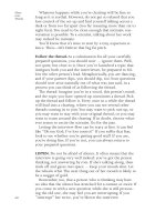
Chemical energy 40%
Heat energy
60%
Fuel
molecules
CO
2
+ H
2
O + NH
3
ADP + P
i
Catabolism
FIGURE 4–17
Flow of chemical energy from fuel molecules to ATP and
heat, and from ATP to energy-requiring cell functions.
Vander et al.: Human
Physiology: The
Mechanism of Body
Function, Eighth Edition
I. Basic Cell Functions 4. Protein Activity and
Cellular Metabolism
© The McGraw−Hill
Companies, 2001
_
vitamin rate-limiting reaction
NAD
ϩ
end-product inhibition
FAD adenosine triphosphate
enzyme activity (ATP)
metabolic pathway
1. How do molecules acquire the activation energy
required for a chemical reaction?
2. List the four factors that influence the rate of a
chemical reaction and state whether increasing the
factor will increase or decrease the rate of the
reaction.
3. What characteristics of a chemical reaction make it
reversible or irreversible?
SECTION B REVIEW QUESTIONS
4. List five characteristics of enzymes.
5. What is the difference between a cofactor and a
coenzyme?
6. From what class of nutrients are coenzymes derived?
7. Why are small concentrations of coenzymes
sufficient to maintain enzyme activity?
8. List three ways in which the rate of an enzyme-
mediated reaction can be altered.
9. How can an irreversible step in a metabolic pathway
be reversed?
10. What is the function of ATP in metabolism?
11. Approximately how much of the energy released
from the catabolism of fuel molecules is transferred
to ATP? What happens to the rest?
70
PART ONE Basic Cell Functions
METABOLIC PATHWAYS
SECTION C
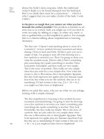
Three distinct but linked metabolic pathways are used
by cells to transfer the energy released from the break-
down of fuel molecules of ATP. They are known as gly-
colysis, the Krebs cycle, and oxidative phosphorylation
(Figure 4–18). In the following section, we will describe
the major characteristics of these three pathways in
terms of the location of the pathway enzymes in a cell,
the relative contribution of each pathway to ATP pro-
duction, the sites of carbon dioxide formation and oxy-
gen utilization, and the key molecules that enter and
leave each pathway.
In this last regard, several facts should be noted in
Figure 4–18. First, glycolysis operates only on carbo-
hydrates. Second, all the categories of nutrients—
carbohydrates, fats, and proteins—contribute to ATP
production via the Krebs cycle and oxidative phos-
phorylation. Third, mitochondria are essential for the
Krebs cycle and oxidative phosphorylation. Finally
one important generalization to keep in mind is that
glycolysis can occur in either the presence or absence
of oxygen, whereas both the Krebs cycle and oxidative
phosphorylation require oxygen as we shall see.
Cellular Energy Transfer
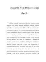
Glycolysis
Glycolysis (from the Greek glycos, sugar, and lysis,
breakdown) is a pathway that partially catabolizes car-
bohydrates, primarily glucose. It consists of 10 enzy-
matic reactions that convert a six-carbon molecule of
glucose into two three-carbon molecules of pyruvate,
the ionized form of pyruvic acid (Figure 4–19). The
Glycolysis
Carbohydrates
Pyruvate
Lactate
CO
2
H
2
O
Fats and
proteins
Energy
ADP
+ P
i
AT P
Krebs cycle
Coenzyme—2H
O
2
Fats
Oxidative
phosphorylation
Cytosol
Mitochondria
Mitochondria
FIGURE 4–18
Pathways linking the energy released from the catabolism of
fuel molecules to the formation of ATP.
Vander et al.: Human
Physiology: The
Mechanism of Body
Function, Eighth Edition
I. Basic Cell Functions 4. Protein Activity and
Cellular Metabolism
© The McGraw−Hill
Companies, 2001
71
Protein Activity and Cellular Metabolism CHAPTER FOUR
CH
2
OH
O
HO
OH
H
H
2
C
P
O
O
O
–
–
O
CH
2
AT P
ADP
O
H
OH
H
HO
OH
H
H
H
P
i
OH
H
P
O
O
O
–
O
–
CH
2
OH
O
OH
H
OH
H
HO
OH
H
H
H
8
4
6
Glucose Glucose 6-phosphate
Dihydroxyacetone
phosphate
HO
HH
H
2
CP
O
O
O
–
–
O
O
OH H
H
Fructose 6-phosphate
3
CH
2
P
O
O
O
–
O
–
OH
CH
2
P
O
C
O
–
OH
CH
2
O
P
O
O
–
O
–
CH
O
CO
CH
2
OH
H
2
H
P
O
O
O
–
O
–
CH
2
O
C
CH
P
O
O
O
–
–
O
AT P
ADP
O
–
H
2
O
AT P
ADP
1
P
O
COO
–
CH
3
OO
–
O
Fructose 1,6-bisphosphate
2-Phosphoglycerate Phosphoenolpyruvate
3-Phosphoglyceraldehyde
5
COOH
P
O
OH
CH
2
O
CH
O
–
O
–
O
–
10
AT P
ADP
7
P
O
COO
–
OHCH
2
OCH O
–
O
–
C
OC
CH
2
COO
–
NADH + H
+
NAD
+
OHCH
COO
–
CH
3
Pyruvate
To Krebs cycle
(anaerobic)
(aerobic)
3-Phosphoglycerate 1,3-Bisphosphoglycerate
Lactate
9
NAD
+
OH
NADH
+ H
+
FIGURE 4–19
Glycolytic pathway. Under anaerobic conditions, there is a net synthesis of two molecules of ATP for every molecule of glucose
that enters the pathway. Note that at the pH existing in the body, the products produced by the various glycolytic steps exist in
the ionized, anionic form (pyruvate, for example). They are actually produced as acids (pyruvic acid, for example) that then ionize.
reactions produce a net gain of two molecules of ATP
and four atoms of hydrogen, two of which are trans-
ferred to NAD
ϩ
and two are released as hydrogen ions:
Glucose ϩ 2 ADP ϩ 2 P
i
ϩ 2 NAD
ϩ
88n (4–1)
2 Pyruvate ϩ 2 ATP ϩ 2 NADH ϩ 2 H
ϩ
ϩ 2 H
2
O
These 10 reactions, none of which utilizes molecular oxy-
gen, take place in the cytosol. Note (Figure 4–19) that
all the intermediates between glucose and the end
product pyruvate contain one or more ionized phos-
phate groups. As we shall learn in Chapter 6, plasma
membranes are impermeable to such highly ionized
Vander et al.: Human
Physiology: The
Mechanism of Body
Function, Eighth Edition
I. Basic Cell Functions 4. Protein Activity and
Cellular Metabolism
© The McGraw−Hill
Companies, 2001
72
PART ONE Basic Cell Functions
C
COO
–
CH
3
O
OH
C
LactatePyruvate
COO
–
cycle
Reaction 6
(anaerobic)
(aerobic)
H
2NADH
+ 2H
+
2NAD
+
Glucose
CH
3
Krebs
22
FIGURE 4–20
Under anaerobic conditions, the coenzyme NAD
ϩ
utilized in
the glycolytic reaction 6 (see Figure 4–19) is regenerated
when it transfers its hydrogen atoms to pyruvate during the
formation of lactate.
molecules, and thus these molecules remain trapped
within the cell.
Note that the early steps in glycolysis (reactions 1
and 3) each use, rather than produce, one molecule of
ATP, to form phosphorylated intermediates. In addi-
tion, note that reaction 4 splits a six-carbon intermedi-
ate into two three-carbon molecules, and reaction 5
converts one of these three-carbon molecules into the
other so that at the end of reaction 5 we have two mol-
ecules of 3-phosphoglyceraldehyde derived from one
molecule of glucose. Keep in mind, then, that from this
point on, two molecules of each intermediate are
involved.
The first formation of ATP in glycolysis occurs dur-
ing reaction 7 when a phosphate group is transferred
to ADP to form ATP. Since, as stressed above, two in-
termediates exist at this point, reaction 7 produces two
molecules of ATP, one from each of them. In this reac-
tion, the mechanism of forming ATP is known as
substrate-level phosphorylation since the phosphate
group is transferred from a substrate molecule to ADP.
As we shall see, this mechanism is quite different from
that used during oxidative phosphorylation, in which
free inorganic phosphate is coupled to ADP to form ATP.
A similar substrate-level phosphorylation of ADP
occurs during reaction 10, where again two molecules
of ATP are formed. Thus, reactions 7 and 10 generate a
total of four molecules of ATP for every molecule of glu-
cose entering the pathway. There is a net gain, however,
of only two molecules of ATP during glycolysis because
two molecules of ATP were used in reactions 1 and 3.
The end product of glycolysis, pyruvate, can pro-
ceed in one of two directions, depending on the avail-
ability of molecular oxygen, which, as we stressed ear-
lier, is not utilized in any of the glycolytic reactions
themselves. If oxygen is present—that is, if aerobic
conditions exist—pyruvate can enter the Krebs cycle
and be broken down into carbon dioxide, as described
in the next section. In contrast, in the absence of oxygen
(anaerobic conditions), pyruvate is converted to lac-
tate (the ionized form of lactic acid) by a single enzyme-
mediated reaction. In this reaction (Figure 4–20) two
hydrogen atoms derived from NADH ϩ H
ϩ
are trans-
ferred to each molecule of pyruvate to form lactate,
and NAD
ϩ
is regenerated. These hydrogens had orig-
inally been transferred to NAD
ϩ
during reaction 6 of
glycolysis, so the coenzyme NAD
ϩ
shuttles hydrogen
between the two reactions during anaerobic glycoly-
sis. The overall reaction for anaerobic glycolysis is
Glucose ϩ 2 ADP ϩ 2 P
i
88n
(4–2)
2 Lactate ϩ 2 ATP ϩ 2 H
2
O
As stated in the previous paragraph, under aerobic
conditions pyruvate is not converted to lactate but
rather enters the Krebs cycle. Therefore, the mechanism
just described for regenerating NAD
ϩ
from NADH ϩ
H
ϩ
by forming lactate does not occur. (Compare Equa-
tions 4–1 and 4–2.) Instead, as we shall see, H
ϩ
and the
hydrogens of NADH are transferred to oxygen during
oxidative phosphorylation, regenerating NAD
ϩ
and
producing H
2
O.
In most cells, the amount of ATP produced by gly-
colysis from one molecule of glucose is much smaller
than the amount formed under aerobic conditions by
the other two ATP-generating pathways—the Krebs
cycle and oxidative phosphorylation. There are special
cases, however, in which glycolysis supplies most, or
even all, of a cell’s ATP. For example, erythrocytes con-
tain the enzymes for glycolysis but have no mito-
chondria, which, as we have said, are required for the
other pathways. All of their ATP production occurs,
therefore, by glycolysis. Also, certain types of skeletal
muscles contain considerable amounts of glycolytic en-
zymes but have few mitochondria. During intense
muscle activity, glycolysis provides most of the ATP in
these cells and is associated with the production of
large amounts of lactate. Despite these exceptions,
most cells do not have sufficient concentrations of gly-
colytic enzymes or enough glucose to provide, by gly-
colysis alone, the high rates of ATP production neces-
sary to meet their energy requirements and thus are
unable to function for long under anaerobic conditions.
Our discussion of glycolysis has focused upon glu-
cose as the major carbohydrate entering the glycolytic
pathway. However, other carbohydrates such as fruc-
tose, derived from the disaccharide sucrose (table
sugar), and galactose, from the disaccharide lactose
Vander et al.: Human
Physiology: The
Mechanism of Body
Function, Eighth Edition
I. Basic Cell Functions 4. Protein Activity and
Cellular Metabolism
© The McGraw−Hill
Companies, 2001
(milk sugar), can also be catabolized by glycolysis since
these carbohydrates are converted into several of the
intermediates that participate in the early portion of
the glycolytic pathway.
In some microoganisms (yeast cells, for example),
pyruvate is converted under anaerobic conditions to
carbon dioxide and alcohol (CH
3
CH
2
OH) rather than
to lactate. This process is known as fermentation and
forms the basis for the production of alcohol from ce-
real grains rich in carbohydrates.
Table 4–5 summarizes the major characteristics of
glycolysis.
Krebs Cycle
The Krebs cycle, named in honor of Hans Krebs, who
worked out the intermediate steps in this pathway
(also known as the citric acid cycle or tricarboxylic
acid cycle), is the second of the three pathways in-
volved in fuel catabolism and ATP production. It uti-
lizes molecular fragments formed during carbohy-
drate, protein, and fat breakdown, and it produces
carbon dioxide, hydrogen atoms (half of which are
bound to coenzymes), and small amounts of ATP. The
enzymes for this pathway are located in the inner mi-
tochondrial compartment, the matrix.
The primary molecule entering at the beginning of
the Krebs cycle is acetyl coenzyme A (acetyl CoA):
Coenzyme A (CoA) is derived from the B vitamin pan-
tothenic acid and functions primarily to transfer acetyl
groups, which contain two carbons, from one molecule
to another. These acetyl groups come either from
pyruvate, which, as we have just seen, is the end prod-
CoA
S
O
CH
3
C
uct of aerobic glycolysis, or from the breakdown of
fatty acids and some amino acids, as we shall see in a
later section.
Pyruvate, upon entering mitochondria from the
cytosol, is converted to acetyl CoA and CO
2
(Figure
4–21). Note that this reaction produces the first mole-
cule of CO
2
formed thus far in the pathways of fuel
catabolism, and that hydrogen atoms have been trans-
ferred to NAD
ϩ
.
The Krebs cycle begins with the transfer of the
acetyl group of acetyl CoA to the four-carbon mole-
cule, oxaloacetate, to form the six-carbon molecule, ci-
trate (Figure 4–22). At the third step in the cycle a mol-
ecule of CO
2
is produced, and again at the fourth step.
Thus, two carbon atoms entered the cycle as part of
the acetyl group attached to CoA, and two carbons (al-
though not the same ones) have left in the form of CO
2
.
Note also that the oxygen that appears in the CO
2
is
not derived from molecular oxygen but from the car-
boxyl groups of Krebs-cycle intermediates.
In the remainder of the cycle, the four-carbon mol-
ecule formed in reaction 4 is modified through a series
of reactions to produce the four-carbon molecule ox-
aloacetate, which becomes available to accept another
acetyl group and repeat the cycle.
73
Protein Activity and Cellular Metabolism CHAPTER FOUR
Entering substrates Glucose and other monosaccharides
Enzyme location Cytosol
Net ATP production 2 ATP formed directly per molecule of glucose entering pathway
Can be produced in the absence of oxygen (anaerobically)
Coenzyme production 2 NADH ϩ 2 H
ϩ
formed under aerobic conditions
Final products Pyruvate—under aerobic conditions
Lactate—under anaerobic conditions
Net reaction
Aerobic: Glucose ϩ 2 ADP ϩ 2 P
i
ϩ 2 NAD
ϩ
88n
2 pyruvate ϩ 2 ATP ϩ 2 NADH ϩ 2 H
ϩ
ϩ 2 H
2
O
Anaerobic: Glucose ϩ 2 ADP ϩ 2 P
i
88n 2 lactate ϩ 2 ATP ϩ 2 H
2
O
TABLE 4–5
Characteristics of Glycolysis
NAD
+
NADH + H
+
Pyruvic acid Acetyl coenzyme A
OC
COOH
CH
3
OC
CH
3
CO
2
CoA
CoAS
+ SH +
FIGURE 4–21
Formation of acetyl coenzyme A from pyruvic acid with the
formation of a molecule of carbon dioxide.
Vander et al.: Human
Physiology: The
Mechanism of Body
Function, Eighth Edition
I. Basic Cell Functions 4. Protein Activity and
Cellular Metabolism
© The McGraw−Hill
Companies, 2001
74
PART ONE Basic Cell Functions
Now we come to a crucial fact: In addition to pro-
ducing carbon dioxide, intermediates in the Krebs cy-
cle generate hydrogen atoms, most of which are trans-
ferred to the coenzymes NAD
ϩ
and FAD to form
NADH and FADH
2
. This hydrogen transfer to NAD
ϩ
occurs in each of steps 3, 4, and 8, and to FAD in re-
action 6. These hydrogens will be transferred from the
coenzymes, along with the free H
ϩ
, to oxygen in the
next stage of fuel metabolism—oxidative phosphory-
lation. Since oxidative phosphorylation is necessary for
regeneration of the hydrogen-free form of these coen-
zymes, the Krebs cycle can operate only under aerobic con-
ditions. There is no pathway in the mitochondria that
can remove the hydrogen from these coenzymes un-
der anaerobic conditions.
So far we have said nothing of how the Krebs cy-
cle contributes to the formation of ATP. In fact, the
Krebs cycle directly produces only one high-energy nu-
cleotide triphosphate. This occurs during reaction 5 in
which inorganic phosphate is transferred to guanosine
6
H
CH
3
CoA SH
S
O
C
2
CH
2
Oxidative
phosphorylation
Malate
C
H
CH
2
CoA
HO
COO
–
COO
–
COO
–
3
Acetyl coenzyme A
Oxaloacetate
α-Ketoglutarate
Citrate
CO
2
O
CH
2
COO
–
C
COO
–
OH
CH
2
COO
–
H
COO
–
OHC
COO
–
CH
2
COO
–
C
1
8
7
4
C
OC
COO
–
COO
–
CH
2
CH
2
NADH + H
+
H
2
O
NADH + H
+
COO
–
NADH + H
+
CO
2
Isocitrate
OC
AT P
GDP
Fumarate
CoA
FADH
2
COO
–
P
i
COO
–
COO
–
5
CH
2
CH
2
CH
Succinyl coenzyme A
CH
Succinate
ADP
GTP
H
2
O
CoA
CoA
COO
–
COO
–
CH
2
CH
2
H
2
O
S
FIGURE 4–22
The Krebs-cycle pathway. Note that the carbon atoms in the two molecules of CO
2
produced by a turn of the cycle are not
the same two carbon atoms that entered the cycle as an acetyl group (identified by the dashed boxes in this figure).
Vander et al.: Human
Physiology: The
Mechanism of Body
Function, Eighth Edition
I. Basic Cell Functions 4. Protein Activity and
Cellular Metabolism
© The McGraw−Hill
Companies, 2001
diphosphate (GDP) to form guanosine triphosphate
(GTP). The hydrolysis of GTP, like that of ATP, can pro-
vide energy for some energy-requiring reactions. In ad-
dition, the energy in GTP can be transferred to ATP by
the reaction
This reaction is reversible, and the energy in ATP can
be used to form GTP from GDP when additional GTP
is required for protein synthesis (Chapter 5) and sig-
nal transduction (Chapter 7).
To reiterate, the formation of ATP from GTP is the
only mechanism by which ATP is formed within the
Krebs cycle. Why, then, is the Krebs cycle so impor-
tant? Because the hydrogen atoms transferred to coen-
zymes during the cycle (plus the free hydrogen ions
generated) are used in the next pathway, oxidative
phosphorylation, to form large amounts of ATP.
The net result of the catabolism of one acetyl
group from acetyl CoA by way of the Krebs cycle can
be written:
Acetyl CoA ϩ 3 NAD
ϩ
ϩ FAD ϩ GDP ϩ P
i
ϩ 2H
2
O 88n
2 CO
2
ϩ CoA ϩ 3 NADH ϩ 3H
ϩ
ϩ FADH
2
ϩ GTP (4–3)
One more point should be noted: Although the ma-
jor function of the Krebs cycle is to provide hydrogen
atoms to the oxidative-phosphorylation pathway,
some of the intermediates in the cycle can be used to
synthesize organic molecules, especially several types
of amino acids, required by cells. Oxaloacetate is one
of the intermediates used in this manner. When a mol-
ecule of oxaloacetate is removed from the Krebs cycle
in the process of forming amino acids, however, it is
not available to combine with the acetate fragment of
acetyl CoA at the beginning of the cycle. Thus, there
must be a way of replacing the oxaloacetate and other
Krebs-cycle intermediates that are consumed in syn-
GTP ϩ ADP GDP ϩ ATP
thetic pathways. Carbohydrates provide one source of
oxaloacetate replacement by the following reaction,
which converts pyruvate into oxaloacetate.
Pyruvate ϩ CO
2
ϩ ATP 88n
Oxaloacetate ϩ ADP ϩ P
i
(4–4)
Certain amino acid derivatives, as we shall see, can
also be used to form oxaloacetate and other Krebs-
cycle intermediates.
Table 4–6 summarizes the characteristics of the
Krebs cycle reactions.
Oxidative Phosphorylation
Oxidative phosphorylation provides the third, and
quantitatively most important, mechanism by which
energy derived from fuel molecules can be transferred
to ATP. The basic principle behind this pathway is sim-
ple: The energy transferred to ATP is derived from the
energy released when hydrogen ions combine with
molecular oxygen to form water. The hydrogen comes
from the NADH ϩ H
ϩ
and FADH
2
coenzymes gener-
ated by the Krebs cycle, by the metabolism of fatty
acids (see below), and, to a much lesser extent, during
aerobic glycolysis. The net reaction is
ᎏ
1
2
ᎏ
O
2
ϩ NADH ϩ H
ϩ
8n H
2
O ϩ NAD
ϩ
ϩ 53 kcal/mol
The proteins that mediate oxidative phosphorylation
are embedded in the inner mitochondrial membrane
unlike the enzymes of the Krebs cycle, which are sol-
uble enzymes in the mitochondrial matrix. The pro-
teins for oxidative phosphorylation can be divided
into two groups: (1) those that mediate the series of
reactions by which hydrogen ions are transferred
to molecular oxygen, and (2) those that couple the
energy released by these reactions to the synthesis
of ATP.
75
Protein Activity and Cellular Metabolism CHAPTER FOUR
Entering substrate Acetyl coenzyme A—acetyl groups derived from pyruvate, fatty acids, and amino acids
Some intermediates derived from amino acids
Enzyme location Inner compartment of mitochondria (the mitochondrial matrix)
ATP production 1 GTP formed directly, which can be converted into ATP
Operates only under aerobic conditions even though molecular oxygen is not used directly
in this pathway
Coenzyme production 3 NADH ϩ 3 H
ϩ
and 2 FADH
2
Final products 2 CO
2
for each molecule of acetyl coenzyme A entering pathway
Some intermediates used to synthesize amino acids and other organic molecules required for special
cell functions
Net reaction Acetyl CoA ϩ 3 NAD
ϩ
ϩ FAD ϩ GDP ϩ P
i
ϩ 2 H
2
O 88n
2 CO
2
ϩ CoA ϩ 3 NADH ϩ 3 H
ϩ
ϩ FADH
2
ϩ GTP
TABLE 4–6
Characteristics of the Krebs Cycle
Vander et al.: Human
Physiology: The
Mechanism of Body
Function, Eighth Edition
I. Basic Cell Functions 4. Protein Activity and
Cellular Metabolism
© The McGraw−Hill
Companies, 2001
Most of the first group of proteins contain iron and
copper cofactors, and are known as cytochromes (be-
cause in pure form they are brightly colored). Their
structure resembles the red iron-containing hemoglo-
bin molecule, which binds oxygen in red blood cells.
The cytochromes form the components of the electron
transport chain, in which two electrons from the hy-
drogen atoms are initially transferred either from
NADH ϩ H
ϩ
or FADH
2
to one of the elements in this
chain. These electrons are then successively transferred
to other compounds in the chain, often to or from
an iron or copper ion, until the electrons are finally
transferred to molecular oxygen, which then combines
with hydrogen ions (protons) to form water. These
hydrogen ions, like the electrons, come from the
free hydrogen ions and the hydrogen-bearing co-
enzymes, having been released from them early in the
transport chain when the electrons from the hydrogen
atoms were transferred to the cytochromes.
Importantly, in addition to transferring the coen-
zyme hydrogens to water, this process regenerates the
hydrogen-free form of the coenzymes, which then be-
come available to accept two more hydrogens from in-
termediates in the Krebs cycle, glycolysis, or fatty acid
pathway (as described below). Thus, the electron trans-
port chain provides the aerobic mechanism for regen-
erating the hydrogen-free form of the coenzymes,
whereas, as described earlier, the anaerobic mechanism,
which applies only to glycolysis, is coupled to the for-
mation of lactate.
At each step along the electron transport chain,
small amounts of energy are released, which in total
account for the full 53 kcal/mol released from a direct
reaction between hydrogen and oxygen. Because this
energy is released in small steps, it can be linked to the
synthesis of several molecules of ATP, each of which
requires only 7 kcal/mol.
ATP is formed at three points along the electron
transport chain. The mechanism by which this occurs
is known as the chemiosmotic hypothesis. As elec-
trons are transferred from one cytochrome to another
along the electron transport chain, the energy released
is used to move hydrogen ions (protons) from the ma-
trix into the compartment between the inner and outer
mitochondrial membranes (Figure 4–23), thus pro-
ducing a source of potential energy in the form of a
hydrogen-ion gradient across the membrane. At three
points along the chain, a protein complex forms a chan-
nel in the inner mitochondrial membrane through
which the hydrogen ions can flow back to the matrix
side and in the process transfer energy to the forma-
tion of ATP from ADP and P
i
. FADH
2
has a slightly
lower chemical energy content than does NADH ϩ H
ϩ
and enters the electron transport chain at a point
76
PART ONE Basic Cell Functions
Cytochromes in electron transport chain
NADH + H
+
FADH
2
NAD
+
+ 2H
+
FAD + 2H
+
Matrix
H
2
O
H
+
2
e
–
2
e
–
2
e
–
Inner mitochondrial
membrane
Outer mitochondrial
membrane
1
2
O
2
+2
ADP
P
i
H
+
ATP ADP
P
i
H
+
ATP
H
+
H
+
H
+
ADP
P
i
H
+
ATP
FIGURE 4–23
ATP is formed during oxidative phosphorylation by the flow of hydrogen ions across the inner mitochondrial membrane. Two
or three molecules of ATP are produced per pair of electrons donated, depending on the point at which a particular
coenzyme enters the electron transport chain.
Vander et al.: Human
Physiology: The
Mechanism of Body
Function, Eighth Edition
I. Basic Cell Functions 4. Protein Activity and
Cellular Metabolism
© The McGraw−Hill
Companies, 2001
beyond the first site of ATP generation (Figure 4–23).
Thus, the transfer of its electrons to oxygen produces
only two ATP rather than the three formed from
NADH ϩ H
ϩ
.
To repeat, the majority of the ATP formed in the
body is produced during oxidative phosphorylation as
a result of processing hydrogen atoms that originated
largely from the Krebs cycle, during the breakdown of
carbohydrates, fats, and proteins. The mitochondria,
where the oxidative phosphorylation and the Krebs-
cycle reactions occur, are thus considered the power-
houses of the cell. In addition, as we have just seen, it
is within these organelles that the majority of the oxy-
gen we breathe is consumed, and the majority of the
carbon dioxide we expire is produced.
Table 4–7 summaries the key features of oxidative
phosphorylation.
Reactive Oxygen Species
As we have just seen, the formation of ATP by oxida-
tive phosphorylation involves the transfer of electrons
and hydrogen to molecular oxygen. Several highly
reactive transient oxygen derivatives can also be formed
during this process—hydrogen peroxide and the free
radicals superoxide anion and hydroxyl radical.
Although most of the electrons transferred along
the electron transport chain go into the formation of
water, small amounts can combine with oxygen to
O
2
O
2
–
•
OH
–
H
2
O
2
+
OH • 2 OH
–
2 H
2
O
2 H
+
2 H
+
e
–
e
–
e
–
e
–
Superoxide
anion
Hydrogen
peroxide
Hydroxyl
radical
form reactive oxygen species. These species can react
with and damage proteins, membrane phospholipids,
and nucleic acids. Such damage has been implicated
in the aging process and in inflammatory reactions to
tissue injury. Some cells use these reactive molecules
to kill invading bacteria, as described in Chapter 20.
Reactive oxygen molecules are also formed by the
action of ionizing radiation on oxygen and by reactions
of oxygen with heavy metals such as iron. Cells con-
tain several enzymatic mechanisms for removing these
reactive oxygen species and thus providing protection
from their damaging effects.
Carbohydrate, Fat, and Protein
Metabolism
Having described the three pathways by which energy
is transferred to ATP, we now consider how each of the
three classes of fuel molecules—carbohydrates, fats,
and proteins—enters the ATP-generating pathways.
We also consider the synthesis of these fuel molecules
and the pathways and restrictions governing their con-
version from one class to another. These anabolic path-
ways are also used to synthesize molecules that have
functions other than the storage and release of energy.
For example, with the addition of a few enzymes, the
pathway for fat synthesis is also used for synthesis of
the phospholipids found in membranes.
Carbohydrate Metabolism
Carbohydrate Catabolism In the previous sections,
we described the major pathways of carbohydrate ca-
tabolism: the breakdown of glucose to pyruvate or lac-
tate by way of the glycolytic pathway, and the metab-
olism of pyruvate to carbon dioxide and water by way
of the Krebs cycle and oxidative phosphorylation.
77
Protein Activity and Cellular Metabolism CHAPTER FOUR
Entering substrates Hydrogen atoms obtained from NADH ϩ H
ϩ
and FADH
2
formed (1) during glycolysis,
(2) by the Krebs cycle during the breakdown of pyruvate and amino acids, and
(3) during the breakdown of fatty acids
Molecular oxygen
Enzyme location Inner mitochondrial membrane
ATP production 3 ATP formed from each NADH ϩ H
ϩ
2 ATP formed from each FADH
2
Final products H
2
O—one molecule for each pair of hydrogens entering pathway.
Net reaction
ᎏ
1
2
ᎏ
O
2
ϩ NADH ϩ H
ϩ
ϩ 3 ADP ϩ 3 P
i
88n H
2
O ϩ NAD
ϩ
ϩ 3 ATP
TABLE 4–7
Characteristics of Oxidative Phosphorylation
Vander et al.: Human
Physiology: The
Mechanism of Body
Function, Eighth Edition
I. Basic Cell Functions 4. Protein Activity and
Cellular Metabolism
© The McGraw−Hill
Companies, 2001
The amount of energy released during the catabo-
lism of glucose to carbon dioxide and water is 686
kcal/mol of glucose:
C
6
H
12
O
6
ϩ 6 O
2
88n 6 H
2
O ϩ 6 CO
2
ϩ 686 kcal/mol
As noted earlier, about 40 percent of this energy is
transferred to ATP. Figure 4–24 illustrates the points at
which ATP is formed during glucose catabolism. As
we have seen, a net gain of two ATP molecules occurs
by substrate-level phosphorylation during glycolysis,
and two more are formed during the Krebs cycle from
GTP, one from each of the two molecules of pyruvate
entering the cycle. The major portion of ATP molecules
produced in glucose catabolism—34 ATP per mole-
cule—is formed during oxidative phosphorylation
from the hydrogens generated at various steps during
glucose breakdown.
To reiterate, in the absence of oxygen, only 2 mol-
ecules of ATP can be formed by the breakdown of glu-
cose to lactate. This yield represents only 2 percent of
the energy stored in glucose. Thus, the evolution of
aerobic metabolic pathways greatly increased the
amount of energy available to a cell from glucose ca-
tabolism. For example, if a muscle consumed 38 mol-
ecules of ATP during a contraction, this amount of ATP
could be supplied by the breakdown of 1 molecule of
glucose in the presence of oxygen or 19 molecules of
glucose under anaerobic conditions.
It is important to note, however, that although only
2 molecules of ATP are formed per molecule of glu-
cose under anaerobic conditions, large amounts of ATP
can still be supplied by the glycolytic pathway if large
amounts of glucose are broken down to lactate. This is
not an efficient utilization of fuel energy, but it does
permit continued ATP production under anaerobic
conditions, such as occur during intense exercise
(Chapter 11).
Glycogen Storage A small amount of glucose can be
stored in the body to provide a reserve supply for use
when glucose is not being absorbed into the blood
from the intestinal tract. It is stored as the polysac-
charide glycogen, mostly in skeletal muscles and the
liver.
Glycogen is synthesized from glucose by the
pathway illustrated in Figure 4–25. The enzymes for
both glycogen synthesis and glycogen breakdown are
located in the cytosol. The first step in glycogen
synthesis, the transfer of phosphate from a molecule
of ATP to glucose, forming glucose 6-phosphate, is
the same as the first step in glycolysis. Thus, glucose
6-phosphate can either be broken down to pyruvate or
used to form glycogen.
Note that, as indicated by the bowed arrows in Fig-
ure 4–25, different enzymes are used to synthesize and
break down glycogen. The existence of two pathways
78
PART ONE Basic Cell Functions
Glycolysis
Glucose
2 Pyruvate
2 ATP
Krebs cycle
(mitochondria)
2 Acetyl coenzyme A
6 H
2
O
4 CO
2
2 ATP
Oxidative phosphorylation
(mitochondria)
2 FADH
2
2
(
NADH + H
+
)
10 ATP 12 ATP 12 ATP
ATP ATP ATP
6 O
2
Cytochromes
34 ATP
6
(
NADH + H
+
)
2 CO
2
12 H
2
O
2
(
NADH + H
+
)
C
6
H
12
0
6
+ 6 O
2
+ 38 ADP + 38 P
i
6 CO
2
+ 6 H
2
O + 38 ATP
(cytosol)
FIGURE 4–24
Pathways of aerobic glucose catabolism and their linkage to ATP formation.
Vander et al.: Human
Physiology: The
Mechanism of Body
Function, Eighth Edition
I. Basic Cell Functions 4. Protein Activity and
Cellular Metabolism
© The McGraw−Hill
Companies, 2001
amino acids. This process of generating new molecules
of glucose is known as gluconeogenesis. The major
substrate in gluconeogenesis is pyruvate, formed from
lactate and from several amino acids during protein
breakdown. In addition, as we shall see, glycerol de-
rived from the hydrolysis of triacylglycerols can be
converted into glucose via a pathway that does not in-
volve pyruvate.
The pathway for gluconeogenesis in the liver and
kidneys (Figure 4–26) makes use of many but not all
of the enzymes used in glycolysis because most of
these reactions are reversible. However, reactions 1, 3
79
Protein Activity and Cellular Metabolism CHAPTER FOUR
Glucose 6-phosphate
Pyruvate
(all tissues)
(liver and
kidneys)
Glucose
P
i
P
i
Glycogen
FIGURE 4–25
Pathways for glycogen synthesis and breakdown. Each
bowed arrow indicates one or more irreversible reactions
that requires different enzymes to catalyze the reaction in
the forward and reverse direction.
containing enzymes that are subject to both covalent
and allosteric modulation provides a mechanism for
regulating the flow of glucose to and from glycogen.
When an excess of glucose is available to a liver or
muscle cell, the enzymes in the glycogen synthesis
pathway are activated by the chemical signals de-
scribed in Chapter 18, and the enzyme that breaks
down glycogen is simultaneously inhibited. This com-
bination leads to the net storage of glucose in the form
of glycogen.
When less glucose is available, the reverse com-
bination of enzyme stimulation and inhibition occurs,
and net breakdown of glycogen to glucose 6-
phosphate occurs. Two paths are available to this glu-
cose 6-phosphate: (1) In most cells, including skeletal
muscle, it enters the glycolytic pathway where it is
catabolized to provide the energy for ATP formation;
(2) in liver (and kidney cells), glucose 6-phosphate
can be converted to free glucose by removal of the
phosphate group, and glucose is then able to pass out
of the cell into the blood, for use as fuel by other cells
(Chapter 18).
Glucose Synthesis In addition to being formed in the
liver from the breakdown of glycogen, glucose can be
synthesized in the liver and kidneys from intermedi-
ates derived from the catabolism of glycerol and some
Glycerol
Glucose
Triacylglycerol
metabolism
Phosphoenolpyruvate
Glucose 6-phosphate
Amino acid
intermediates
Lactate
CO
2
Citrate
Krebs
cycle
CO
2
CO
2
Oxaloacetate
Amino acid
intermediates
CO
2
CO
2
Pyruvate
Acetyl coenzyme A
FIGURE 4–26
Gluconeogenic pathway by which pyruvate, lactate, glycerol,
and various amino acid intermediates can be converted into
glucose in the liver. Note the points at which each of these
precursors, supplied by the blood, enters the pathway.
Vander et al.: Human
Physiology: The
Mechanism of Body
Function, Eighth Edition
I. Basic Cell Functions 4. Protein Activity and
Cellular Metabolism
© The McGraw−Hill
Companies, 2001
and 10 (see Figure 4–19) are irreversible, and addi-
tional enzymes are required, therefore, to form glucose
from pyruvate. Pyruvate is converted to phospho-
enolpyruvate by a series of mitochondrial reactions in
which CO
2
is added to pyruvate to form the four-
carbon Krebs-cycle intermediate oxaloacetate. [In ad-
dition to being an important intermediary step in glu-
coneogenesis, this reaction (Equation 4–4) provides a
pathway for replacing Krebs-cycle intermediates, as
described earlier.] An additional series of reactions
leads to the transfer of a four-carbon intermediate de-
rived from oxaloacetate out of the mitochondria and
its conversion to phosphoenolpyruvate in the cytosol.
Phosphoenolpyruvate then reverses the steps of gly-
colysis back to the level of reaction 3, in which a dif-
ferent enzyme from that used in glycolysis is required
to convert fructose 1,6-bisphosphate to fructose 6-
phosphate. From this point on, the reactions are again
reversible, leading to glucose 6-phosphate, which can
be converted to glucose in the liver and kidneys or
stored as glycogen. Since energy is released during the
glycolytic breakdown of glucose to pyruvate in the
form of heat and ATP generation, energy must be
added to reverse this pathway. A total of six ATP are
consumed in the reactions of gluconeogenesis per mol-
ecule of glucose formed.
Many of the same enzymes are used in glycolysis
and gluconeogenesis, so the question arises: What con-
trols the direction of the reactions in these pathways?
What conditions determine whether glucose is broken
down to pyruvate or whether pyruvate is converted
into glucose? The answer lies in the concentrations of
glucose or pyruvate in a cell and in the control of the
enzymes involved in the irreversible steps in the path-
way, a control exerted via various hormones that alter
the concentrations and activities of these key enzymes
(Chapter 18).
Fat Metabolism
Fat Catabolism Triacylglycerol (fat) consists of three
fatty acids linked to glycerol (Chapter 2). Fat accounts
for the major portion (approximately 80 percent) of the
energy stored in the body (Table 4–8). Under resting
conditions, approximately half the energy used by
such tissues as muscle, liver, and kidneys is derived
from the catabolism of fatty acids.
Although most cells store small amounts of fat, the
majority of the body’s fat is stored in specialized cells
known as adipocytes. Almost the entire cytoplasm of
these cells is filled with a single large fat droplet. Clus-
ters of adipocytes form adipose tissue, most of which
is in deposits underlying the skin. The function of
adipocytes is to synthesize and store triacylglycerols
during periods of food uptake and then, when food is
not being absorbed from the intestinal tract, to release
fatty acids and glycerol into the blood for uptake and
use by other cells to provide the energy for ATP for-
mation. The factors controlling fat storage and release
from adipocytes will be described in Chapter 18. Here
we will emphasize the pathway by which fatty acids
are catabolized by most cells to provide the energy for
ATP synthesis, and the pathway for the synthesis of
fatty acids from other fuel molecules.
Figure 4–27 shows the pathway for fatty acid ca-
tabolism, which is achieved by enzymes present in
the mitochondrial matrix. The breakdown of a fatty
acid is initiated by linking a molecule of coenzyme A
to the carboxyl end of the fatty acid. This initial step
is accompanied by the breakdown of ATP to AMP and
two P
i
.
The coenzyme-A derivative of the fatty acid then
proceeds through a series of reactions, known as beta
oxidation, which split off a molecule of acetyl coen-
zyme A from the end of the fatty acid and transfer two
pairs of hydrogen atoms to coenzymes (one pair to
FAD and the other to NAD
ϩ
). The hydrogen atoms
from the coenzymes then enter the oxidative-
phosphorylation pathway to form ATP.
When an acetyl coenzyme A is split from the end
of a fatty acid, another coenzyme A is added (ATP is
not required for this step), and the sequence is re-
peated. Each passage through this sequence shortens
the fatty acid chain by two carbon atoms until all the
carbon atoms have been transferred to coenzyme A
molecules. As we saw, these molecules then enter the
Krebs cycle to produce CO
2
and ATP via the Krebs cy-
cle and oxidative phosphorylation.
How much ATP is formed as a result of the total
catabolism of a fatty acid? Most fatty acids in the body
contain 14 to 22 carbons, 16 and 18 being most com-
mon. The catabolism of one 18-carbon saturated fatty
acid yields 146 ATP molecules. In contrast, as we have
seen, the catabolism of one glucose molecule yields a
maximum of 38 ATP molecules. Thus, taking into ac-
count the difference in molecular weight of the fatty
acid and glucose, the amount of ATP formed from the
catabolism of a gram of fat is about 2
ᎏ
1
2
ᎏ
times greater
80
PART ONE Basic Cell Functions
TABLE 4–8
Fuel Content of a 70-kg Person
Total-Body
Total-Body Energy Energy
Content, Content, Content
kg kcal/g kcal %
Triacylglycerols 15.6 9 140,000 78
Proteins 9.5 4 38,000 21
Carbohydrates 0.5 4 2,000 1
Vander et al.: Human
Physiology: The
Mechanism of Body
Function, Eighth Edition
I. Basic Cell Functions 4. Protein Activity and
Cellular Metabolism
© The McGraw−Hill
Companies, 2001
than the amount of ATP produced by catabolizing 1
gram of carbohydrate. If an average person stored
most of his or her fuel as carbohydrate rather than fat,
body weight would have to be approximately 30 per-
cent greater in order to store the same amount of
usable energy, and the person would consume more
energy moving this extra weight around. Thus, a ma-
jor step in fuel economy occurred when animals
evolved the ability to store fuel as fat. In contrast,
plants store almost all their fuel as carbohydrate
(starch).
Fat Synthesis The synthesis of fatty acids occurs by
reactions that are almost the reverse of those that de-
grade them. However, the enzymes in the synthetic
pathway are in the cytosol, whereas (as we have just
seen) the enzymes catalyzing fatty acid breakdown are
in the mitochondria. Fatty acid synthesis begins with
cytoplasmic acetyl coenzyme A, which transfers its
acetyl group to another molecule of acetyl coenzyme
A to form a four-carbon chain. By repetition of this
process, long-chain fatty acids are built up two carbons
at a time, which accounts for the fact that all the fatty
acids synthesized in the body contain an even number
of carbon atoms.
Once the fatty acids are formed, triacylglycerol can
be synthesized by linking fatty acids to each of the
three hydroxyl groups in glycerol, more specifically, to
a phosphorylated form of glycerol called
␣
-glycerol
phosphate. The synthesis of triacylglycerol is carried
out by enzymes associated with the membranes of the
smooth endoplasmic reticulum.
Compare the molecules produced by glucose ca-
tabolism with those required for synthesis of both fatty
81
Protein Activity and Cellular Metabolism CHAPTER FOUR
C
18
Fatty acid
O
AMP
+2P
i
2
CH
2
CoA SH
(CH
2
)
14
CoAS
H
2
O
CH
3
CH
2
COOH(CH
2
)
14
CH
3
C
+
H
2
O
O
CH
2
(CH
2
)
14
CoA
S
CH
3
CCH
2
CoA SH
CH
3
(CH
2
)
14
S
CoAC
O
C
O
CoA
S
C
+
CH
3
Acetyl CoA
9 ATP
H
2
O
O
2
CO
2
CH
2
FADH
2
AT P
FAD
NAD
+
NADH H
+
O
139 ATP
Coenzyme—2H
cycle
Krebs
Oxidative
phosphorylation
FIGURE 4–27
Pathway of fatty acid catabolism, which takes place in the mitochondria. The energy equivalent of two ATP is consumed at
the start of the pathway.
Vander et al.: Human
Physiology: The
Mechanism of Body
Function, Eighth Edition
I. Basic Cell Functions 4. Protein Activity and
Cellular Metabolism
© The McGraw−Hill
Companies, 2001
acids and
␣
-glycerol phosphate. First, acetyl coenzyme
A, the starting material for fatty acid synthesis, can be
formed from pyruvate, the end product of glycolysis.
Second, the other ingredients required for fatty acid
synthesis—hydrogen-bound coenzymes and ATP—
are produced during carbohydrate catabolism. Third,
␣
-glycerol phosphate can be formed from a glucose in-
termediate. It should not be surprising, therefore, that
much of the carbohydrate in food is converted into fat
and stored in adipose tissue shortly after its absorp-
tion from the gastrointestinal tract. Mass action result-
ing from the increased concentration of glucose inter-
mediates, as well as the specific hormonal regulation
of key enzymes, promotes this conversion, as will be
described in Chapter 18.
It is very important to note that fatty acids, or more
specifically the acetyl coenzyme A derived from fatty
acid breakdown, cannot be used to synthesize new mol-
ecules of glucose. The reasons for this can be seen by
examining the pathways for glucose synthesis (see Fig-
ure 4–26). First, because the reaction in which pyru-
vate is broken down to acetyl coenzyme A and carbon
dioxide is irreversible, acetyl coenzyme A cannot be
converted into pyruvate, a molecule that could lead to
the production of glucose. Second, the equivalent of
the two carbon atoms in acetyl coenzyme A are con-
verted into two molecules of carbon dioxide during
their passage through the Krebs cycle before reaching
oxaloacetate, another takeoff point for glucose synthe-
sis, and therefore cannot be used to synthesize net
amounts of oxaloacetate.
Thus, glucose can readily be converted into fat, but
the fatty acid portion of fat cannot be converted to glu-
cose. However, the three-carbon glycerol backbone of
fat can be converted into an intermediate in the glu-
coneogenic pathway and thus give rise to glucose, as
mentioned earlier.
Protein and Amino Acid Metabolism
In contrast to the complexities of protein synthesis, de-
scribed in Chapter 5, protein catabolism requires only
a few enzymes, termed proteases, to break the peptide
bonds between amino acids. Some of these enzymes
split off one amino acid at a time from the ends of the
protein chain, whereas others break peptide bonds be-
tween specific amino acids within the chain, forming
peptides rather than free amino acids.
Amino acids can be catabolized to provide energy
for ATP synthesis, and they can also provide interme-
diates for the synthesis of a number of molecules other
than proteins. Since there are 20 different amino acids,
a large number of intermediates can be formed, and
there are many pathways for processing them. A few
basic types of reactions common to most of these path-
ways can provide an overview of amino acid catabo-
lism.
Unlike most carbohydrates and fats, amino acids
contain nitrogen atoms (in their amino groups) in ad-
dition to carbon, hydrogen, and oxygen atoms. Once
the nitrogen-containing amino group is removed, the
remainder of most amino acids can be metabolized to
intermediates capable of entering either the glycolytic
pathway or the Krebs cycle.
The two types of reactions by which the amino
group is removed are illustrated in Figure 4–28. In the
first reaction, oxidative deamination, the amino group
gives rise to a molecule of ammonia (NH
3
) and is re-
placed by an oxygen atom derived from water to form
82
PART ONE Basic Cell Functions
NH
3
COOHR
1
CH
Amino acid 2
coenzyme
2H
Oxidative deamination
Transamination
COOH
O
C
NH
2
coenzyme
Keto acid 1
Ammonia
COOHRCH
Amino acid
COOHR
O
C
NH
2
Keto acid
H
2
O
R
2
COOHR
2
O
C
Keto acid 2
CH
Amino acid 1
COOH
NH
2
R
1
+ +
+ +
+ +
FIGURE 4–28
Oxidative deamination and transamination of amino acids.
Vander et al.: Human
Physiology: The
Mechanism of Body
Function, Eighth Edition
I. Basic Cell Functions 4. Protein Activity and
Cellular Metabolism
© The McGraw−Hill
Companies, 2001
a keto acid, a categorical name rather than the name
of a specific molecule. The second means of removing
an amino group is known as transamination and in-
volves transfer of the amino group from an amino acid
to a keto acid. Note that the keto acid to which the
amino group is transferred becomes an amino acid.
The nitrogen derived from amino groups can also be
used by cells to synthesize other important nitrogen-
containing molecules, such as the purine and pyrimi-
dine bases found in nucleic acids.
Figure 4–29 illustrates the oxidative deamination
of the amino acid glutamic acid and the transamina-
tion of the amino acid alanine. Note that the keto acids
formed are intermediates either in the Krebs cycle
(
␣
ketoglutaric acid) or glycolytic pathway (pyruvic
acid). Once formed, these keto acids can be metabo-
lized to produce carbon dioxide and form ATP, or they
can be used as intermediates in the synthetic pathway
leading to the formation of glucose. As a third alter-
native, they can be used to synthesize fatty acids af-
ter their conversion to acetyl coenzyme A by way of
pyruvic acid. Thus, amino acids can be used as a
source of energy, and some can be converted into car-
bohydrate and fat.
As we have seen, the oxidative deamination of
amino acids yields ammonia. This substance, which is
highly toxic to cells if allowed to accumulate, readily
passes through cell membranes and enters the blood,
which carries it to the liver (Figure 4–30). The liver
contains enzymes that can link two molecules of
ammonia with carbon dioxide to form urea. Thus,
urea, which is relatively nontoxic, is the major ni-
trogenous waste product of protein catabolism. It en-
ters the blood from the liver and is excreted by the kid-
neys into the urine. Two of the 20 amino acids also
contain atoms of sulfur, which can be converted to sul-
fate, SO
4
2Ϫ
, and excreted in the urine.
Thus far, we have discussed mainly amino acid ca-
tabolism; now we turn to amino acid synthesis. The keto
acids pyruvic acid and
␣
-ketoglutaric acid can be de-
rived from the breakdown of glucose; they can then be
83
Protein Activity and Cellular Metabolism CHAPTER FOUR
CH
2
COOHCH
Coenzyme 2H
Oxidative
deamination
Transamination
COOH O
C
NH
2
Coenzyme
H
2
O
NH
3
CH
3
COOH
O
C
CH
2
COOH CH
2
COOH
CH
2
CH
NH
2
CH
3
COOH
Glutamic acid
α
-Ketoglutaric acid
Pyruvic acid
Alanine
FIGURE 4–29
Oxidative deamination and transamination of the amino
acids glutamic acid and alanine lead to keto acids that can
enter the carbohydrate pathways.
Blood
Oxidative deamination
Amino acids Keto acids
NH
3
Ammonia
Liver
Blood
Ammonia
CO
2
O
NH
2
—C—NH
2
Urea
Kidneys
Urea
Urine
Cells
FIGURE 4–30
Formation and excretion of urea, the major nitrogenous
waste product of protein catabolism.
Vander et al.: Human
Physiology: The
Mechanism of Body
Function, Eighth Edition
I. Basic Cell Functions 4. Protein Activity and
Cellular Metabolism
© The McGraw−Hill
Companies, 2001
transaminated, as described above, to form the amino
acids glutamate and alanine. Thus glucose can be used
to produce certain amino acids, provided other amino
acids are available in the diet to supply amino groups
for transamination. However, only 11 of the 20 amino
acids can be formed by this process because 9 of the
specific keto acids cannot be synthesized from other
intermediates. The 9 amino acids corresponding to
these keto acids must be obtained from the food we
eat and are known as essential amino acids.
Figure 4–31 provides a summary of the multiple
routes by which amino acids are handled by the body.
The amino acid pools, which consist of the body’s to-
tal free amino acids, are derived from (1) ingested pro-
tein, which is degraded to amino acids during diges-
tion in the intestinal tract, (2) the synthesis of
nonessential amino acids from the keto acids derived
from carbohydrates and fat, and (3) the continuous
breakdown of body proteins. These pools are the
source of amino acids for the resynthesis of body pro-
tein and a host of specialized amino acid derivatives,
as well as for conversion to carbohydrate and fat. A
very small quantity of amino acids and protein is lost
from the body via the urine, skin, hair, fingernails, and
in women, the menstrual fluid. The major route for the
loss of amino acids is not their excretion but rather
their deamination, with ultimate excretion of the ni-
trogen atoms as urea in the urine. The terms negative
nitrogen balance and positive nitrogen balance refer
to whether there is a net loss or gain, respectively, of
amino acids in the body over any period of time.
If any of the essential amino acids are missing from
the diet, a negative nitrogen balance—that is, loss
greater than gain—always results. The proteins that
require a missing essential amino acid cannot be syn-
thesized, and the other amino acids that would have
been incorporated into these proteins are metabolized.
This explains why a dietary requirement for protein
cannot be specified without regard to the amino acid
composition of that protein. Protein is graded in terms
of how closely its relative proportions of essential
amino acids approximate those in the average body
protein. The highest quality proteins are found in an-
imal products, whereas the quality of most plant pro-
teins is lower. Nevertheless, it is quite possible to ob-
tain adequate quantities of all essential amino acids
from a mixture of plant proteins alone.
Fuel Metabolism Summary
Having discussed the metabolism of the three major
classes of organic molecules, we can now briefly re-
view how each class is related to the others and to the
process of synthesizing ATP. Figure 4–32, which is an
expanded version of Figure 4–18, illustrates the major
pathways we have discussed and the relations of the
common intermediates. All three classes of molecules
can enter the Krebs cycle through some intermediate,
and thus all three can be used as a source of energy
for the synthesis of ATP. Glucose can be converted into
fat or into some amino acids by way of common in-
termediates such as pyruvate, oxaloacetate, and acetyl
coenzyme A. Similarly, some amino acids can be
84
PART ONE Basic Cell Functions
Excretion as
sloughed hair,
skin, etc.
(very small)
Dietary
proteins and
amino acids
Body
proteins
Amino acid
pools
Urinary
excretion
(very small)
Nitrogen-containing
derivatives of
amino acids
Nucleotides, hormones,
creatine, etc.
Carbohydrate
and fatNH
3
NH
3
Urea
Urinary
excretion
FIGURE 4–31
Pathways of amino acid metabolism.
Vander et al.: Human
Physiology: The
Mechanism of Body
Function, Eighth Edition
I. Basic Cell Functions 4. Protein Activity and
Cellular Metabolism
© The McGraw−Hill
Companies, 2001
converted into glucose and fat. Fatty acids cannot be
converted into glucose because of the irreversibility of
the reaction converting pyruvate to acetyl coenzyme
A, but the glycerol portion of triacylglycerols can be
converted into glucose. Fatty acids can be used to syn-
thesize portions of the keto acids used to form some
amino acids. Metabolism is thus a highly integrated
process in which all classes of molecules can be used,
if necessary, to provide energy, and in which each class
of molecule can provide the raw materials required to
synthesize most but not all members of other classes.
Essential Nutrients
About 50 substances required for normal or optimal
body function cannot be synthesized by the body or
are synthesized in amounts inadequate to keep pace
with the rates at which they are broken down or ex-
creted. Such substances are known as essential nutri-
ents (Table 4–9). Because they are all removed from
the body at some finite rate, they must be continually
supplied in the foods we eat.
It must be emphasized that the term “essential nu-
trient” is reserved for substances that fulfill two crite-
ria: (1) they must be essential for health, and (2) they
must not be synthesized by the body in adequate
amounts. Thus, glucose, although “essential” for
normal metabolism, is not classified as an essential
nutrient because the body normally can synthesize all
it needs, from amino acids, for example. Furthermore,
the quantity of an essential nutrient that must be pres-
ent in the diet in order to maintain health is not a
criterion for determining if the substance is essential.
Approximately 1500 g of water, 2 g of the amino acid
methionine, but only about 1 mg of the vitamin thi-
amine are required per day.
Water is an essential nutrient because far more of
it is lost in the urine and from the skin and respiratory
tract than can be synthesized by the body. (Recall that
water is formed as an end product of oxidative phos-
phorylation as well as from several other metabolic re-
actions.) Therefore, to maintain water balance, water
intake is essential.
The mineral elements provide an example of sub-
stances that cannot be synthesized or broken down but
are continually lost from the body in the urine, feces,
and various secretions. The major minerals must be
supplied in fairly large amounts, whereas only small
quantities of the trace elements are required.
We have already noted that 9 of the 20 amino acids
are essential. Two fatty acids, linoleic and linolenic
85
Protein Activity and Cellular Metabolism CHAPTER FOUR
Amino acids Glucose Glycerol Fatty acids
Protein
Glycogen
Fat
R
NH
2
Glycolysis
Pyruvate
Acetyl coenzyme A
Krebs cycle
Coenzyme 2H
Oxidative
phosphorylation
ATP
O
2
H
2
O
CO
2
CO
2
ATP
ATP
NH
3
Urea
FIGURE 4–32
Interrelations between the pathways for the metabolism of carbohydrate, fat, and protein.
Vander et al.: Human
Physiology: The
Mechanism of Body
Function, Eighth Edition
I. Basic Cell Functions 4. Protein Activity and
Cellular Metabolism
© The McGraw−Hill
Companies, 2001
acid, which contain a number of double bonds and
serve important roles in chemical messenger systems,
are also essential nutrients. Three additional essential
nutrients—inositol, choline, and carnitine—have func-
tions that will be described in later chapters but do not
fall into any common category other than being es-
sential nutrients. Finally, the class of essential nutrients
known as vitamins deserves special attention.
Vitamins
Vitamins are a group of 14 organic essential nutrients
that are required in very small amounts in the diet. The
exact chemical structures of the first vitamins to be dis-
covered were unknown, and they were simply identi-
fied by letters of the alphabet. Vitamin B turned out to
be composed of eight substances now known as the
vitamin B complex. Plants and bacteria have the en-
zymes necessary for vitamin synthesis, and it is by eat-
ing either plants or meat from animals that have eaten
plants that we get our vitamins.
The vitamins as a class have no particular chemi-
cal structure in common, but they can be divided into
the water-soluble vitamins and the fat-soluble vita-
mins. The water-soluble vitamins form portions of
coenzymes such as NAD
ϩ
, FAD, and coenzyme A. The
fat-soluble vitamins (A, D, E, and K) in general do not
function as coenzymes. For example, vitamin A
(retinol) is used to form the light-sensitive pigment in
the eye, and lack of this vitamin leads to night blind-
ness. The specific functions of each of the fat-soluble
vitamins will be described in later chapters.
The catabolism of vitamins does not provide chem-
ical energy, although some of them participate as coen-
zymes in chemical reactions that release energy from
other molecules. Increasing the amount of vitamins in
the diet beyond a certain minimum does not neces-
sarily increase the activity of those enzymes for which
the vitamin functions as a coenzyme. Only very small
quantities of coenzymes participate in the chemical
reactions that require them and increasing the con-
centration above this level does not increase the reac-
tion rate.
The fate of large quantities of ingested vitamins
varies depending upon whether the vitamin is water-
soluble or fat-soluble. As the amount of water-soluble
vitamins in the diet is increased, so is the amount ex-
creted in the urine; thus the accumulation of these vi-
tamins in the body is limited. On the other hand, fat-
soluble vitamins can accumulate in the body because
they are poorly excreted by the kidneys and because
they dissolve in the fat stores in adipose tissue. The in-
take of very large quantities of fat-soluble vitamins can
produce toxic effects.
A great deal of research is presently being done
concerning the health consequences of taking large
amounts of different vitamins, amounts much larger
than one would ever normally ingest in food. Many
claims have been made for the beneficial effects of this
practice—the use of vitamins as drugs—but most of
these claims remain unsubstantiated. On the other
hand, it is now clear that ingesting large amounts of
certain vitamins does indeed have proven health-
promoting effects; most notably, the ingestion of large
amounts of vitamin E (400 International Units per day)
is protective against both heart disease and multiple
forms of cancer, the most likely explanation of these
effects being that vitamin E is an antioxidant and thus
scavenges toxic free radicals. (See also the section on
aging in Chapter 7.)
86
PART ONE Basic Cell Functions
Water
Mineral Elements
7 major mineral elements (see Table 2–1)
13 trace elements (see Table 2–1)
Essential Amino Acids
Isoleucine
Leucine
Lysine
Methionine
Phenylalanine
Threonine
Tryptophan
Tyrosine
Valine
Essential Fatty Acids
Linoleic
Linolenic
Vitamins
Water-soluble vitamins
B
1
: thiamine
B
2
: riboflavin
B
6
: pyridoxine
B
12
: cobalamine
Niacin
Pantothenic acid
Folic acid
Biotin
Lipoic acid
Vitamin C
Fat-soluble vitamins
Vitamin A
Vitamin D
Vitamin E
Vitamin K
Other Essential Nutrients
Inositol
Choline
Carnitine
TABLE 4–9
Essential Nutrients
Vitamin B complex
Vander et al.: Human
Physiology: The
Mechanism of Body
Function, Eighth Edition
I. Basic Cell Functions 4. Protein Activity and
Cellular Metabolism
© The McGraw−Hill
Companies, 2001
Cellular Energy Transfer
I. The end products of glycolysis under aerobic
conditions are ATP and pyruvate, whereas ATP and
lactate are the end products under anaerobic
conditions.
a. Carbohydrates are the only major fuel molecules
that can enter the glycolytic pathway, enzymes for
which are located in the cytosol.
b. During anaerobic glycolysis, hydrogen atoms are
transferred to NAD
ϩ
, which then transfers them
to pyruvate to form lactate, thus regenerating the
original coenzyme molecule.
c. During aerobic glycolysis, NADH ϩ H
ϩ
transfers
hydrogen atoms to the oxidative-phosphorylation
pathway.
d. The formation of ATP in glycolysis is by substrate-
level phosphorylation, a process in which a
phosphate group is transferred from a
phosphorylated metabolic intermediate directly
to ADP.
II. The Krebs cycle, the enzymes of which are in the
matrix of the mitochondria, catabolizes molecular
fragments derived from fuel molecules and produces
carbon dioxide, hydrogen atoms, and ATP.
a. Acetyl coenzyme A, the acetyl portion of which is
derived from all three types of fuel molecules, is
the major substrate entering the Krebs cycle.
Amino acids can also enter at several sites in the
cycle by being converted to cycle intermediates.
b. During one rotation of the Krebs cycle, two
molecules of carbon dioxide are produced, and
four pairs of hydrogen atoms are transferred to
coenzymes. Substrate-level phosphorylation
produces one molecule of GTP, which can be
converted to ATP.
III. Oxidative phosphorylation forms ATP from ADP and
P
i
, using the energy released when molecular oxygen
ultimately combines with hydrogen atoms to form
water.
a. The enzymes for oxidative phosphorylation are
located on the inner membrane of mitochondria.
b. Hydrogen atoms derived from glycolysis, the
Krebs cycle, and the breakdown of fatty acids are
delivered, most bound to coenzymes, to the
electron transport chain, which regenerates the
hydrogen-free forms of the coenzymes NAD
ϩ
and
FAD by transferring the hydrogens to molecular
oxygen to form water.
c. The reactions of the electron transport chain
produce a hydrogen-ion gradient across the inner
mitochondrial membrane. The flow of hydrogen
ions back across the membrane provides the
energy for ATP synthesis.
d. Small amounts of reactive oxygen species, which
can damage proteins, lipids, and nucleic acids, are
formed during electron transport.
SECTION C SUMMARY
Carbohydrate, Fat, and Protein
Metabolism
I. The aerobic catabolism of carbohydrates proceeds
through the glycolytic pathway to pyruvate, which
enters the Krebs cycle and is broken down to carbon
dioxide and to hydrogens, which are transferred to
coenzymes.
a. About 40 percent of the chemical energy in
glucose can be transferred to ATP under aerobic
conditions; the rest is released as heat.
b. Under aerobic conditions, 38 molecules of ATP
can be formed from 1 molecule of glucose: 34
from oxidative phosphorylation, 2 from
glycolysis, and 2 from the Krebs cycle.
c. Under anaerobic conditions, 2 molecules of ATP
are formed from 1 molecule of glucose during
glycolysis.
II. Carbohydrates are stored as glycogen, primarily in
the liver and skeletal muscles.
a. Two different enzymes are used to synthesize and
break down glycogen. The control of these
enzymes regulates the flow of glucose to and
from glycogen.
b. In most cells, glucose 6-phosphate is formed by
glycogen breakdown and is catabolized to
produce ATP. In liver and kidney cells, glucose
can be derived from glycogen and released from
the cells into the blood.
III. New glucose can be synthesized (gluconeogenesis)
from some amino acids, lactate, and glycerol via the
enzymes that catalyze reversible reactions in the
glycolytic pathway. Fatty acids cannot be used to
synthesize new glucose.
IV. Fat, stored primarily in adipose tissue, provides
about 80 percent of the stored energy in the body.
a. Fatty acids are broken down, two carbon atoms at
a time, in the mitochondrial matrix by beta
oxidation, to form acetyl coenzyme A and
hydrogen atoms, which combine with coenzymes.
b. The acetyl portion of acetyl coenzyme A is
catabolized to carbon dioxide in the Krebs cycle,
and the hydrogen atoms generated there, plus
those generated during beta oxidation, enter the
oxidative-phosphorylation pathway to form ATP.
c. The amount of ATP formed by the catabolism of 1
g of fat is about 2
ᎏ
1
2
ᎏ
times greater than the amount
formed from 1 g of carbohydrate.
d. Fatty acids are synthesized from acetyl coenzyme
A by enzymes in the cytosol and are linked to
␣
-
glycerol phosphate, produced from carbohydrates,
to form triacylglycerols by enzymes in the smooth
endoplasmic reticulum.
V. Proteins are broken down to free amino acids by
proteases.
a. The removal of amino groups from amino acids
leaves keto acids, which can either be catabolized
via the Krebs cycle to provide energy for the
synthesis of ATP or be converted into glucose and
fatty acids.
87
Protein Activity and Cellular Metabolism CHAPTER FOUR
Vander et al.: Human
Physiology: The
Mechanism of Body
Function, Eighth Edition
I. Basic Cell Functions 4. Protein Activity and
Cellular Metabolism
© The McGraw−Hill
Companies, 2001
b. Amino groups are removed by (1) oxidative
deamination, which gives rise to ammonia, or by
(2) transamination, in which the amino group is
transferred to a keto acid to form a new amino
acid.
c. The ammonia formed from the oxidative
deamination of amino acids is converted to urea
by enzymes in the liver and then excreted in the
urine by the kidneys.
VI. Some amino acids can be synthesized from keto
acids derived from glucose, whereas others cannot
be synthesized by the body and must be provided in
the diet.
Essential Nutrients
I. Approximately 50 essential nutrients, listed in Table
4–9, are necessary for health but cannot be
synthesized in adequate amounts by the body and
must therefore be provided in the diet.
II. A large intake of water-soluble vitamins leads to
their rapid excretion in the urine, whereas large
intakes of fat-soluble vitamins lead to their
accumulation in adipose tissue and may produce
toxic effects.
glycolysis glycogen
pyruvate gluconeogenesis
substrate-level phosphorylation adipocyte
aerobic adipose tissue
anaerobic beta oxidation
lactate
␣
-glycerol phosphate
Krebs cycle protease
citric acid cycle oxidative deamination
tricarboxylic acid cycle keto acid
acetyl coenzyme A (acetyl CoA) transamination
oxidative phosphorylation urea
cytochrome essential amino acid
electron transport chain negative nitrogen balance
chemiosmotic hypothesis positive nitrogen balance
hydrogen peroxide essential nutrient
superoxide anion water-soluble vitamin
hydroxyl radical fat-soluble vitamin
1. What are the end products of glycolysis under
aerobic and anaerobic conditions?
2. To which molecule are the hydrogen atoms in
NADH ϩ H
ϩ
transferred during anaerobic
glycolysis? During aerobic glycolysis?
3. What are the major substrates entering the Krebs
cycle, and what are the products formed?
4. Why does the Krebs cycle operate only under
aerobic conditions even though molecular oxygen is
not used in any of its reactions?
SECTION C REVIEW QUESTIONS
SECTION C KEY TERMS
5. Identify the molecules that enter the oxidative-
phosphorylation pathway and the products that are
formed.
6. Where are the enzymes for the Krebs cycle located?
The enzymes for oxidative phosphorylation? The
enzymes for glycolysis?
7. How many molecules of ATP can be formed from
the breakdown of one molecule of glucose under
aerobic conditions? Under anaerobic conditions?
8. Describe the origin and effects of reactive oxygen
molecules.
9. Describe the pathways by which glycogen is
synthesized and broken down by cells.
10. What molecules can be used to synthesize glucose?
11. Why can’t fatty acids be used to synthesize glucose?
12. Describe the pathways used to catabolize fatty acids
to carbon dioxide.
13. Why is it more efficient to store fuel as fat than as
glycogen?
14. Describe the pathway by which glucose is converted
into fat.
15. Describe the two processes by which amino groups
are removed from amino acids.
16. What can keto acids be converted into?
17. What is the source of the nitrogen atoms in urea, and
in what organ is urea synthesized?
18. Why is water considered an essential nutrient
whereas glucose is not?
19. What is the consequence of ingesting large quantities
of water-soluble vitamins? Fat-soluble vitamins?
(Answers are given in Appendix A.)
1. A variety of chemical messengers that normally
regulate acid secretion in the stomach bind to
proteins in the plasma membranes of the acid-
secreting cells. Some of these binding reactions lead
to increased acid secretion, and others to decreased
secretion. In what ways might a drug that causes
decreased acid secretion be acting on these cells?
2. In one type of diabetes, the plasma concentration of
the hormone insulin is normal, but the response of
the cells to which insulin usually binds is markedly
decreased. Suggest a reason for this in terms of the
properties of protein binding sites.
3. Given the following substances in a cell and their
effects on each other, predict the change in
compound H that will result from an increase in
compound A, and diagram this sequence of changes.
Compound A is a modulator molecule that
allosterically activates protein B.
Protein B is a protein kinase enzyme that activates
protein C.
Protein C is an enzyme that converts substrate D
to product E.
Compound E is a modulator molecule that
allosterically inhibits protein F.
CHAPTER 4 THOUGHT QUESTIONS
88
PART ONE Basic Cell Functions
Vander et al.: Human
Physiology: The
Mechanism of Body
Function, Eighth Edition
I. Basic Cell Functions 4. Protein Activity and
Cellular Metabolism
© The McGraw−Hill
Companies, 2001
Protein F is an enzyme that converts substrate G
to product H.
4. Shown below is the relation between the amount of
acid secreted and the concentration of compound X,
which stimulates acid secretion in the stomach by
binding to a membrane protein.
At a plasma concentration of 2 pM, compound X
produces an acid secretion of 20 mmol/h.
a. Specify two ways in which acid secretion by
compound X could be increased to 40 mmol/h.
b. Why will increasing the concentration of
compound X to 28 pM not produce more acid
secretion than increasing the concentration of X to
18 pM.
5. How would protein regulation be affected by a
mutation that causes the loss of phosphoprotein
phosphatase from cells?
20
40
60
0
Acid secretion (mmol/h)
48
Plasma concentration of compound X (pM)
12 16 20 24 28
6. How much energy is added to or released from a
reaction in which reactants A and B are converted to
products C and D if the energy content, in
kilocalories per mole, of the participating molecules
is: A ϭ 55, B ϭ 93, C ϭ 62, and D ϭ 87? Is this
reaction reversible or irreversible? Explain.
7. In the following metabolic pathway, what is the rate
of formation of the end product E if substrate A is
present at a saturating concentration? The maximal
rates (products formed per second) of the individual
steps are indicated.
30 5 20 40
A 88n B 88n C 88n D 88n E
8. If the concentration of oxygen in the blood delivered
to a muscle is increased, what effect will this have on
the rate of ATP production by the muscle?
9. During prolonged starvation, when glucose is not
being absorbed from the gastrointestinal tract, what
molecules can be used to synthesize new glucose?
10. Why does the catabolism of fatty acids occur only
under aerobic conditions?
11. Why do certain forms of liver disease produce an
increase in the blood levels of ammonia?
89
Protein Activity and Cellular Metabolism CHAPTER FOUR
Vander et al.: Human
Physiology: The
Mechanism of Body
Function, Eighth Edition
I. Basic Cell Functions 5. Genetic Information and
Protein Synthesis
© The McGraw−Hill
Companies, 2001
91
chapter
CHAPTER
_
91
Protein Secretion
Replication and Expression of
Genetic Information
Replication of DNA
Cell Division
Mutation
Genetic Code
Protein Synthesis
Transcription: mRNA Synthesis
Translation: Polypeptide Synthesis
Regulation of Protein Synthesis
Protein Degradation
Cancer
Genetic Engineering
SUMMARY
KEY TERMS
REVIEW QUESTIONS
CLINICAL TERMS
THOUGHT QUESTIONS
5
Genetic Information and
Protein Synthesis
Vander et al.: Human
Physiology: The
Mechanism of Body
Function, Eighth Edition
I. Basic Cell Functions 5. Genetic Information and
Protein Synthesis
© The McGraw−Hill
Companies, 2001
W
Whether an organism is a human being or a mouse, has blue
eyes or black, has light skin or dark, is determined by the
types of proteins the organism synthesizes. Moreover, the
properties of muscle cells differ from those of nerve cells and
epithelial cells because of the types of proteins present in
each cell type and the functions performed by these proteins.
The information for synthesizing the cell’s proteins is
contained in the hereditary material in each cell coded into
DNA molecules. Given that different cell types synthesize
different proteins and that the specifications for these
proteins are coded in DNA, one might be led to conclude that
different cell types contain different DNA molecules. However,
this is not the case. All cells in the body, with the exception of
sperm or egg cells, receive the same genetic information
when DNA molecules are duplicated and passed on to
daughter cells at the time of cell division. Therefore, cells
differ in structure and function because only a portion of the
total genetic information common to all cells is used by any
given cell to synthesize proteins.
This chapter describes: (1) how genetic information is
used to synthesize proteins, (2) some of the factors that
govern the selective expression of genetic information, (3) the
process by which DNA molecules are replicated and their
genetic information passed on to daughter cells during cell
division, and (4) how altering the genetic message—
mutation—can lead to the class of diseases known as
inherited disorders as well as to cancers.
Genetic Code
Molecules of DNA contain information, coded in the
sequence of nucleotides, for the synthesis of proteins.
A sequence of DNA nucleotides containing the infor-
mation that specifies the amino acid sequence of a sin-
gle polypeptide chain is known as a gene. A gene is
thus a unit of hereditary information. A single mole-
cule of DNA contains many genes.
The total genetic information coded in the DNA of
a typical cell in an organism is known as its genome.
The human genome contains between 50,000 and
100,000 genes, the information required for producing
50,000 to 100,000 different proteins. Currently, scien-
tists from around the world are collaborating in the
Human Genome Project to determine the nucleotide
sequence of the human genome that will involve lo-
cating the position of the approximately 3 billion nu-
cleotides that make up the human genome.
It is easy to misunderstand the relationship be-
tween genes, DNAmolecules, and chromosomes. In all
human cells (other than the eggs or sperm), there are
46 separate DNA molecules in the cell nucleus, each
molecule containing many genes. Each DNA molecule
is packaged into a single chromosome composed of
DNA and proteins, so there are 46 chromosomes in
each cell. A chromosome contains not only its DNA
molecule, but a special class of proteins called histone
proteins, or simply histones. The cell’s nucleus is a
marvel of packaging; the very long DNA molecules,
having lengths a thousand times greater than the di-
ameter of the nucleus, fit into the nucleus by coiling
around clusters of histones at frequent intervals to
form complexes known as nucleosomes. There are
about 25 million of these complexes on the chromo-
somes, resembling beads on a string.
Although DNA contains the information specify-
ing the amino acid sequences in proteins, it does not
itself participate directly in the assembly of protein
molecules. Most of a cell’s DNA is in the nucleus (a
small amount is in the mitochondria), whereas most
protein synthesis occurs in the cytoplasm. The trans-
fer of information from DNA to the site of protein syn-
thesis is the function of RNA molecules, whose syn-
thesis is governed by the information coded in DNA.
Genetic information flows from DNA to RNA and then
to protein (Figure 5–1). The process of transferring ge-
netic information from DNA to RNA in the nucleus is
known as transcription; the process that uses the
coded information in RNA to assemble a protein in the
cytoplasm is known as translation.
transcription translation
DNA 888888888n RNA 88888888n Protein
As described in Chapter 2, a molecule of DNA con-
sists of two chains of nucleotides coiled around each
other to form a double helix (see Figure 2–24). Each
DNA nucleotide contains one of four bases—adenine
(A), guanine (G), cytosine (C), or thymine (T)—and
each of these bases is specifically paired by hydrogen
bonds with a base on the opposite chain of the double
helix. In this base pairing, A and T bond together and
G and C bond together. Thus, both nucleotide chains
contain a specifically ordered sequence of bases, one
chain being complementary to the other. This speci-
ficity of base pairing, as we shall see, forms the basis
of the transfer of information from DNA to RNA and
of the duplication of DNA during cell division.
92
Vander et al.: Human
Physiology: The
Mechanism of Body
Function, Eighth Edition
I. Basic Cell Functions 5. Genetic Information and
Protein Synthesis
© The McGraw−Hill
Companies, 2001
corresponding to the bases A, G, C, and T. The genetic
words are three-base sequences that specify particular
amino acids—that is, each word in the genetic lan-
guage is only three letters long. This is termed a triplet
code. The sequence of three-letter code words (triplets)
along a gene in a single strand of DNA specifies the
sequence of amino acids in a polypeptide chain (Fig-
ure 5–2). Thus, a gene is equivalent to a sentence, and
the genetic information in the human genome is equiv-
alent to a book containing 50,000 to 100,000 sentences.
Using a single letter (A, T, C, G) to specify each of the
four bases in the DNA nucleotides, it will require about
550,000 pages, each equivalent to this text page to print
the nucleotide sequence of the human genome.
The four bases in the DNA alphabet can be
arranged in 64 different three-letter combinations to
form 64 code words (4 ϫ 4 ϫ 4 ϭ 64). Thus, this code
actually provides more than enough words to code for
the 20 different amino acids that are found in proteins.
This means that a given amino acid is usually speci-
fied by more than one code word. For example, the
four DNA triplets C–C–A, C–C–G, C–C–T, and C–C–
C all specify the amino acid glycine. Only 61 of the 64
possible code words are used to specify amino acids.
The code words that do not specify amino acids are
known as “stop” signals. They perform the same func-
tion as does a period at the end of a sentence—they
indicate that the end of a genetic message has been
reached.
The genetic code is a universal language used by
all living cells. For example, the code words for the
amino acid tryptophan are the same in the DNA of a
bacterium, an amoeba, a plant, and a human being. Al-
though the same code words are used by all living
cells, the messages they spell out—the sequences of
code words that code for a specific protein—vary from
gene to gene in each organism. The universal nature
of the genetic code supports the concept that all forms
of life on earth evolved from a common ancestor.
93
Genetic Information and Protein Synthesis CHAPTER FIVE
DNA
RNA
Transcription
Translation
RNA
Nucleus
Cytoplasm
Proteins having
other functions
ProteinsAmino acids
Enzymes
Substrates Products
FIGURE 5–1
The expression of genetic information in a cell occurs
through the transcription of coded information from DNA to
RNA in the nucleus, followed by the translation of the RNA
information into protein synthesis in the cytoplasm. The
proteins then perform the functions that determine the
characteristics of the cell.
T A C A A A C C A A G G C C A A C C G T A A A G
Met Phe Gly Ser Gly Trp His
Phe
Portion of
a gene in one
strand of DNA
Amino acid
sequence coded
by gene
FIGURE 5–2
The sequence of three-letter code words in a gene determines the sequence of amino acids in a polypeptide chain. The
names of the amino acids are abbreviated. Note that more than one three-letter code sequence can indicate the same amino
acid; for example, the amino acid phenylalanine (Phe) is coded by two triplet codes, A–A–A and A–A–G.
The genetic language is similar in principle to a
written language, which consists of a set of symbols,
such as A, B, C, D, that form an alphabet. The letters
are arranged in specific sequences to form words, and
the words are arranged in linear sequences to form sen-
tences. The genetic language contains only four letters,