Pediatric PET Imaging - part 2 ppsx
Bạn đang xem bản rút gọn của tài liệu. Xem và tải ngay bản đầy đủ của tài liệu tại đây (646.49 KB, 59 trang )
and frequency of each emission of the radionuclide are known. If we
designate E
i
as the energy of the i
th
emission, n
i
is the frequency of that
emission. The amount of radiation energy emitted per unit of accu-
mulated radioactivity can then be described as
D
i
= 2.13n
i
E
i
(6)
where E
i
is in MeV and D
i
is in (g-rad/mCi-hr). D
i
is defined as equilib-
rium absorbed dose constant of the i
th
emitter. The energy emitted from
the i
th
emission of the radionuclide in the source organ is a product of
the equilibrium absorbed dose constant, D
i
, and accumulated radioac-
tivity, Ã. If a radionuclide deposited in the source organ has more than
one emission, the equilibrium absorbed dose constant should be cal-
culated for each emission and summated.
Total Energy Absorbed by Target Organ, D
Due to the distance and attenuation between the source organ and
target oranges, only a fraction of the energy emitted by the source organ
is absorbed by the target organ. This fraction factor needs to be
quantified so that the total absorbed dose by the target organ can be
estimated.
Absorbed Fraction f
The absorbed fraction depends on the geometric relationship of the
source and target organ, the emission energy of the radionuclide, and
the composition of the source organs, the target organ, and those
organs in between. Mathematically, the absorbed fraction of the i
th
emission of the radionuclide can be expressed as f
i
(t
k
¨ s
j
). The energy
absorbed by the target organ, t
k
, from the i
th
emission of the radionu-
clide in source organ, s
j
, is equal to Ã
j
f
i
(t
k
¨ s
j
)D
i
. So the total energy
absorbed by target organ, t
k
, from all emissions in the source organ,
s
j
, is
(7)
Because the absorbed dose is defined as energy absorbed in unit mass,
the dose delivered from the source organ, s
j
, to the target organ, t
k
, is
(8)
where Ã
j
is the cumulated activity in source organ, s
j
, and m
k
is the mass
of the target organ, t
k
. The total dose to the target organ can be obtained
by summing the doses from all the source organ of the body:
The calculation of absorbed fraction, f, for each penetrating emis-
sion, for example, photons, is very complicated, as it is highly depen-
dent on the energy of the radiation emission, the geometry between the
target and source organs, and the characteristics of the tissue and
organ. The range of f is between 0 and 1 from the source organ to target
D t rad D ts
kk
j
j
()
()
=¨
()
Â
.
D tsrad
A
m
ts
k
j
j
k
i
k
ji
i
¨
()
()
=
Ê
Ë
Á
ˆ
¯
˜
¨
()
Â
˜
fD
Energy Absorbed (g - rad) =¨
()
Â
˜
.Ats
ji
k
ji
i
fD
40 Chapter 4 Dosage of Radiopharmaceuticals and Internal Dosimetry
organ (target organ can be the source organ itself) for photons
with emitting energy >10 keV. When the target organ is the same as the
source organ, and electron or photon energy is <10keV, f=1. If the
target organ is a different organ, then f=0. This assumes that the source
organ will attenuate and absorb within itself the entire radiation
energy when the radiation emission is a low-energy photon or a non-
penetrating particle, such as an electron.
Specific Absorbed Dose Fraction, F
Arearrangement of equation 8, gives us
(9)
The term
, is defined as the specific absorbed fraction,
F
i
(t
k
¨ s
j
). This is the fraction of the i
th
radiation emitter that is given
off by the radionuclide in the source organ, s
j
, and absorbed, per unit
mass, by target organ t
k
. Equation 9 can then be written as
(10)
The specific absorbed fraction has been calculated using mathematical
phantom models based on different age groups with complex mathe-
matical simulations for source-target pairs. The results are a set of com-
prehensive tables of specific absorbed fractions for each reference age
group. Table 4.1 is an example that was formulated by Oak Ridge
National Lab (1). This example involves a 500keV photon, the specific
absorbed fraction from the kidney (source organ) to what could be con-
sidered the average liver of a 10-year-old (2.35E-2/kg or 2.35E-5/g).
A simplified quantity, dose per cumulated activity, or S value, has been
calculated for the source-target organs for many radionuclides of inter-
est. The S value of the source-target organs, pair j and k, is defined
as
.
This is calculated in the conventional
units of rad/mCi-hr. Medical Internal Radiation Dose (MIRD) com-
mittee pamphlet No. 11 tabulated many of the most commonly used
radionuclides for the standard adult phantom (2). Now Equation 10
can be rewritten as
D(t
k
¨ s
j
)(rad) = Ã
j
S(t
k
¨ s
j
). (11)
The total dose D(t
k
) to target organ k is then described as
(12)
If the accumulated radioactivity in each source organ is known, one
can calculate the total dose to the target organ by using the S-value
table and summing up the dose delivered to the target organ from each
D tASts
k
j
k
j
j
()
=¨
()
Â
˜
.
St s t s
k
ji
k
j
i
i
¨
()
=¨
()
Â
FD.
D tsradA t s
k
jji
k
j
i
i
¨
()
()
=¨
()
Â
˜
.FD
f
i
k
j
k
ts
m
¨
()
D tsradA
ts
m
k
jj
i
k
j
k
i
i
¨
()
()
=
¨
()
Â
˜
.
f
D
X. Zhu 41
42 Chapter 4 Dosage of Radiopharmaceuticals and Internal Dosimetry
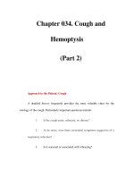
Table 4.1. Specific absorbed fraction of photon energy in kg-1: recommended values for a 10-year-old
Source = Energy (MeV)
Kidneys
Target 0.010 0.015 0.020 0.030 0.0500.100 0.200 0.500 1.000 1.500 2.000 4.000
Adrenals 4.84E - 03 5.22E - 021.29E - 011.57E - 011.06E - 01 7.46E - 02 6.89E - 02 6.69E - 02 6.74E - 02 6.21E - 025.67E - 02 4.40E - 02
UB Wall 0.0 0.0 5.85E - 07 2.95E - 04 2.08E - 03 2.49E - 03 2.91E - 03 3.29E - 03 3.33E - 03 3.23E - 03 3.09E - 03 2.64E - 03
Bone Sur 1.39E - 04 3.49E - 03 1.40E - 02 3.67E - 02 3.90E - 021.90E - 021.06E - 02 8.00E - 03 7.26E - 03 6.82E - 03 6.45E - 03 5.42E - 03
Brain 0.0 0.0 0.0 5.72E - 08 9.29E - 06 5.32E - 05 7.84E - 051.31E - 04 1.91E - 04 2.33E - 04 2.68E - 04 3.65E - 04
Breasts 0.0 0.0 3.85E - 07 7.36E - 04 2.82E - 03 2.59E - 03 3.19E - 03 3.63E - 03 3.53E - 03 3.36E - 03 3.21E - 03 2.82E - 03
St Wall 0.0 2.18E - 051.78E - 03 1.94E - 022.80E - 022.27E - 022.08E - 022.06E - 021.92E - 021.70E - 021.53E - 021.25E - 02
SI Wall 5.55E - 10 1.06E - 04 3.50E - 03 1.77E - 022.70E - 022.20E - 022.06E - 021.90E - 021.73E - 021.62E - 021.52E - 021.25E - 02
ULI Wall 0.0 1.70E - 051.61E - 03 1.43E - 022.70E - 022.13E - 021.76E - 021.79E - 021.64E - 021.54E - 021.45E - 021.17E - 02
LLI Wall 0.0 4.04E - 07 1.18E - 04 2.68E - 03 5.43E - 03 6.63E - 03 6.04E - 03 5.70E - 03 5.63E - 03 5.34E - 03 5.05E - 03 4.27E - 03
Kidneys 5.37E + 00 4.43E + 00 3.21E + 00 1.54E + 00 5.95E - 01 3.46E - 01 3.56E - 01 3.74E - 01 3.54E - 01 3.29E - 01 3.06E - 012.39E - 01
Liver 8.35E - 05 3.55E - 03 1.58E - 02 3.85E - 02 3.75E - 022.74E - 022.49E - 022.35E - 022.19E - 022.04E - 021.91E - 021.62E - 02
Lng Tiss 0.0 5.05E - 06 5.96E - 04 5.35E - 03 9.10E - 03 7.87E - 03 7.35E - 03 6.81E - 03 7.09E - 03 6.34E - 03 5.74E - 03 5.15E - 03
Muscle 3.13E - 03 8.91E - 03 1.43E - 021.59E - 021.16E - 02 8.53E - 03 8.27E - 03 8.39E - 03 8.04E - 03 7.58E - 03 7.14E - 03 5.88E - 03
Ovaries 0.0 1.55E - 08 4.97E - 052.30E - 03 7.47E - 03 8.85E - 03 8.20E - 03 8.49E - 03 7.53E - 03 6.96E - 03 6.66E - 03 6.24E - 03
Pancreas 5.22E - 10 3.99E - 04 1.26E - 02 6.20E - 02 6.58E - 02 4.69E - 02 3.98E - 02 4.04E - 02 3.44E - 02
3.02E - 022.77E - 022.36E - 02
R Marrow 6.38E - 051.33E - 03 4.91E - 03 1.23E - 021.51E - 021.38E - 021.37E - 021.35E - 021.23E - 021.14E - 021.08E - 02 8.81E - 03
Skin 1.14E - 04 7.68E - 04 2.97E - 03 5.28E - 03 4.27E - 03 3.41E -
03 3.68E - 03 4.08E - 03 3.89E - 03 3.94E - 03 3.93E - 03 3.31E - 03
Spleen 2.92E - 03 3.80E - 021.13E - 011.51E - 01 9.95E - 02 6.31E - 025.57E - 025.76E - 025.31E - 02 4.89E - 02 4.55E - 02 3.69E - 02
Testes 0.0 0.0 1.53E - 09 1.76E - 05 3.60E - 04 6.40E - 04 8.50E - 04 1.05E - 03 1.16E - 03 1.18E - 03 1.20E - 03 1.12E - 03
Thymus 0.0 0.0 7.94E - 08 1.13E - 04 7.80E - 04 1.89E - 03 2.30E - 03 2.50E - 03 2.60E - 03 2.64E - 03 2.57E - 03 2.21E - 03
Thyroid 0.0 0.0 1.36E - 10 5.08E - 06 2.18E - 04 6.20E - 04 7.04E - 04 7.71E - 04 8.36E - 04 9.48E - 04 1.01E - 03 9.37E - 04
GB Wall 0.0 3.31E - 05 3.69E - 03 2.55E - 025.26E - 02 3.56E - 03 2.48E - 022.40E - 022.00E - 021.86E - 021.80E - 021.66E - 02
Ht Wall 0.0 2.51E - 08 4.81E - 052.89E - 03 7.30E - 03 8.58E - 03 7.65E - 03 7.51E - 03 7.66E - 03 6.95E - 03 6.32E - 03 5.20E - 03
Uterus 0.0 1.26E - 09 1.72E - 051.78E - 03 6.39E - 03 8.05E - 03 6.87E - 03 6.99E - 03 7.56E - 03 7.10E - 03 6.57E - 03 5.48E - 03
Cristy M, Eckerman KF, Specific absorbed fraction of energy at various ages from internal photon source. IV. Ten-year-old. Oak Ridge National Laboratory Report
ORNL/TM-8381:Vol. 4, 1987
Bone Sur: Bone Surface; GBWall: Gall Bladder Wall; Ht Wall: Heart Wall; LLI Wall: Lower Large Intestine Wall; Long Tiss: Lung Tissue; R Marrow: Red Marrow; SI Wall:
Small Intestine Wall; St. Wall: Stomach Wall; UB wall: Urinary Bladder Wall; ULI Wall: Upper Large Intestine Wall.
X. Zhu 43
source organ. In absence of the S-value tables for other age groups, the
S value can be calculated using tabulated F and D values, as discussed
earlier.
Pediatric Dose Estimate
For pediatric patients, radiopharmaceutical dosages are based on
a pediatric dosing schedule. There are many different dosing sche-
dules. The most common ones are those using body weight or body
surface areas as guides to scale the dose. Pediatric dose schedules
consider many factors to scale down the dosage from that
of adult to child, including organ doses, effective dose, and image
quality.
However, absorbed radiation dose and effective dose to pediatric
patients are not as simple as the dosing schedule. They are not just
simple linear scaled-down doses of those for adult patients. As we dis-
cussed before, radiation doses to patients depend on geometric and
anatomic relationships of source to target organs. Differences in pedi-
atric organ size, density, and composition significantly change the geo-
metric and anatomic relationships that were established for adult
patient (or phantom). Differences of biokinetics, due to age-related dif-
ferences in uptakes (e.g., thyroid uptake of iodine), and excretion (e.g.,
bladder voiding interval), must be considered when estimate radiation
doses for pediatric patients.
Mathematical phantoms for age groups considering the geometric
and anatomic variables have been well developed. They are typically
for infants, and 1-, 5-, 10-, and 15-year-olds. Specific absorbed fraction
has been calculated and tabulated (e.g., Table 4.1) for each age-specific
phantom group. Combined with dose schedule, age-adjusted uptake
and excretion parameters, pediatric radiation doses can then be
estimated according to Equation 10.
Practical Approach to Internal Dose Estimate
The estimation of internal dose from a radionuclide in a human is
rather a complicated process. Studies of biokinetic models of a partic-
ular radiopharmaceutical normally begin through investigations of the
model in animals. Modeling data are collected starting with the initial
amount of the radiopharmaceutical of interest that is injected into
the animal. The percentage of the radionuclide that is taken up by
the source organ is determined through imaging. Other pertinent data
are collected through assays of blood and urine. These data points
are then carefully plotted or fitted to an established mathematical
model that describes the biokinetics of the radionuclides in each source
organ. Complex regulatory requirements regarding human research
subjects dictate that dose estimates in human subjects should con-
ducted after successful animal studies. Many radiopharmaceuticals are
not directly studied for pediatric applications because of complicated
social and ethical issues related to conducting radiation research in
children.
A wealth of information concerning internal dosimetry for the most
commonly used radionuclides in nuclear medicine has been estab-
lished and published, including dosimetry data for radionuclides used
in positron emission tomography (PET) scanning (3–6). Pediatric dose
estimates have also been calculated for different age groups based on
adult biokinetics of radiopharmaceuticals and anatomic phantom
models. Researchers have observed the differences between pediatric
biokinetic models and those of an adult, especially in regard to infants,
and so improvements in dosimetry data for pediatric patients continue.
The Annals of International Commission on Radiological Protection
(ICRP) Publication 53 provides biokinetic models and lists radiation
doses to patients from the most commonly used radiopharmaceuticals
in nuclear medicine (7). ICRP Publication 80 recalculated 19 of the most
frequently used radiopharmaceuticals from ICRP 53 and added 10
more new radiopharmaceuticals (8). Tables 4.2 to 4.4 are absorbed-dose
tables of several radiopharmaceuticals used for PET imaging, adapted
from ICRP80.
44 Chapter 4 Dosage of Radiopharmaceuticals and Internal Dosimetry
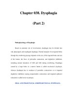
Table 4.2. Absorbed dose of
18
F-FDG (2-fluoro-2-deoxy-D-glucose)
Absorbed dose per unit activity administered
18
F 109.77min
(mGy/MBq)
Organ Adult 15 years 10 years 5 years 1 year
Adrenals 1.2E - 021.5E - 022.4E - 02 3.8E - 02 7.2E - 02
Bladder 1.6E - 012.1E - 012.8E - 01 3.2E - 015.9E - 01
Bone surfaces 1.1E - 021.4E - 022.2E - 02 3.5E - 02 6.6E - 02
Brain 2.8E - 022.8E - 02 3.0E - 02 3.4E - 02 4.8E - 02
Breast 8.6E - 03 1.1E - 021.8E - 022.9E - 025.6E - 02
Gall bladder 1.2E - 021.5E - 022.3E - 02 3.5E - 02 6.6E - 02
GI-tract
Stomach 1.
1E - 021.4E - 022.2E - 02 3.6E - 02 6.8E - 02
SI 1.3E - 021.7E - 022.7E - 02 4.1E - 02 7.7E - 02
Colon 1.3E - 021.7E - 022.7E - 02 4.0E - 02 7.4E - 02
(ULI 1.2E - 021.6E - 022.5E - 02 3.9E - 02 7.2E - 02)
(LLI 1.5E - 021.9E - 022.9E - 02 4.2E - 02 7.6E - 02)
Heart 6.2E - 02 8.1E - 021.2E - 012.0E - 01 3.5E - 01
Kidneys 2.1E - 022.5E - 02 3.6E - 025.4E - 02 9.6E - 02
Liver 1.1E - 021.4E - 022.2E - 02 3.7E - 02 7.0E - 02
Lungs 1.0E - 021.4E - 022.1E - 02 3.4E - 02 6.5E - 02
Muscles 1.1E - 021.4E - 022.1E - 02 3.4E - 02 6.5E - 02
Oesophagus 1.1E - 021.5E - 022.2E - 02 3.5E - 02 6.8E - 02
Ovaries 1.5E - 022.0E - 02 3.0E - 02 4.4E - 02 8.2E - 02
Pancreas 1.2E - 021.6E - 022.5E - 02 4.0E - 02 7.6E - 02
Red marrow 1.1E - 021.4E - 022.2E - 02 3.2E - 02 6.1E - 02
Skin 8.0E - 03 1.0E - 021.6E - 022.7E - 025.2E -
02
Spleen 1.1E - 021.4E - 022.2E - 02 3.6E - 02 6.9E - 02
Testes 1.2E - 021.6E - 022.6E - 02 3.8E - 02 7.3E - 02
Thymus 1.1E - 021.5E - 022.2E - 02 3.5E - 02 6.8E - 02
Thyroid 1.0E - 021.3E - 022.1E - 02 3.5E - 02 6.8E - 02
Uterus 2.1E - 022.6E - 02 3.9E - 025.5E - 021.0E - 01
Remaining organs 1.1E - 021.4E - 022.2E - 02 3.4E - 02 6.3E - 02
Effective dose 1.9E - 022.5E - 02 3.6E - 025.0E - 02 9.5E - 02
(mSv/MBq)
Source: ICRP Publication 80 Radiation Dose to Patients from Radiopharmaceutical.
Annals of ICRP 1998;28(3):10–49, with permission from the ICRP.
X. Zhu 45
Table 4.3. Absorbed dose [methyl-
11
C]thymidine
Absorbed dose per unit activity administered
11
C 20.38min
(mGy/MBq)
Organ Adult 15 years 10 years 5 years 1 year
Adrenals 2.9E - 03 3.7E - 03 5.8E - 03 9.3E - 03 1.7E - 02
Bladder 2.3E - 03 2.7E - 03 4.3E - 03 7.1E - 03 1.3E - 02
Bone surfaces 2.4E - 03 3.0E - 03 4.7E - 03 7.6E - 03 1.5E - 02
Brain 1.9E - 03 2.4E - 03 4.0E - 03 6.7E - 03 1.3E - 02
Breast 1.8E - 03 2.3E - 03 3.6E - 03 5.9E - 03 1.1E - 02
Gall bladder 2.8E - 03 3.4E - 03 5.2E - 03 7.9E - 03 1.5E - 02
GI-tract
Stomach 2.4E - 03 2.9E - 03 4.6E - 03 7.3E - 03 1.4E - 02
SI 2.4E - 03 3.1E - 03 4.9E - 03 7.8E - 03 1.5E - 02
Colon
2.4E - 03 2.9E - 03 4.7E - 03 7.4E - 03 1.4E - 02
(ULI 2.4E - 03 3.0E - 03 4.8E - 03 7.7E - 03 1.4E - 02)
(LLI 2.3E - 03 2.7E - 03 4.5E - 03 7.1E - 03 1.3E - 02)
Heart 3.4E - 03 4.3E - 03 6.8E - 03 1.1E - 022.0E - 02
Kidneys 1.1E - 021.3E - 021.9E - 022.8E - 025.1E -
02
Liver 5.2E - 03 6.8E - 03 1.0E - 021.6E - 022.9E - 02
Lungs 3.0E - 03 3.9E - 03 6.2E - 03 9.9E - 021.9E - 02
Muscles 2.1E - 03 2.6E - 03 4.1E - 03 6.6E - 03 1.3E - 02
Oesophagus 2.2E - 03 2.8E - 03 4.3E - 03 6.9E - 03 1.3E - 02
Ovaries 2.4E - 03 3.0E - 03 4.8E - 03 7.6E - 03 1.4E - 02
Pancreas 2.7E - 03 3.4E - 03 5.3E - 03 8.3E - 03 1.6E - 02
Red marrow 2.5E - 03 3.1E - 03 4.8E - 03 7.6E - 03 1.4E - 02
Skin 1.7E - 03 2.1E - 03 3.4E - 03 5.6E - 03 1.1E - 02
Spleen 3.0E - 03 3.7E - 03 5.9E - 03 9.6E - 03 1.8E - 02
Testes 2.0E - 03 2.5E - 03 3.9E - 03 6.2E - 03 1.2E - 02
Thymus 2.2E - 03 2.8E - 03 4.3E - 03 6.9E - 03 1.3E - 02
Thyroid 2.3E - 03 2.9E - 03 4.7E - 03 7.8E - 03 1.5E - 02
Uterus 2.4E - 03 3.0E - 03 4.8E - 03 7.6E - 03 1.4E - 02
Remaining organs 2.1E - 03 2.6E - 03 4.2E - 03 6.8E - 03 1.3E - 02
Effective dose 2.7E - 03 3.4E - 03 5
.3E - 03 8.4E - 03 1.6E - 02
(mSv/MBq)
Source: ICRP Publication 80 Radiation Dose to Patients from Radiopharmaceutical.
Annals of ICRP 1998;28(3):10–49, with permission from the ICRP.
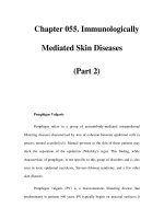
Table 4.4. Absorbed dose
15
O-abeled water
Absorbed dose per unit activity administered
15
O 2.04min
(mGy/MBq)
Organ Adult 15 years 10 years 5 years 1 year
Adrenals 1.4E - 03 2.2E - 03 3.1E - 03 4.3E - 03 6.6E - 03
Bladder 2.6E - 04 3.1E - 04 5.0E - 04 8.4E - 04 1.5E - 03
Bone surfaces 6.2E - 04 8.0E - 04 1.3E - 03 2.3E - 03 5.5E - 03
Brain 1.3E - 03 1.3E - 03 1.4E - 03 1.6E - 03 2.2E - 03
Breast 2.8E - 04 3.5E - 04 6.0E - 04 9.9E - 04 2.0E - 03
Gall bladder 4.5E - 04 5.5E - 04 8.6E - 04 1.4E - 03 2.7E - 03
GI-tract
Stomach 7.8E - 04 2.2E - 03 3.1E - 03 5.3E - 03 1.2E - 02
SI 1.3E - 03 1.7E - 03 3.0E - 03
5.0E - 03 9.9E - 03
Colon 1.0E - 03 2.1E - 03 3.7E - 03 6.2E - 03 1.2E - 02
(ULI 1.0E - 03 2.1E - 03 3.7E - 03 6.2E - 03 1.2E - 02)
(LLI 1.1E - 03 2.1E - 03 3.7E - 03 6.2E - 03 1.2E - 02)
Heart 1.9E - 03 2.4E - 03 3.8E - 03 6.0E - 03 1.1E
- 02
Kidneys 1.7E - 03 2.1E - 03 3.0E - 03 4.5E - 03 8.1E - 03
References
1. Cristy M, Eckerman KF. Specific absorbed fraction of energy at various ages
from internal photon source. IV. Ten-year-old. Oak Ridge National Labora-
tory Report ORNL/TM-8381, vol. 4, 1987.
2. Snyder WS, Ford MR, Warner GG, et al. “S” absorbed dose per unit cumu-
lated activity. Nm/MIRD Pamphlet No. 11. New York: Society of Nuclear
Medicine, 1975.
3. Ruotsalainen U, S
uhonen-Polvi H, Eronen E, et al. Estimated radiation dose
to the newborn in FDG-PET studies. J Nucl Med 1996;37:387–393.
4.Hays MT, Watson EE, Stabin M, et al. MIRD dose estimate report No. 19:
radiation absorbed dose estimates from 18F-FDG. J Nucl Med 2002;43:210–
214.
5. Sorenson JA, Phelps ME. Physics in Nuclear Medicine. New York: Harcourt
Brace Jovanovich, 1987.
6. Stabin MG, Stabbs JB, Toohey RE, et al. Radiation Dose for Radiopharma-
ceuticals, NEREG/CR. Radiation Internal Dose Center, Oak Ridge Institute
of Science and Education, 1996.
7. ICRP Publication 53, Radiation Dose to Patient from Radiopharmaceutucal,
Annals of ICRP, vol. 18, pp. 1–4. New York: Elsevier, 1988.
8. ICRP Publication 80, Radiation Dose to Patients from Radiopharmaceutical,
Annals of ICRP, vol. 28, p. 3. New York: Elsevier, 1998.
46 Chapter 4 Dosage of Radiopharmaceuticals and Internal Dosimetry
Table 4.4. Absorbed dose
15
O-abeled water (Continued)
Absorbed dose per unit activity administered
15
O 2.04min
(mGy/MBq)
Organ Adult 15 years 10 years 5 years 1 year
Liver 1.6E - 03 2.1E - 03 3.2E - 03 4.8E - 03 9.3E - 03
Lungs 1.6E - 03 2.4E - 03 3.4E - 03 5.2E - 03 1.0E - 02
Muscles 2.9E - 04 3.7E - 04 6.1E - 04 1.0E - 03 2.0E - 03
Oesophagus 3.3E - 04 4.2E - 04 6.7E - 04 1.1E - 03 2.1E - 03
Ovaries 8.5E - 04 1.1E - 03 1.8E - 03 2.8E - 03 5.8E - 03
Pancreas 1.4E - 03 2.0E - 03 4.2E - 03 5.4E - 03 1.2E - 02
Red marrow 8.5E - 04 9.7E - 04 1.6E - 03 3.0E - 03 6.1E - 03
Skin 2.5E - 04 3.1E - 04 5.2E - 04 8.8E - 04 1.8E - 03
Spleen 1
.6E - 03 2.3E - 03 3.7E - 03 5.8E - 03 1.1E - 02
Testes 7.4E - 04 9.3E - 04 1.5E - 03 2.6E - 03 5.1E - 03
Thymus 3.3E - 04 4.2E - 04 6.7E - 04 1.1E - 03 2.1E - 03
Thyroid 1.5E - 03 2.5E - 03 3.8E - 03 8.5E - 03 1.6E - 02
Uterus 3.5E - 04 4.4E - 04 7.2E - 04 1.2E - 03 2.3E - 03
Remaining organs 4.0E - 04 5.6E - 04 9.4E - 04 1.7E - 03 2.9E - 03
Effective dose 9.3E - 04 1.4E - 03 2.3E - 03 3.8E - 03 7.7E - 03
(mSv/MBq)
Source: ICRP Publication 80 Radiation Dose to Patients from Radiopharmaceutical.
Annals of ICRP 1998;28(3):10–49, with permission from the ICRP.
5
Pediatric PET Research Regulations
Geoffrey Levine
Good intentions are necessary, but not sufficient, to conduct pediatric
positron emission tomography (PET) research. This chapter provides
direction to guide the process of conducting PET research in children.
Code of Federal Regulations (CFR)
When the executive rule-making voice of the government speaks, it
does so officially through the Code of Federal Regulations (1). These
are not the laws, per se, but rather the nitty gritty rules necessary to
carry out the laws that are made by Congress. For example, Congress
may pass a law to provide for a safe drug supply; the executive branch
(e.g., the Food and Drug Administration, FDA) carries out the intent of
the law and writes the rules (e.g., “Intravenous products shall be sterile
and pyrogen-free”).
Reading 21 CFR (Title 21 of the CFR, where the FDArules are
located) is about as exciting as reading the telephone book or the Inter-
nal Revenue Service regulations for preparing tax returns (until you
come to that one paragraph that appears to justify your objective), but
it is necessary. The judicial system interprets the regulations and may
enforce compliance. Each agency of the executive branch of the gov-
ernment or each specific purpose for a set of regulations has a partic-
ular location. Title 10, for example, is where one finds radiation safety
and safe use of radiopharmaceutical use in humans. Table 5.1 provides
an example of several other locations within the CFR that may be of
interest to the reader (3). In addition to the CFR, the various agencies
issue letters, guidelines, interpretations, descriptions of courses, com-
ments, request for comments, etc., in an effort to communicate with the
public and research investigators, among others. And, like cement, the
rules become more solidified with time. Occasionally, the book is
opened for a rewrite, providing a glimpse into the “mind” of the gov-
ernment. One such opportunity appeared on November 16, 2004, in an
open meeting at the FDAheadquarters in which an update of the
Radioactive Drug Research Committee (RDRC) regulations was being
47
48 Chapter 5Pediatric PET Research Regulations
Table 5.1. Some additional examples of codified federal policy
07 CFR Part 1C Department of Agriculture
10 CFR Part 35 Human Use of Radiopharmaceuticals
10 CFR Part 745 Department of Energy
15 CFR Part 27 Department of Commerce
16 CFR Part 1028 Consumer Product Safety Commission
21 CFR Part 361.1 Radiopharmaceutical Use in Humans
40 CFR Part 26 Environmental Protection Agency
45 C
FR Part 46 Public Welfare, Protection of Human Subjects
45 CFR Part 690 National Science Foundation
Note:There are source documents, regulations, amendments to regulations, Web sites,
parts, subparts, preliminary documents for review, rewrites, updates, clarifications, and
numerous other forms of communication.
Source: Data from ref. 2.
considered (4). The regulations will be examined shortly, particularly
as they relate to PET research in children. Table 5.2 provides a resource
list to facilitate communication (4,5,14).
Pathways Allowed by the Federal Regulatory System
There are three major routes to conduct research that are allowed by
the federal regulatory system: (1) an investigational new drug (IND)
application, (2) a physician-sponsored IND, and (3) the RDRC mecha-
nism (6–8,15–21).
The full IND approach is the one taken by drug manufacturers who
intend to obtain FDA approval to market a pharmaceutical to the
general public, usually for commercial purposes. The manufacturer
conducts physical, chemical, and biologic studies in vitro and then in
animals prior to studies in humans (clinical trials, phases I, II, III
described below), followed by postmarketing studies (phase IV),
post–new drug approval. The pharmaceutical house has sufficient
talent, expertise, and staff in its regulatory and medical departments to
know how to proceed on its own.
Asecond pathway is the physician-sponsored IND, which usually
involves studies with more than 30 subjects, can be conducted at one
or multiple sites, and can involve agents that are new entities, new
routes of administration, new dosage forms for existing or new drugs,
new populations (including children) or disease states, new indica-
tions, etc. The physician or other qualified investigator (with a physi-
cian as co-investigator) is usually medical center or hospital based and
will be required to fill out FDAforms 1571, 1572, and 1573 among pos-
sibly others. This process of how to compile, assemble, complete and
submit the physician-sponsored IND has been reviewed broadly and
in detail elsewhere (15).
A third pathway is the RDRC approach. Using this mechanism, the
FDA delegates authority to a local committee to approve research
studies (usually up to 30 patients, although the number can be higher
under certain circumstances, for example, if FDAform 2915 is com-
pleted). The composition of the membership of that committee has
FDA prior approval. Authority is given by this committee to investi-
gators to conduct only phase I and phase II clinical trials, meeting very
strict and specific criteria (see below). Under no circumstances are the
results from such studies to be used to make clinical decisions for any
of the participants in the study until the study is completed and the
data are analyzed. In theory, the findings are investigational and
remain unproven at this point. It is possible that approved clinical
methods used to validate the research finding may be clinically helpful
or of benefit to a study participant. For example, the findings from a
computed tomography (CT) scan used to study the metabolism and
distribution of a new diagnostic radiopharmaceutical such as a radio-
labeled monoclonal antibody that was designed to locate a tumor, may
find their way to the patient’s or subject’s medical record, but not infor-
mation provided by the radiolabeled monoclonal antibody. This RDRC
G. Levine 49
Table 5.2. Selected reference sites and sources relative to pediatric
PET research
Food and Drug Administration (December, 2004)
Main telephone number 1-888-INFO-FDA
Drug information telephone number 1-301-827-4570
Pediatric Drug Development (PDD) 1-301-594-PEDS (7337)
Division of Drug Imaging and DMIRPD, RDRC Drug Program
Radiopharmaceutical Drug
Products (DMIRPD)
E-mail .
gov/cder/
regulatory/RDRC/default.htm.
Radioactive Drug Research Program
Address Food and Drug Administration
Center for Drug Evaluation and
Research
Division of Medical Imaging
and Radiopharmaceutical
Drug Products HFD-160
Parklawn Building, Room
18R-45 5600 Fishers Lane
Rockville, MD 20852
Attention: RDRC Team
Director Geor
ge Mills, MD
Senior manager Capt. Richard Fejka, USPHS,
RPh, BCNP
Clinical trials
Government
United Healthcare Foundation tedhealth-
carefoundation.org/emb.html
Books
Kowalsky RJ, Falen SW. Radiopharmaceuticals in Nuclear Pharmacy, 2nd
ed. Available from the American Pharmaceutical and Nuclear Medicine
Association, Washington, D.C. />Clinical evidence by the evidence-based update on more tha
n 1000 medical
conditions including clinical trials. British Medical Journal. Free of
charge to healthcare professionals.
/>Legislative Information Gateway to the Congressional Record and
Congressional Committee Information.
Source: Data from refs. 4–13.
approach to conduct PET research in children is the one on which we
concentrate in this chapter (6–8,16–18,21).
The Clinical Trial Process
The clinical trial is a biomedical or behavioral research study of human
subjects that is designed to answer specific questions about biomedical
or behavioral interventions (drugs, treatments, devices, or new ways
of using known drugs, treatments, or devices). Clinical trials are used
to determine whether new biomedical or behavioral interventions are
safe, efficacious, and effective (17,18). Trials of an experimental drug,
device, treatment, or intervention may proceed through four distinct
phases. Sometimes more than one phase can be conducted at the same
time. The actual number of subjects studied in each phase may depend
in part on the incidence or prevalence of the disease state or condition
being investigated.
Phase I
This phase entails testing in a small group of people (e.g., 20 to 80 sub-
jects) to determine efficacy and evaluate safety (e.g., determine a safe
dosage range) and identify side effects. A typical phase I trial of a new
drug agent frequently involves relatively high risk to a small number
of participants. The investigator and occasionally others have the only
relevant knowledge regarding the treatment because these are the first
human uses. The study investigator may be required to perform con-
tinuous monitoring on participant safety with frequent reporting to
institute and center staff with oversight responsibility.
Phase II
This phase entails a study of a larger group of people (several hundred)
to determine the efficacy and further evaluate safety. A typical phase II
study follows phase I studies, and there is more information regarding
risks, benefits, and monitoring procedures. However, more participants
are involved, and the disease process confounds the toxicity and out-
comes. An institute or center may require monitoring similar to that of
a phase I trial or may supplement that level of monitoring with indi-
viduals with expertise relevant to the study who might assist in inter-
preting the data to ensure patient safety (17,18).
Phase III
This phase entails a study to determine the efficacy in large groups of
people (from several hundred to several thousand) by comparing the
intervention to other standard or experimental interventions, to
monitor adverse effects, and to collect information to allow safe use.
The definition includes pharmacologic, nonpharmacologic, and behav-
ioral interventions given for disease prevention, prophylaxis, diagno-
sis, or therapy. Community-based trials and other population-based
trials are also included. A phase III trial frequently compares a new
50Chapter 5Pediatric PET Research Regulations
treatment to a standard treatment or to no treatment, and treatment
allocation may be randomly assigned and the data masked. These
studies frequently involve a large number of participants followed
for longer periods of treatment exposure. Although short-term risk is
usually slight, one must consider the long-term effects of a study agent
or achievement of significant safety or efficacy differences between
the control and the study groups for the masked study. An institute
or center may require a data safety monitoring board (DSMB) to
perform monitoring functions. This DSMB would be composed of
experts relevant to the study and would regularly assess the trial
and offer recommendations to the institute or center concerning its
continuation.
Phase IV
This phase entails studies done after the intervention has been mar-
keted. These studies are designed to monitor the effectiveness of the
approved intervention in the general population and to collect infor-
mation about any adverse effects associated with widespread use. The
controversy that appeared in the lay media in December 2004 as well
as in medical publications (22) concerning adverse events associated
with Vioxx and Celebrex is an example of a postmarketing discovery
following new drug approval.
Radioactive Drug Research Committee Update
Meeting and Transition
After more than a quarter of a century, it became obvious that techno-
logic progress and events had surpassed the intent of the original 1975
FDA, RDRC regulations (6–8,16). During the current transition period
(June 2005) and until the updated RDRC regulations are finalized, the
1997 FDAModernization Act (FDAMA) provides a mechanism for the
uninterrupted production of PET radiopharmaceutical by specifying
that they should meet United States Pharmacopoeia (USP) monograph
standards (23,24). An example of a PET radiopharmaceutical coming
through that process was
18
F-fluorodeoxyglucose (FDG) injection,
which received a new drug approval in less than 6 months after sub-
mission on August 5, 2004 (25).
RDRC Update Issues
Six issues or areas of concern, proposed by the FDA/RDRC, were
placed on the agenda for discussion (4,5):
1.Pharmacologic issues
2. Radiation dose limits for adult subjects
3. Assurance of safety for pediatric subjects
4.Quality and purity
5. Exclusion of pregnant women
6. RDRC membership
G. Levine 51
As this chapter is being written, participants at the open meeting and
other interested parties and organizations are submitting written com-
ments for the record and for consideration regarding the updated reg-
ulations. Who could have predicted in 1975 how to best conduct
research or manufacture pharmaceuticals (including radiopharmaceu-
ticals), given the advent of monoclonal antibodies, cloning, stem cells,
gene therapy, biologic response modifiers, and the growth of PET and
other imaging modalities?
Vulnerable Populations
There are four populations addressed specifically in Title 45 part 46 of
the Code of Federal Regulations, which deals with public welfare pro-
tection of human subjects (2,19–21):
Subpart A: Human subjects, research subjects, and volunteers as con-
trols or normals
Subpart B: Additional protections for pregnant women, human fetuses,
and neonates
Subpart C: Additional protections pertaining to biomedical and behav-
ioral research in prisoners
Subpart D: Additional protections for children as subjects in research
(21).
Assurance of Safety for Pediatric Subjects
Currently 21 CFR 361.1 (that FDAsection of the code that deals with
radiopharmaceutical research in humans) allows the study of radioac-
tive drugs in subjects less than 18 years of age without an IND appli-
cation, if the following conditions are met:
1.The study presents a unique opportunity to gain information not
currently available, requires the use of research subjects less than 18
years of age, is without significant risk, and is supported with
review by qualified consultants to the RDRC.
2.The radiation dose does not exceed 10% of the adult radiation dose
as specified in 21 CFR 361.1 (b)(i) and, as with adult subjects, the fol-
lowing additional requirements are met:
3. The study is approved by an institutional review board (IRB) that
conforms to the requirements of 21 CFR part 56.
4. Informed consent of the subject’s legal representative is obtained in
accordance with 21 CFR part 50.
5.The study is approved by the RDRC, which assures all other require-
ments of 21 CFR 361.1 are met (5,16).
Alternatively, when a study is conducted under an IND (as com-
pared to a RDRC) in accordance with part 312 (21 CFR part 312), the
sponsor must submit to the FDAthe study protocol, protocol changes
and information amendments, pharmacology/toxicology and chem-
istry information, and information regarding prior human experience
with the same or similar drugs (see 21 CFR 312.22, 312.33, 312.30 and
312.31). Additionally, 21 CFR 32 requires that sponsors (of the IND)
promptly review all information relevant to the safety of the drug
obtained or otherwise received by the sponsor by any source, foreign
52 Chapter 5Pediatric PET Research Regulations
or domestic. This includes information derived from any clinical or epi-
demiologic experience, reports in the scientific literature and unpub-
lished scientific papers, as well as reports from foreign regulatory
authorities. 21 CFR part 32 also requires that sponsors submit IND
safety reports to the FDA(4,5).
Pediatric Concerns Considered for Update
Does 21 CFR 361.1 provide adequate safeguards for pediatric subjects
during the course of a research project intended to obtain basic infor-
mation about a radioactive drug, or should these studies be conducted
only under an IND?
If we assume that 21 CFR 361.1 provides adequate safeguards for
pediatric studies during such studies, given our present knowledge
about radiation and its effects, can we conclude that the current dose
limits would be appropriate to ensure no significant risk for pediatric
participants? Should there be different dose limits for different pedi-
atric groups (5)? At present, it is estimated that only about half of
the RDRCs in conjunction with their IRBs consider approval of radioac-
tive drug research in children. The operative phrase appears to be
minimal risk.
Protections for Children Involved as Subjects of PET Research
There are three basic areas of concern in using children as PET research
subjects: (1) conformity with IRB requirements, (2) radiation dosime-
try of not more than 10% of the adult dose and in conformity with
ALARA(as low as reasonably achievable) considerations, and (3)
special considerations relevant to vulnerable populations (2,5,16,21).
Under certain circumstances, the secretary of the Department of Health
and Human Services (HHS) may waive some or all of the requirements
of these regulations for research of this type (2,21).
Some Additional Protections Addressed in 45 CFR
Part 46, Subpart D
To whom do the requirements to carry out the regulations apply?
To whom do the requirements apply as subjects, and who may give
assent and grant permission for the children?
What are the IRB responsibilities related to children?
What protections are appropriate for research not involving greater
than minimal risk?
What protections are appropriate for research involving greater than
minimal risk but presenting the prospect of direct benefit to the indi-
vidual subjects?
What protections should be required for research involving greater
than minimal risk and no prospect of direct benefit to individual sub-
jects but likely to yield generalizable knowledge about the disorder
or condition?
What protections should be required for research not otherwise
approvable that presents an opportunity to understand, prevent, or
alleviate a serious problem affecting the health or welfare of children?
What is the requirement for permission by parents or guardians and
for assent by children?
G. Levine 53
What protections should be required and who grants permission for
children who are wards of the State? (21).
RDRC Specific Responsibilities Abstracted
from the CFR
This section is taken directly from the minutes of the University of
Pittsburgh Medical Center (UPMC) RDRC and Human Use Subcommit-
tee (HUSC), Radiation Safety Committee, Dennis Swanson, M.S., Chair-
man (26).
In taking this action, the RDRC considered and assured that each of
the following criteria were met:
1.The research study is intended to obtain basic information regard-
ing the metabolism (including kinetics, distribution, and localization)
of a radioactively labeled drug or regarding human physiology, patho-
physiology or biochemistry. The research study is not intended for
immediate therapeutic, diagnostic, or similar purposes or to determine
the safety and effectiveness of the drug in humans for such purposes.
2.The research study involves the use of a radioactive drug(s), which
will be prepared in accordance with a RDRC-approved drug master file
or HUSC/RDRC Form 1002. The drug master file of HUSC/RDRC
Form 1002 documents:
a.that the amount of active ingredient or combination of
active ingredient shall not cause any clinically detectable phar-
macologic effect in humans as known based on pharmacologic
dose calculations derived from data available published or
other valid human studies;
b. absorbed dose calculations based on the MIRD formalism and
biologic distribution data available from the published litera-
ture or from other valid studies;
c. that an acceptable method will be used to radioassay the drug
prior to its use;
d.that adequate and appropriate instrumentation will be utilized
for the detection and measurement of the specific radionuclide;
e.that the radioactive drug meets appropriate chemical, phar-
maceutical, and radionuclidic standards of identity, strength,
quality, and purity as determined by suitable testing proce-
dures;
f. that, for parenteral use, the radioactive drug is prepared in a
sterile and pyrogen free form; and
g. that the package and labeling of the radioactive drug is in com-
pliance with the requirements of 21 CFR 361.1 and NRC (if
applicable) and Commonwealth of Pennsylvania regulations
regarding radioactive drugs.
3. For this specific research protocol:
a. Scientific knowledge and benefit is likely to result from this
study;
—The proposed research is based on sound rationale derived
from the published literature or other valid studies.
—The proposed research is of sound design.
54Chapter 5Pediatric PET Research Regulations
b. The radiation dose is sufficient and no greater than necessary
to obtain valid data.
— In consideration of available radioactive drugs, the radioac-
tive drug used in the study has the combination of half-life,
type of radiation, radiation energy, metabolism, and chem-
ical properties that results in the lowest radiation dosime-
try as needed to obtain the necessary information.
—For adult subjects: the projected radiation dose to the
whole body effective dose equivalent (EDE), active blood-
forming organs, lens of eye, and gonads does not exceed 3
rem (single study) or 5 rem (annual and total dose), and
the projected radiation dose to any other organ does not
exceed 5 rem (single study) or 15rem (annual and total
dose).
—For subjects under the age of 18 (if applicable), the projected
radiation dose does not exceed 10% of the adult limits.
—The projected radiation dose commitments address
expected radionuclidic contaminants and x-ray and other
radiation-emitting procedures performed as part of the
research study.
c. The projected number of subjects is sufficient and no greater
than necessary for the purpose of the study as supported by a
statistical or other valid justification;
d.The proposed population is appropriate to the purpose of the
study; and
—The involvement of subjects less than 18 years of age, if
applicable, is justified as (1) presenting a unique opportu-
nity to gain information not currently available; and (2)
necessitating the use of such subjects. The scientific review
of research involving subjects less than 18 years of age is
supported by qualified pediatric consultants to the RDRC.
—Pregnancy testing, to confirm absence of pregnancy prior to
administration of the radioactive drug(s), is performed on
female subjects of childbearing potential.
e.The investigators are qualified by training and experience to
conduct the proposed research study.
—The research study involves, as a listed co-investigator, a
physician “authorized user” recognized by the Radiation
Safety Committee, University of Pittsburgh, as qualified to
oversee the preparation, handling and use of the radioac-
tive drug (26).
Illustrative Examples that Have Come to
the UPMC-RDRC Requiring Directed Change,
Correction, or Reconsideration
1.Not including the gallium-68 rod transmission scan to calibrate
the PET scanner as part of the radiation dosimetry.
2. Submitting a phase III clinical trial to the RDRC.
G. Levine 55
3. Submitting an appropriate research protocol and informed
consent for a study using
18
F-FDG to the IRB, but not the RDRC.
4.Inappropriate expression of radiation dose and risk to the patient
in the informed consent. The UPMC has adopted a uniform radiation
risk statement model which it recommends be used in both the consent
and protocol, although other statements are also acceptable, for
example, “Participation in this research study involves exposure to
radiation from the two PET transmission scans, the one 12mCi (a unit
of radioactivity dosage) injection of [15-O] water, one 15-mCi dose of
[11-C]WAY, and one 10-mCi injection of [11-C]raclopride. The amount
of radiation exposure you will receive from these procedures is
equivalent to a whole-body radiation dose of 0.47rem (a unit of
radiation exposure). This is less than 10% of the annual whole-body
radiation exposure (5 rem) permitted to radiation workers by federal
regulations. There is no minimum level of radiation exposure that is
recognized as being totally free of the risk of causing genetic defects
(cell abnormalities) or cancer. However, the risk associated with the
amount of radiation exposure that you will receive from this study is
considered to be low and similar to other everyday risks” (26).
5. While using magnetic resonance imaging (MRI) for co-registration
with PET, performing the PET scan before MRI. A certain number of
MRI subjects will be eliminated or withdrawn due to claustrophobia.
If this is the case, then they have been exposed to the radiation dose
unnecessarily.
6.Apatient has a pregnancy test at a screening session 1 month prior
to a research PET scan. The pregnancy test is due to the research nature
of the PET scan. The pregnancy test should be conducted as close as pos-
sible to the time that the PET scan is scheduled; within 48 hours of PET.
7. A patient has a pacemaker and is going to have an MRI prior to
a PET study. If there is a question of metal or metal fragment being
attracted by the magnets, then an x-ray may be required. The x-ray is
required as part of the research and thus should be included as part of
the dosimetry table and consent.
8. A new drug that has been tested in thousands of mice to treat
memory loss is to be trace radiolabeled and administered to humans
as part of a multicenter trial of 50 patients at each site. Because the drug
has never been given to a human (lack of a pharmacologic effect cannot
be substantiated), and is a multicenter study with over 30 patients, it
is best conducted under an IND. Even for a radiopharmaceutical, the
mass of the administered radiolabeled compound currently must be
quantified.
9.Aphysician wants to test a brachytherapy unit on his patients
who have a tumor different from the one for which the FDAgave initial
approval. There are 10 patients and he is comparing two types of seeds
in two different cell types. This should not be submitted to the RDRC,
but should be reviewed by the Human Use Subcommittee. The holder
of the IND is a manufacturer of a radiation device.
10. A study comes before the RDRC that is so complicated that the
members of the committee don’t believe it can be carried out without
losing data. The project is sent back for reconsideration because if the
56Chapter 5Pediatric PET Research Regulations
data cannot be analyzed in a meaningful way, then subjects will have
been exposed unnecessarily.
References
1.Fostering a culture of compliance. National Institutes of Health education
and outreach seminar. Pittsburgh, July 15, 2004.
2.Administering and overseeing clinical research. Title 45 Public welfare. Part
46 Protection of human subjects. Revised November 13, 2001.
Effective December 13, 2001. Subpart A—Federal policy for the protection
of human subjects. Basic DHHS policy for the protection of human
research subjects. In: Fostering a Culture of Complia
nce. National Institutes
of Health education and outreach seminar. Pittsburgh, PA, July 15,
2004. />htm.
3. Fostering a culture of compliance. National Institutes of Health education
an
d outreach seminar. Code of Federal Regulations. The common rule
(Federal Regulations). Pittsburgh, PA, July 15, 2004. phs.
dhhs.gov/ human subjects/guidance/45cfr46.htm.
4.Notice of public meeting—radioactive drugs for certain research uses.
Radioactive Drug Research Committee (RDRC) program. Rockville,
MD, November 16,2004. />default.htm.
5.Agenda of public meeting—radioactive drugs for certain research uses.
Radioactive Drug Re
search Committee (RDRC) program minutes.
Rockville, MD, November 16, 2004. meeting/
clinicalresearch/default.htm.
6.Positron emission tomography (PET) related documents. http://www.
fda.gov/cder/regulatory/PET/default.htm.
7. What information does the RDRC review? Radioactive Drug Research Com-
mittee (RDRC) program. regulatory/RDRC/
review.htm.
8. What are the responsibilities of the RDRC? Radioactive d
rug research com-
mittee (RDRC) program. regulatory/RDRC/
Responsibilities.htm.
9. />10.Having trouble keeping up with clinical trials? APhA-AAPM news you can
use 4(2), October 28, 2004. . Info-center@
apha.org.
11.Kowalsky RJ, Falen SW. Radiopharmaceuticals in Nuclear Pharmacy and
Nuclear Medicine, 2nd ed. Washington, DC: APhA, 2004. http://www.
Pharmacist.com/store.cfm.
12. Clinical evidence to help support the clinician’s skillful use of
scientifically valid and evidence based information. http://Unitedhealth
carefoundation.org.ebm.html.
13. How do I find and track bills? Health Physics News 2005;33(1):3. http://
www.hps.org.
14.FDAmeeting to focus on radioactive drugs for basic research. APhA-
AAPM electronic newsletter. .
15. Levine G, Abel N. Considerations in the assembly and submission of the
physician sponsored investigational new drug application. In: Hladik WB,
Saha GB, Study KT, eds. Essentials of Nuclear Medicine Science. Baltimore:
Williams & Wilkins, 1987:357–386.
G. Levine 57
16.Pediatric drug development. pediatrics/index.
htm.
17. NIH grants-general information glossary (NIH-grants policy statement,
revised 12/01/03. In: Fostering a Culture of Compliance. National Insti-
tutes of H
ealth education and outreach seminar. Pittsburgh, PA, July 15,
2004:6–15. />18. NIH guide: NIH policy for data and safety monitoring, release date June
10, 1998. In: Fostering a Culture of Compliance. National Institutes of
Health education and outreach seminar. Pittsburgh, PA, July 15, 2004.
/>19.Administering and overseeing clinical research. Title 45 Public welfare. Part
46 Prote
ction of human subjects. Revised November 13,2001. Effective
December 13, 2001. Subpart B—additional protections for pregnant
women, human fetuses and neonates involved in research. In: Fostering a
Culture of Complia
nce. National Institutes of Health education and out-
reach seminar. Pittsburgh, PA, July 15, 2004. s.
gov./humansubjects/guidance/45cfr46.htm.
20.Administering and overseeing clinical research. Title 45 Public welfare. Part
46 Prot
ection of human subjects. Revised November 13, 2001. Effective
December 13, 2001. Subpart C—additional protections pertaining to bio-
medical and behavioral research involving prisoners as subjects in
research. In: Fost
ering a Culture of Compliance. National Institutes of
Health education and outreach seminar. Pittsburgh, PA. July 15, 2004.
/>21.Administering and overseeing clinical research. Title 45 Public welfare. Part
46 Protection of human subjects. Revised November 13, 2001. Effective
December 13, 2001. Subpart D—additional DHHS protections for children
involved as subjects in research. In: Fostering a Culture of Compli
ance.
National Institutes of Health education and outreach seminar. Pittsburgh,
PA, July 15, 2004. />guidance/45cfr46.htm.
22. COX-2 inhibitors under scrutiny in wake of Rofecoxib withdrawal. APhA
Drug Info Line 2004;1–2.
23. Food and Drug Administration Modernization Act of 1997. Title 21.
Section 121. Positron emission tomography. />regulatory/Pet/petlaw.html.
24. Radiopharmaceuticals for positron emissi
on tomography-compounding.
Chapter 823. US Pharmacopeia 20/National Formulary 25, 2002.
25.Update—new fludeoxyglucose F-18 injection PET drug approved in less
than 6 months. />htm.
26. Swanson DP. Radioactive drug research committee/human use subcom-
mittee meeting minutes. University of Pittsburgh. Pittsburgh, PA, Novem-
ber 17, 2004.
58Chapter 5Pediatric PET Research Regulations
6
Issues in the Institutional Review
Board Review of PET Scan Protocols
Robert M. Nelson
The lack of reliable information on the use of medications for children
has been addressed in the United States through two legislative initia-
tives: the Best Pharmaceuticals for Children Act (BPCA) of 2002 (1) and
the Pediatric Research Equity Act (PREA) of 2003 (2). These two ini-
tiatives have stimulated pediatric pharmaceutical research, resulting in
valuable information to guide the appropriate use of many medications
(3). In addition, the National Institutes of Health now requires (as of
1998) that children be included in research unless there are scientific
and ethical reasons not to include them (4). The resulting increase in
pediatric research has led to concerns that the regulations governing
pediatric research provide insufficient protection. This chapter refers to
only the Food and Drug Administration (FDA) regulations governing
research with children (21 CFR 50 and 56), as the use of radiopharma-
ceuticals in PET scanning is regulated by the FDA. Comparable regu-
lations are found in 45 CFR 46, subparts A and D.
The FDA did not adopt additional safeguards for children in research
(referred to as subpart D) until April 2001 (5). In passing the BPCA, the
U.S. Congress also commissioned the Institute of Medicine (IOM) to
review the adequacy of subpart D; their report was issued in March
2004. The IOM found that there are problems in the application of
subpart D due to insufficient guidance and thus variable interpretation
of key concepts (6).
The additional safeguards for children in research found in subpart
D can be viewed as a further specification of the general requirement
that the “risks to subjects are reasonable in relation to anticipated ben-
efits, if any, to subjects, and the importance of the knowledge that may
be expected to result” (21 CFR 56.111.a.2). Absent the prospect of direct
benefit, the research risks to which children may be exposed must be
restricted to either minimal risk (21 CFR 50.51) or a minor increase over
minimal risk (21 CFR 50.53), depending on whether the children have
the disorder or condition under investigation (5). If there is a prospect
of direct benefit from the research intervention, the research risk must
be justified by the anticipated benefit to the enrolled children (rather
than by any knowledge that may result) (21 CFR 50.52) (5,7). Thus, to
59
determine whether a research protocol involving children may
proceed, an institutional review board (IRB) must assess (1) the level
of risk, and (2) the prospect of direct benefit to the child presented by
each research intervention or procedure (7).
This chapter examines the use of positron emission tomography
(PET) scanning in research involving children from the perspective of
the additional safeguards found in subpart D. The risks of the two
major components of PET scanning (i.e., administration of the radio-
pharmaceutical tracer and procedural sedation) are discussed within
this regulatory framework governing pediatric research. In the course
of the analysis, key concepts from the pediatric research regulations
that will be discussed include the component analysis of risk, minimal
risk, minor increase over minimal risk, and disorder or condition (6).
Finally, the relationship between subpart D(5) and other FDAregula-
tions concerning the investigational use of radiopharmaceuticals (21
CFR 312 and 21 CFR 361.1) is discussed.
Component Analysis of Risk
The risks (i.e., potential harms) and benefits of each intervention or pro-
cedure included in a research protocol must be assessed independently.
The potential benefits from one procedure should not be used to offset
or justify the risks of another (IOM recommendation 4.6) (6). The appli-
cation of this principle is fairly straightforward when the performance
of one procedure does not depend on or require the performance of the
other procedure. However, when the two procedures are dependent on
each other, the analysis is more complex. In the case of a PET scan, the
key procedural components for the purpose of risk analysis are the
administration of the radioactive tracer and the necessary procedural
sedation. Other risks such as the physical environment (e.g., an
enclosed space and the possibility of claustrophobia) are less than those
associated with computed tomography (CT) or magnetic resonance
imaging (MRI) scans, as the child can be accompanied (and reassured)
by a parent during the entire procedure. All of the other necessary pro-
cedures (e.g., venipuncture, placement of a peripheral intravenous
catheter) are appropriately considered minimal risk given the limited
duration (i.e., less than 2 hours) of a PET scan. Thus, the following dis-
cussion is limited to the risks of the radiotracer administration and pro-
cedural sedation.
Procedural sedation is usually required for the successful completion
of the PET scan, given the need to reduce motion artifact. Thus, for the
purpose of IRB analysis, the administration of the radiotracer, and the
risk or benefit of radiation exposure, is the key component of the PET
scan. If the PET scan, and thus the radiotracer administration, offers
the prospect of direct benefit to the child undergoing the procedure,
the radiation risks to which the child may be exposed can be greater
than minimal risk assuming that the balance of potential harms and
anticipated benefits is justified and comparable to any available alter-
natives (21 CFR 50.52) (5). As such, the risks of any procedural seda-
60 Chapter 6 Issues in the Institutional Review Board Review of PET Scan Protocols
tion necessary to complete the PET scan become part of this balancing
of risks and benefits. However, if the PET scan does not offer the
prospect of direct benefit to the child undergoing the procedure, the
risks of the radiation exposure and any necessary procedural sedation
must be no more than a minor increase over minimal risk for children
with a disorder or condition (21 CFR 50.53) or no more than minimal
risk for children without a disorder or condition (21 CFR 50.51) (5). In
effect, the level of appropriate (and allowable) risk exposure associated
with the procedural sedation depends on whether or not the results of
the PET scan offer the child a prospect of direct benefit. A common
mistake is to determine that the risk of a procedure that does not offer
any prospect of direct benefit is no more than a minor increase over
minimal risk but to fail to appreciate that the risks of any associated
procedures must also be similarly restricted.
Administration of Radioactive Tracers
The risks of administering a radiopharmaceutical tracer can be divided
into two aspects: (1) the risk from the compound to which the radioac-
tive tracer is attached, and (2) the risk from the level of radiation expo-
sure associated with the tracer. The risk from the compound itself is
independent of the radiation risk and are discussed below (see
Research Under an Investigational New Drug Application). The dis-
cussion here focuses on the general risks of radiation, and not on how
one would determine the actual effective dose (ED) of radiation expo-
sure to any given organ from individual radiopharmaceuticals. The sci-
entific determination of the level of radiation exposure for any given
radiopharmaceutical depends on such factors as the targeted receptor,
blood flow to the area of interest, isotope and carrier compound half-
life, mechanisms of metabolism and excretion, and so forth (8–10).
The Risks of Radiation Exposure
The data derived from atomic bomb survivors in Japan are the best
available on the effects of ionizing radiation on a large human popu-
lation (11). These data support the view that “the risk of solid cancers
appears to be a linear function of dose” (12), perhaps down to a dose
of about 5 rad (i.e., 5 rem) (12,13). Some argue that there is direct evi-
dence of risk at low-level radiation exposure in the range of 600 mrem
to 10rem (13,14). Others place the lower limit of the range at which
low-level ionizing radiation increases the risk of some cancers at 1 rem
for acute exposure and 5rem for protracted exposure (15). However,
the risk of cancer is probably overestimated using these data, as “cancer
rates may vary due to other risk factors correlated with the expo-
sure under investigation” (13).
The predominant model for describing the risks of low-level radia-
tion (i.e., less than 10 rem) is the linear no-threshold (LNT) model. This
theoretical model is based on two assumptions: “(a) any radiation dose
can produce adverse effects such as cancer or genetic damage; [and]
(b) the severity of adverse effects is directly proportional to the
R.M. Nelson 61
radiation dose received” (16). In support of this model, the dose-
response relationship between low-level radiation and “the biological
alterations that are precursors to cancer, such as mutations and chro-
mosome aberrations,” appears to be linear (17). Although the LNT
model is the customary approach, “existing data do not exclude the
possibility that there may be thresholds for such effects in the low-dose
domain” (17).
The dose-response relationship between low-level radiation expo-
sure and the risk of developing cancer cannot be precisely defined by
extrapolating from observations at moderate-to-high doses (15,17). As
a result, there is considerable debate about whether low-level radiation
(i.e., less than 10rem) increases the risk of developing cancer, with the
data concerning the risk of low-level radiation exposure subject to wide
interpretation (19,20). In addition, some data support the view that
low-level radiation exposure may be protective (12,16,18–20). This pos-
sibility of “adaptive responses” (i.e., hormesis) further complicates the
“assessment of the dose-response relationships for the genetic and car-
cinogenic effects of low-level irradiation” (17).
Critics argue that the LNT theory “grossly overestimates the risk
from low-level radiation”. In addition, no “statistically sound well-
designed studies” (20) support the use of the LNT model at low-level
radiation doses (16,20). The confidence limits from epidemiologic
studies of the dose-response relationship of low-level radiation expo-
sure are sufficiently wide “to be consistent with an increased effect, a
decreased effect, or no effect” (20). Overall, “the health risk from low-
level doses could not be detected above the ‘noise’ of adverse events
of everyday life” (16). Proponents of the LNT theory, however,
point out that the failure to find an increase in cancer, and the obser-
vation of a reduction in some instances, among populations exposed
to low-level radiation does not contradict the LNT theory given the
small increase that would be expected and the methodologic limita-
tions of the studies. These limits are such that “it may never be possi-
ble to prove or disprove the validity of the LNT hypothesis” (17).
However, there are no data that “suggest a threshold dose below which
radiation exposure does not cause cancer” (21) nor “reliable data
proving that radiation doses as used in diagnostic x-rays do induce
cancer” (11).
In summary, there are three general views of the risk of low-level
radiation exposure: (1) the relationship between potential harm and
effective radiation dose is linear, with no level of radiation exposure
being nonharmful (i.e., LNT model); (2) there is a threshold level of
radiation below which there is no harm, with a linear relationship
between potential harm and effective radiation dose above this thresh-
old (i.e., threshold model); and (3) there is a threshold level of radia-
tion below which there is benefit from enhanced cellular repair (i.e.,
hormesis model), with a linear relationship above this threshold. Below
1rem effective radiation dose, there are no data that will discriminate
among these three models. Between 1 and 5rem effective radiation
dose, the data are controversial, with the LNT model being the more
62 Chapter 6 Issues in the Institutional Review Board Review of PET Scan Protocols
favored approach. Above 5 to 10 rem, the linear relationship between
potential harms and ED is generally accepted (with some difference of
opinion on the lower limit of the range of this linear relationship).
Characterizing the Risks of Radiation
What level of radiation exposure should be considered “minimal risk”
in light of the above data? Minimal risk is defined as follows: “The
probability and magnitude of harm or discomfort anticipated in the
research are not greater in and of themselves than those ordinarily
encountered in daily life or during the performance of routine
physical or psychological examinations or tests” (21 CFR 56.102i).
Given the variability in the interpretation of minimal risk (22), the IOM
recommended that minimal risk be interpreted “in relation to the
normal experiences of average, healthy, normal children” (recom-
mendation 4.1) (6). Children may be exposed to ionizing radiation
during diagnostic radiologic studies; however, no such studies are per-
formed as part of routine physical examinations of healthy children.
Absent a disorder or condition, such as an injury, the interpretive
standard of a healthy child appears to exclude diagnostic radiation
exposure. However, children are exposed to background radiation
from natural sources that ranges from 300 to 450mrem per year
depending on the altitude at which they live (19). Children are also
exposed to additional radiation during such normal activities as air
travel. Given the absence of data suggesting an increase in cancer at
altitude, a one-time exposure to ionizing radiation that falls in the
range of yearly environmental exposure would appear to qualify as
minimal risk.
The IOM also recommended that the risks of research could be con-
sidered minimal if they were equivalent to the risks “that average,
healthy, normal children may encounter in their daily lives or experi-
ence in routine physical or psychological examinations or tests” (rec-
ommendation 4.1) (6). Studies of radiation exposure from “background
radiation, radon in homes, medical procedures, and occupational radi-
ation in large population samples” have not demonstrated any addi-
tional health risks “above the ‘noise’ of adverse events of everyday life”
(16). This conclusion is supported by the observation that “exposure to
1rem [only] adds about 100 more genetic mutations” to the “average
of 240,000 genetic mutations [that] occur spontaneously every day in
the human body” (16). Although younger children are thought to be
more susceptible to radiation-induced cancer (23), two reviews con-
cluded that there are no data demonstrating higher risk to children of
exposure to low-level radiation (14,16). What is the threshold level of
radiation exposure which, if one remains below, could be considered
minimal risk?
Proponents of the LNT interpretation of low-level radiation risk
express concern that adopting the view of a radiation threshold below
which the risk is zero may undermine efforts to minimize radiation
exposure (12,19). Others argue that the LNT model imposes an undue
R.M. Nelson 63
regulatory burden that “is detrimental to the welfare of our society”
(20). The minimal-risk standard does not require that the risks of the
research be zero but rather that the risks be no different from those
that are experienced by healthy children in the course of everyday life.
One possible choice for the level of radiation exposure that presents no
more than minimal risk can be taken from the 1996 Health Physics
Society statement that the health risks from exposure up to 10 rem
is “either too small to be observed or nonexistent” (24). A more con-
servative approach, taking into account more recently published data
(12), would reduce the radiation level at which there is unobservable,
and thus minimal, risk to 1rem exposure (25). This approach is con-
sistent with published research studies involving the exposure of
healthy children to ionizing radiation that have been approved by an
IRB (16).
Allowable Research Risk for Children with Conditions
Subpart D allows researchers to expose children with a disorder or
condition to more than minimal risk, provided (among other condi-
tions) that “the risk represents a minor increase over minimal risk” and
“the intervention or procedure is likely to yield generalizable
knowledge that is of vital importance for the understanding or ame-
lioration of the subjects’ disorder or condition” (5). The IOM report rec-
ommends that a “minor increase over minimal risk” be interpreted “to
mean a slight increase in the potential for harms or discomfort beyond
minimal risk” (recommendation 4.2, emphasis added) (6). Based on the
above discussion of the risks of radiation exposure, one could consider
low-level radiation exposure falling between 1 and 5rem as presenting
only a minor increase over minimal risk. Even so, exposure to this level
of radiation during research that does not offer the prospect of direct
benefit is only justified if (a) the child has a disorder or condition, and
(b) the research is likely to yield knowledge that is of “vital im-
portance” for understanding or ameliorating the child’s disorder or
condition.
There are no guidelines on how to interpret the phrase “vital impor-
tance.” At a minimum, the enrollment of children should be necessary
(i.e., vital) to answer the research question (26). In addition, the require-
ment of having a disorder or condition should not be interpreted so
broadly as to encompass all children. The IOM report recommends that
“the term condition should be interpreted as referring to a specific (or
a set of specific) physical, psychological, neurodevelopmental, or social
characteristic(s) that an established body of scientific evidence or clinical
knowledge has shown to negatively affect children’s health and well-being
or to increase their risk of developing a health problem in the future”
(recommendation 4.3, emphasis added) (6). A normal stage of child
development could be considered a condition provided that evidence
exists that our lack of understanding of this condition may negatively
affect children’s health and well-being, perhaps through the use of an
inappropriate medication dose. However, the inclusion of healthy chil-
64 Chapter 6 Issues in the Institutional Review Board Review of PET Scan Protocols