Báo cáo y học: " Elevated endotoxin levels in non-alcoholic fatty liver disease" pdf
Bạn đang xem bản rút gọn của tài liệu. Xem và tải ngay bản đầy đủ của tài liệu tại đây (799.77 KB, 10 trang )
Harte et al. Journal of Inflammation 2010, 7:15
/>Open Access
RESEARCH
BioMed Central
© 2010 Harte et al; licensee BioMed Central Ltd. This is an Open Access article distributed under the terms of the Creative Commons
Attribution License ( which permits unrestricted use, distribution, and reproduction in
any medium, provided the original work is properly cited.
Research
Elevated endotoxin levels in non-alcoholic fatty
liver disease
Alison L Harte*
†1
, Nancy F da Silva
†1
, Steven J Creely
1
, Kirsty C McGee
1
, Thomas Billyard
1
, Elham M Youssef-Elabd
2
,
Gyanendra Tripathi
1
, Esmat Ashour
2
, Mohga S Abdalla
3
, Hayat M Sharada
3
, Ashraf I Amin
4
, Alastair D Burt
5
,
Sudhesh Kumar
1
, Christopher P Day
5
and Philip G McTernan
1
Abstract
Background: Emerging data indicate that gut-derived endotoxin may contribute to low-grade systemic inflammation
in insulin resistant states. This study aimed to examine the importance of serum endotoxin and inflammatory markers
in non-alcoholic fatty liver disease (NAFLD) patients, with and without type 2 diabetes mellitus (T2DM), and to explore
the effect of treatment with a lipase inhibitor, Orlistat, on their inflammatory status.
Methods: Fasted serum from 155 patients with biopsy proven NAFLD and 23 control subjects were analysed for
endotoxin, soluble CD14 (sCD14), soluble tumour necrosis factor receptor II (sTNFRII) and various metabolic
parameters. A subgroup of NAFLD patients were re-assessed 6 and 12 months after treatment with diet alone (n = 6) or
diet plus Orlistat (n = 8).
Results: Endotoxin levels were significantly higher in patients with NAFLD compared with controls (NAFLD: 10.6(7.8,
14.8) EU/mL; controls: 3.9(3.2, 5.2) EU/mL, p < 0.001); NAFLD alone produced comparable endotoxin levels to T2DM
(NAFLD: T2DM: 10.6(5.6, 14.2) EU/mL; non-diabetic: 10.6(8.5, 15.2) EU/mL), whilst a significant correlation between
insulin resistance and serum endotoxin was observed (r = 0.27, p = 0.008). Both sCD14 (p < 0.01) and sTNFRII (p < 0.001)
increased with severity of fibrosis. A positive correlation was also noted between sTNFRII and sCD14 in the NAFLD
subjects (r = 0.29, p = 0.004).
Sub-cohort treatment with Orlistat in patients with NAFLD showed significant decreases in ALT (p = 0.006), weight (p
= 0.005) and endotoxin (p = 0.004) compared with the NAFLD, non-Orlistat treated control cohort at 6 and 12 months
post therapy, respectively.
Conclusions: Endotoxin levels were considerably increased in NAFLD patients, with marked increases noted in early
stage fibrosis compared with controls. These results suggest elevated endotoxin may serve as an early indicator of
potential liver damage, perhaps negating the need for invasive liver biopsy. As endotoxin may promote insulin
resistance and inflammation, interventions aimed at reducing endotoxin levels in NAFLD patients may prove beneficial
in reducing inflammatory burden.
Background
Non-alcoholic fatty liver disease (NAFLD) is a condition
in which triglycerides accumulate within the hepatocytes
of patients with only moderate intake of alcohol or none
at all. NAFLD affects 20-30% of the general Western pop-
ulation [1], and the condition is strongly associated with
insulin resistant states such as obesity [2], metabolic syn-
drome [3] and type 2 diabetes mellitus (T2DM) [4]. In
most cases the liver pathology is non-progressive, how-
ever some patients will develop non-alcoholic steatohep-
atitis (NASH) and fibrosis, which may progress to liver
cirrhosis over time [1].
NAFLD and other insulin resistant states are associated
with activation of the innate immune system resulting in
chronic sub-clinical inflammation, particularly affecting
the adipose tissue [5,6]. However, the underlying mecha-
nisms behind this association remain poorly understood.
* Correspondence:
1
University of Warwick, Unit for Diabetes and Metabolism, Warwick Medical
School, Clinical Sciences Research Institute, UHCW, Clifford Bridge Road,
Coventry, CV2 2DX, UK
†
Contributed equally
Full list of author information is available at the end of the article
Harte et al. Journal of Inflammation 2010, 7:15
/>Page 2 of 10
In recent years, the major outer membrane constituent of
gram-negative bacteria, lipopolysaccharide (LPS), also
referred to as endotoxin, has been implicated as poten-
tially important in this regard - as it is a potent inducer of
inflammation. It activates the innate immune pathway via
stimulation of toll-like receptors (TLRs), enabling a rapid
reaction to infection, and represents the first line of
defence against gram-negative infections [7]. TLRs com-
bine with the pattern recognition molecule CD14 to form
a complex (TLR4-CD14), which activates the NFκB path-
way, thus sCD14 levels are strongly associated with endo-
toxin levels. This, in turn, induces the expression of
inflammatory mediators (adipocytokines) such as leptin,
tumour necrosis factor-α (TNFα) and interleukin-6 (IL-
6), amongst others [7,8]. As a result, an acute phase
response [8] is initiated, in conjunction with the liver, as
the latter is the primary site of endotoxin clearance under
typical physiological conditions.
In normal circumstances, only small amounts of endo-
toxin will cross from the intestinal lumen into the sys-
temic circulation and the absorbed endotoxin will rapidly
be removed by monocytes, particularly resident kupffer
cells within the liver. However, emerging evidence indi-
cates that chronic, low level elevation of endotoxin levels
may play a role in insulin resistant states. Elevated endo-
toxin levels have been noted as an aggravating factor in
alcoholic liver disease [9], whilst Erridge and colleagues
observed that a high-fat meal induces post-prandial low
grade endotoxinaemia [10]. In addition, recent studies by
Ghoshal and colleagues identified a mechanism through
which long chain dietary fats promote the transport of
gut-derived LPS into the bloodstream [11]. Furthermore,
studies have confirmed that intestinal permeability and
small intestinal bacterial overgrowth are increased in
NAFLD patients and that these factors are associated
with the severity of hepatic steatosis [12]. Indeed, our
previous studies in human adipose tissue have shown that
both states of obesity and T2DM induce up-regulation of
TLRs [13], whilst treatment of human subcutaneous adi-
pocytes with endotoxin leads to activation of the NFκB
pathway and subsequent downstream secretion of TNFα
and IL-6 [13]. With chronic low grade endotoxinaemia
also identified within mouse models of obesity/diabetes
[14], as well as NASH [15], many studies support a possi-
ble role for endotoxin in metabolic disease.
For our present studies, we hypothesised that endo-
toxin levels are increased in patients with NAFLD. There-
fore, we 1) examined levels of circulating endotoxin in a
large cohort of patients with NAFLD in comparison with
healthy, control subjects; conducting further sub-analysis
to determine differences in endotoxin levels in NAFLD
and NASH patients 2) assessed whether endotoxin levels
correlate with disease severity and with markers of
inflammation and insulin resistance and 3) explored
whether treatment with Orlistat, a lipase inhibitor used
as a weight-reducing agent, is associated with a reduction
in endotoxin levels.
Materials and methods
Subjects
Fasted human blood was collected from a total of 155
patients (50 ± 12 years, 69 males) with biopsy proven
NAFLD and 23 healthy controls (45 ± 10 years, 8 males).
The sub-categories of NAFLD were determined by liver
biopsies and liver function tests, by ballooning and/or
fibrosis in accordance with the proposals set out by Brunt
et al [16]. Diabetic status was also ascertained by glucose
and insulin levels. The subjects were firstly divided into 2
categories: simple fatty liver disease (NAFLD, n = 63) and
steatohepatosis (NASH, n = 92) in order to determine dif-
ferences between the 'non-progressive' NAFLD and
NASH, which has pathogenic implications. These
cohorts were then further subdivided into fibrosis and
cirrhosis (n = 20) and T2DM (DB, n = 49) to investigate
the influence of these disease states on endotoxin and
inflammatory mediator levels. In a subgroup of 14 non-
diabetic patients with NAFLD, anthropometric and bio-
chemical parameters (BMI, insulin, glucose, lipid profile,
C-peptide and ALT) were assessed at baseline and 6 and
12 months post treatment with Orlistat (120 mg twice
daily, n = 8) or a placebo (n = 6), as part of a randomised
trial. The study was approved by the Local Ethics
Research Committee and informed consent was obtained
from all participants.
Biochemical analyses
In patients with NAFLD, serum levels of lipids, glucose,
ALT and insulin were measured consecutively in the hos-
pital's laboratory. The method for measuring insulin was
the same throughout the study period via routine bio-
chemistry lab protocols. In control subjects, insulin mea-
surements were performed by a solid phase enzyme
amplified sensitivity immunoassay (Linco Research, St
Charles, MO), and glucose was measured by a glucose
oxidase method (YSL 200 STAT plus). Homeostasis
model assessment for insulin resistance (HOMA-IR) was
calculated for all patients using the HOMA formula:
HOMA-IR = Fasting insulin (mU/L) × plasma glucose
(mmol/L)/22.5.
Analysis of circulating endotoxin levels
Serum endotoxin was analysed using a commercially
available QCL-1000 LAL Endpoint Assay (Lonza, New
Jersey, USA). The assay, and the values given by the man-
ufacturer for intra-assay CV (3.9 ± 0.46) and inter-assay
CV (9.6 ± 0.75), have been validated in our laboratory, as
detailed previously [12].
Harte et al. Journal of Inflammation 2010, 7:15
/>Page 3 of 10
Assessment of inflammatory markers
Sera were analysed by enzyme-linked immunosorbent
assay (ELISA) for quantification of the inflammatory
markers, soluble CD14 (sCD14) and soluble tumour
necrosis factor (TNF)-α receptor II (sTNFRII) (R&D Sys-
tems, UK). According to the manufacturers', intra- and
inter-assay coefficients of variation were < 7% for all
assays.
Statistical analysis
Statistical analysis was carried out using SPSS 16.0 for
Windows software (SPSS Inc, Chicago, IL). Variables
were expressed as mean ± standard deviation (SD) or
median (interquartile range), depending on assessment
for Gaussian distribution. Data were analysed by para-
metric or non-parametric tests, accordingly. Multivariate
linear regression analyses were used to explore the effects
of T2DM and fibrosis stage on levels of sCD14. Probabil-
ity values (two-sided) were considered significant at p <
0.05.
Results
Serum levels of endotoxin
Serum endotoxin levels were significantly higher in
patients with NAFLD and NASH compared with healthy
controls, independent of diabetic status (p < 0.001, Table
1, Figure 1A), whilst no significant difference between
endotoxin levels in NAFLD, NASH and cirrhosis subjects
was observed (Figure 1A, Table 2, cirrhosis data not
shown). Further sub-analysis showed endotoxin levels
were comparable, independent of fibrosis score (1-3)
except for stage 4, in which endotoxin levels were signifi-
cantly lower compared with stage 3 (endotoxin, fibrosis
score 0: 11.9 ± 1.1 EU/mL, 1: 12.1 ± 1.1 EU/mL, 2: 11.4 ±
1.6 EU/mL, 3: 12.6 ± 1.8 EU/mL and 4: 8.2 ± 1.3 EU/mL, p
= 0.03 Figure 2A). Endotoxin levels correlated strongly
with insulin levels in the whole cohort (r = 0.31, p = 0.002,
Figure 3A), fasting triglycerides in patients with NAFLD
(r = 0.51, p < 0.0001, Figure 3B) and with HOMA-IR lev-
els in the whole cohort (r = 0.27, p = 0.008, Figure 3C).
Serum levels of sCD14
To further assess the potential role of endotoxin in
NAFLD, we also measured serum levels of sCD14, as ele-
vated levels of this protein may reflect increased endo-
toxin activity in vivo [8]. No significant differences in
sCD14 levels were noted upon analysis of control versus
NAFLD and NASH subjects. However, NASH subjects
did show significantly higher levels of sCD14 than
NAFLD subjects (p = 0.01, Figure 1B, Table 2). Analysis of
fibrotic stage showed that sCD14 levels increased with
severity of fibrosis, with significant differences observed
at stages 2-4 (p < 0.01, Figure 2B). Lastly, an association
between sCD14 and sTNFRII levels was reflected in the
significant, positive correlation present between these
two variables in the NAFLD cohort (r = 0.29, p = 0.004,
Figure 3D), a finding that was absent in the healthy con-
trol cohort (data not shown).
Serum levels of sTNFRII
To assess a potential relationship between endotoxin lev-
els and inflammation, we also measured serum levels of
sTNFRII. TNFRs are released from the cell surface of
monocytes as a result of the same inflammatory media-
tors that are known to induce TNFα [17]. However, due
to the short half-life of TNFα, TNFRs are considered to
be a more accurate reflection of TNFα activity. For
sTNFRII, the results show that serum levels were signifi-
cantly higher in patients with NASH compared with
healthy control and NAFLD subjects, respectively, inde-
pendent of diabetic status (Figure 1C, Table 2, diabetic
data not shown). Further sub-analysis showed sTNFRII
levels were significantly elevated in subjects with cirrho-
sis compared with those subjects with NASH (Figure 2D).
In a similar pattern to that of sCD14, sTNFRII was signif-
icantly elevated with increased fibrosis (stages 3 & 4)
compared with controls (p < 0.001, Figure 2C).
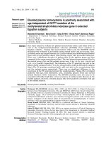
Figure 1 Levels of endotoxin, sCD14 and TNFRII in NAFLD and
NASH subjects compared with controls. The figures show the mean
log endotoxin levels (A), mean log sCD14 levels (B) and mean log
TNFRII levels (C) in control, NAFLD and NASH subjects for the whole co-
hort. The last figure (D) shows the mean log of TNFRII levels in NASH
versus cirrhosis (* p < 0.05, ** p < 0.01, ***p < 0.001). Endotoxin or
sCD14 showed no significant differences between NASH and cirrhosis
in these subjects (data not shown).
Mean log endotoxin levels
Control NAFLD NASH
***
***
0
0.2
0.4
0.6
0.8
1
1.2
Mean log sCD14 levels
6.05
6.1
6.15
6.2
6.25
6.3
6.35
Control NAFLD NASH
3.2
3.25
3.3
3.35
3.4
3.45
3.5
Mean log sTNFRII levels
Control NAFLD NASH
Control Vs
NASH *;
NAFLD Vs
NASH***
3.25
3.3
3.35
3.4
3.45
3.5
3.55
3.6
NASH Cirrhosis
Mean log sTNFRII levels
***
D)
C)
A)
B)
NAFLD Vs
NASH*
Harte et al. Journal of Inflammation 2010, 7:15
/>Page 4 of 10
The effect of diabetic status on levels of endotoxin and
sCD14
Endotoxin levels were similar at all stages of NAFLD,
independent of diabetic status (p = 0.049). In contrast,
serum levels of sCD14 were significantly higher in NASH
patients with T2DM compared with control and NAFLD
subjects with and without T2DM (p < 0.01). This differ-
ence, however, could be due to the stage of liver disease,
as noted by an increased frequency of advanced fibrosis
(i.e. bridging fibrosis or cirrhosis) in T2DM patients
(42%) as compared with those subjects without T2DM
(11%) (p < 0.001). A multivariate regression model
revealed that fibrosis stage (p = 0.003) but not T2DM (p =
0.151) was a significant predictor of sCD14 levels.
Sub-analysis examined the impact of diabetic status on
biochemical factors, which determined that glucose and
ALT were significantly different between NAFLD diabet-
ics when compared with NAFLD non-diabetics (glucose:
p < 0.0001, ALT: p = 0.024).
Therapeutic influence of Orlistat on metabolic markers in
NAFLD patients
No significant changes in body weight and metabolic
markers were observed in six patients treated with diet
for one year (Table 3). In contrast, eight patients that also
received Orlistat exhibited a significant reduction in body
weight post 6 and 12 month treatment (p = 0.001 and p =
0.005, respectively). Furthermore, circulating levels of
endotoxin were significantly reduced in Orlistat treated
patients (p = 0.012) after one year. With regard to serum
ALT, reduced levels were observed in both groups at 6
and 12 months compared with baseline. The reduction at
6 months in Orlistat treated patients was statistically sig-
nificant (p = 0.017), whilst there were no significant
changes in serum levels of sCD14 and lipids. Finally,
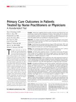
Figure 2 Serum levels of endotoxin (A), sCD14 (B) and sTNFRII (C)
in 23 healthy controls and 155 patients with NAFLD. The horizontal
lines represent the median of the data. Statistical analysis compared
the log mean serum levels of the inflammatory markers at each fibrosis
stage of liver disease against the log mean serum levels of healthy con-
trols subjects, (p < 0.01, p < 0.001).
Figure 3 Correlations between log endotoxin and log fasting in-
sulin, log triglycerides, HOMA-IR, and between sTNFRII and
sCD14. The figures show Pearson correlations between log endotoxin
(EU/mL) and log fasting insulin (μU/mL) in the whole cohort (A) log
triglycerides (mmol/L) in patients with NAFLD (B) log serum levels of
endotoxin and HOMA-IR in the whole cohort (C). The lines of best fit
are also shown: a) r = 0.31, p = 0.02, b) r = 0.51, p < 0.0001 c) r = 0.27, p
= 0.008. A Pearson correlation between log sTNFRII (ng/mL) and sCD14
(μg/mL) in patients with NAFLD is also shown (D). The line of best fit is:
d) r = 0.29, p = 0.004.
Log Endotoxin (Eu/mL)
1.501.000.500.00
Log Triglycerides (mmol/L)
1.00
0.80
0.60
0.40
0.20
0.00
-0.20
r = 0.51
p<0.0001
Log sTNFRII (ng/mL)
3.603.403.20
3.00
sCD14 ( g/mL)
3.0
2.5
2.0
1.5
1.0
0.5
r = 0.29
p=0.004
A
B
C
1.501.000.500.00
Log Endotoxin Eu/mL
2.0
1.5
1.0
0.5
Log Insulin ( U/mL)
r = 0.31
p=0.002
D
1.5
1.0
0.5
0.0
-0.5
Log HOMA-IR
Log Endotoxin (Eu/mL)
1.501.000.50
0.00
r = 0.27
p=0.008
Harte et al. Journal of Inflammation 2010, 7:15
/>Page 5 of 10
changes in endotoxin levels did not correlate with
changes in any other metabolic parameters.
Discussion
In the present study we have identified that patients with
NAFLD are characterised by a significant increase in cir-
culating levels of endotoxin. This finding was indepen-
dent of diabetic status and, as such, suggests that
endotoxin levels may represent an important early
marker of potential liver abnormality. The study also
observed an increase in serum levels of sCD14 and
sTNFRII within the NASH group; both these markers of
inflammation increased as liver disease progressed, as
determined by fibrotic stage, with clear significance
noted co-current with severe stages of fibrosis. Taken
together, these findings are indicative of the severity of
associated inflammation through the progression of
NAFLD.
Specifically, raised levels of endotoxin have been high-
lighted as a secondary insult in patients with alcoholic
liver disease [9] as well as a potential mediator of inflam-
mation in patients with T2DM [13]. Increased levels of
endotoxin have been observed in animal models of
NAFLD [15,18] and manipulation of the gut flora has
been associated with reduced hepatic inflammation, as a
direct result [19,20]. However, the role of endotoxinaemia
in human NAFLD remains unclear. Prior studies have
illustrated that bacterial overgrowth may impact on dis-
ease progression, as examined in 22 patients with NAFLD
[21]; however, serum levels of TNF-α were twice as high
in patients compared with healthy controls, whilst no dif-
ference in endotoxin levels between patients and controls
was observed. In contrast to these findings, recent studies
have shown a five-fold elevation of serum endotoxin lev-
els in 16 patients with NAFLD [22]. This apparent dis-
crepancy may align with the endotoxin assay and how
Table 1: Clinical and biochemical characteristics of NAFLD Compared with Control Subjects.
NAFLD Controls P-value
Age
(yrs)
49.1 ± 12.6 44.6 ± 9.9 NS
BMI
(kg/m2)
34.0 ± 6.0 26.4 ± 4.5 0.0001
Endotoxin #:
(EU/mL)
10.6(7.8, 14.8) 3.9(3.2, 5.2) 0.0001
Fatty Liver # 11.7(7.3, 15.6) n/a 0.0001
NASH # 10.5(8.0, 14.0) n/a 0.0001
Cirrhosis # 7.1(5.2, 11.2) n/a 0.01
Insulin #
(μU/mL)
18.5(10.3, 28.8) 12.9(10.6, 16.0) 0.001
Glucose #
(mmol/L)
5.5(4.8, 6.5) 5.3(4.9, 6.2) NS
HOMA-IR # 4.2(2.5, 7.5) 3.1(2.4, 4.1) 0.001
TNF-α #
(pg/mL)
5.8(4.5, 8.1) 11.2(9.5, 12.2) 0.0001
sCD14 #
(ng/mL)
1623(1370, 2013) 1431.3 ± (1244, 1827) NS
sTNFRII #
(pg/mL)
2229.2(1865.6, 2879.9) 2253.2(1900.4, 2536.5) NS
Data are presented as mean (± SD) unless log transformed (#) in which case they are presented as mean (interquartile range).
Harte et al. Journal of Inflammation 2010, 7:15
/>Page 6 of 10
Table 2: Clinical and biochemical characteristics of NAFLD Compared with NASH Subjects.
NAFLD NASH P-value
NAFLD Vs NASH
Age (yrs) 47.2 ± 11.1 50.4 ± 13.3 NS
BMI (kg/m2)
32.6 ± 5.7 35.0 ± 6.0 p < 0.05
Endotoxin (EU/mL) # 12.3(7.3, 15.6) 10.9(7.8, 13.9) NS
Fibrosis score: 0 12.3(6.6, 17.2) 8.6(5.5, 11.7)
1 13.3(11.1, 15.6) 11.7(8.0, 17.2)
2 7.1(7.1, 7.1) 11.7(8.2, 15.0)
3 NA 12.6(9.4, 14.5)
4 NA 8.2(5.2, 11.2)
Insulin (μU/mL) # 18.3(8.1, 22.3) 30.4(14.6, 36.3) p < 0.001
Fibrosis score: 0 16.7(8.1, 21.1) 29.1(9.8, 26.2)
1 37.2(8.7, 76.3) 26.8(14.2, 33.4)
2 NA 27.4(16.3, 36.9)
3 NA 38.6(18.4, 67.2)
4 NA 37.0(17.6, 54.8)
Glucose (mmol/L) # 5.5(4.8, 6.1) 6.6(4.8, 7.0) p = 0.01
Fibrosis score: 0 5.4(4.8, 5.9) 6.9(4.5, 6.2)
1 6.6(4.8, 8.3) 5.5(4.7, 5.8)
2 6.7(6.7, 6.7) 5.7(4.5, 6.4)
3 NA 8.0(5.5, 9.2)
4 NA 8.2(5.0, 10.8)
HOMA-IR # 4.8(1.7, 5.6) 8.6(3.3, 13.3) p < 0.001
Fibrosis score: 0 4.1(1.7, 5.4) 7.4(1.9, 7.3)
1 12.4(1.8, 27.0) 7.0(3.0, 8.6)
2 NA 6.9(3.3, 9.1)
3 NA 12.8(4.4, 22.5)
4 NA 12.7(7.0, 21.9)
TNF-α (pg/mL) # 8.3(4.2, 7.3) 14.7(5.0, 8.9) p < 0.05
Fibrosis score: 0 8.6(4.2, 7.4) 7.4(4.7, 8.0)
1 6.0(4.0, 8.9) 16.6(5.1, 6.9)
2 6.3(6.3, 6.3) 8.5(4.9, 10.6)
3 NA 43.7(4.4, 21.6)
4 NA 6.7(4.7, 9.0)
sCD14 (ng/mL) # 1575.0(1242.6, 1840.8) 1805.2(1374.8, 2169.6) p = 0.01
Fibrosis score: 0 1533.3(1224.6, 1809.2) 1716.0(1331.1, 2018.2)
Harte et al. Journal of Inflammation 2010, 7:15
/>Page 7 of 10
this is performed, as it requires careful technical execu-
tion as well as ensuring assay comparison of cohorts is
undertaken under the exact same assays conditions, with
appropriate validation [13].
Our present studies report the largest studied cohort of
NAFLD patients' circulating endotoxin levels. The cur-
rent findings clearly indicate that endotoxin levels in the
peripheral circulation are increased in patients with
NAFLD, with no discernible differences between levels in
NAFLD and NASH subjects. However, due to the cross-
sectional nature of the present study, it was not possible
to determine whether increased endotoxin levels are the
cause or consequence of NAFLD. Accumulating evidence
does indicate that elevated levels of endotoxin may,
indeed, play a role in metabolic disease [13,14]. Notably,
endotoxin seems to promote liver fibrogenesis by stimu-
lating TLR4, as elegantly shown in three different mouse
models of liver fibrosis [23]. In the present study, endo-
toxin levels were elevated in all fibrosis stages of liver dis-
ease, although no clear association between stages and
serum endotoxin levels was identified. The sCD14 levels
showed a positive trend with disease progression, which
was noted as significant at fibrosis stages 2-4, compared
with controls, and was also evident by the presence of
higher sCD14 levels in NASH compared with NAFLD
subjects. However, no association between endotoxin and
sCD14 levels was observed, which may be a result of
sCD14's duplicitous function. Soluble CD14 is considered
to enhance endotoxin clearance from serum [24] whilst
also having an active role in endotoxin induced activation
of macrophages, as the TLR and sCD14 complex
responds to an acute phase response, recruiting further
macrophages [8]. Studies by Moreno and colleagues iden-
tified that the inhibition of sCD14, via the administration
of monoclonal antibodies, in a system absent of mem-
brane bound CD14, blocked monocyte activation [25].
Furthermore, that the introduction of sCD14, present in
replacement serum, initiated the LPS/endotoxin response
once more [25]. Similarly, in a study by Lloyd et al, the
application of serum devoid of sCD14 prevented low level
detection of LPS, whilst the introduction of recombinant
sCD14 restored this response [26]. Therefore, the essen-
tial role of sCD14 in the activation of LPS/endotoxin may
explain the lack of any association between endotoxin
and sCD14 levels in our current findings and, perhaps,
the slight decrease in endotoxin levels with increasing
liver damage (between fibrosis stage 3 & 4). It should also
be noted that, as the serum samples were not pre-heated
in this study, the assay measured unbound, accessible
endotoxin. As sCD14 complexes with LPS to activate the
TLR-4 pathway, the increase in sCD14 levels that occurs
with the progression of liver disease might explain the
reduction in endotoxin levels, as more is bound to sCD14
and inaccessible for endotoxin detection. Similar findings
have previously been reported, with no relationship iden-
tified between endotoxin and sCD14 in disease states
including malaria and meningococcal septic shock
[27,28]. In these studies, the results indicate that sCD14
does not provide a useful early marker for disease detec-
tion, which is in accord with our present findings.
No significant difference in endotoxin levels was
observed between patients with simple steatosis
(NAFLD) and patients with NASH, yet the significant
correlation between sTNFRII and sCD14 levels may still
reflect the presence of endotoxin induced inflammation
in patients with NAFLD. Again, sTNFRII is known to
remain elevated for a longer duration than TNFα, thus
proving to be a more indicative measure of activity of the
TNFα system [17]. As a result, previous studies have
noted sTNFRII to correlate with the severity of liver dam-
age - which is confirmed by our present findings [29,30].
Interestingly, TLR4-signaling has recently been shown to
play a crucial role in the development of hepatic inflam-
mation in a mouse model of NASH [18]. Further, mice fed
1 1851.2(1319.3, 2408.3 1642.2(1302.1, 1857.5)
2 1747.1(1747.1, 1747.1) 1829.3(1558.2, 2253.3)
3 NA 1870.0(1680.2, 2080.5)
4 NA 2000.3(1361.0, 2460.1)
sTNFRII (pg/mL) # 2097.3(1642.42453.7) 2676.9(2075.6, 3034.2) p < 0.001
Fibrosis score: 0 2062.9(1621.2, 2445.0) 2491.0(2076.8, 2377.6)
1 2292.8(1729.0, 3067.5) 2330.2(1991.2, 2752.2)
2 2494.9(2494.9, 2494.9) 2597.2(1833.7, 3341.7)
3 NA 2688.9(1962.0, 3047.1)
4 NA 3538.0(2558.3, 4229.2)
Data are presented as mean (± SD) unless log transformed (#) in which case they are presented as mean (interquartile range). NA refers to no
data available as there were no subjects in this group.
Table 2: Clinical and biochemical characteristics of NAFLD Compared with NASH Subjects. (Continued)
Harte et al. Journal of Inflammation 2010, 7:15
/>Page 8 of 10
on a high fructose diet resulted in the development of
NASH co-current with an association with increased
endotoxin concentration in portal blood [20]. Moreover,
in humans, a mutation in the promoter for CD14, which
leads to increased transcriptional activity, is associated
with increased susceptibility for NASH [31].
The possible detrimental effects of endotoxin are not
necessarily restricted to the liver. It is now widely recogn-
ised that insulin resistant states, such as T2DM, cardio-
vascular disease and the metabolic syndrome, are
characterised by a low-grade systemic inflammation, as
well as inflammatory changes in adipose tissue
[6,13,32,33]. Supporting the role for endotoxin in this
context, we have previously reported that endotoxin
exerts proinflammatory effects on human adipocytes in
vitro [13]. Further support for an association between
endotoxin and insulin resistance has been identified by
Cani et al who have shown, using mouse models, that
continuous infusion of endotoxin for four weeks induced
identical metabolic changes as those induced by a high-
Table 3: Clinical/biochemical characteristics of NAFLD patients on diet and Orlistat treatment
Diet alone Diet and Orlistat
Baseline 6 months 12 months Baseline 6 months 12 months
Age (years) 48.8 ± 11.5 53.4 ± 16
Body weight
(kg)
100.0 ± 16.1 100.8 ± 16.3 101.6 ± 16.3 100.9 ± 24.5 95.5 ± 24.4 * 96.4 ± 25.7 *
Endotoxin
(EU/mL)
15.9 ± 7.2 16.7 ± 5.5 14.4 ± 11.0 15.8 ± 4.6 14.4 ± 5.7 11.1 ± 4.0 **
sCD14 (μg/
mL)
1.52 ± 0.40 2.46 ± 0.19 2.25 ± 1.28 1.45 ± 0.60 1.55 ± 0.35 1.59 ± 0.69
Glucose
(mmol/L)
6.5 ± 2.4 6.1 ± 1.7 8.2 ± 3.8 5.6 ± 0.9 5.3 ± 0.7 5.4 ± 1.4
Insulin (μU/
mL)
24.2 ± 20.2 21.2 ± 9.5 22.6 ± 13.5 21.6 ± 13.1
HOMA-IR 3.2 ± 2.6 3.0 ± 1.3 2.9 ± 1.8 2.8 ± 1.6
ALT (U/L) 129 ± 86 90 ± 67 100 ± 68 93 ± 31 60 ± 20 ** 66 ± 41
Cholesterol
(mmol/L)
4.9 ± 0.8 4.8 ± 1.4 4.7 ± 1.1 5.1 ± 1.4 4.6 ± 1.6 4.9 ± 1.7
LDL-
cholesterol
(mmol/L)
2.8 ± 0.8 3.0 ± 1.2 2.8 ± 1.0 3.1 ± 1.3 2.7 ± 1.4 2.8 ± 1.6
HDL-
cholesterol
(mmol/L)
1.09 ± 0.17 1.62 ± 0.58 1.68 ± 1.35 1.16 ± 0.26 1.38 ± 0.39 1.23 ± 0.36
Triglycerides
(mmol/L)
2.4 ± 1.3 2.3 ± 1.0 2.4 ± 1.6 1.9 ± 0.6 1.7 ± 0.6 1.9 ± 0.7
NAFLD patients underwent a 12 month course of intensive diet-treatment with (n = 8) or without) Orlistat (n = 6). Data are mean (± SD). * p
< 0.005 (versus baseline), ** p < 0.05 (versus baseline).
Harte et al. Journal of Inflammation 2010, 7:15
/>Page 9 of 10
fat diet, namely insulin resistance and weight gain [14].
Additionally, the use of CD14 mutant mice caused a
reduction to most of the LPS and high-fat diet-induced
detrimental changes. As such, the authors suggest that
the CD14/LPS system sets the tone for insulin sensitivity.
This is in accord with our current findings, as determined
by the positive correlations identified between endotoxin
and insulin levels, as well as insulin resistance. As insulin
resistance is almost universally present in NAFLD,
chronic endotoxinaemia may be of particular importance
in this condition, not only as a factor that induces hepatic
inflammation and fibrosis, but also as a factor contribut-
ing to insulin resistance.
The causes of increased blood levels of endotoxin in
patients with NAFLD are not clear with several explana-
tions to be considered, such as increased amounts of
endotoxin in the intestinal lumen, increased intestinal
absorption and reduced clearance from the blood. It is
possible, if not likely, that the amount of endotoxin in the
intestinal lumen depends on the type of bacteria present,
thus, obesity-associated changes in the gut flora, as
recently reported in both humans [34] and mice [35],
could have important metabolic consequences. More-
over, intestinal dysmotility and/or bacterial overgrowth
have been reported in diabetic patients [36], as well as in
NASH patients [21,37]. In a recent study by Miele et al,
NAFLD subjects were shown to have both increased gut
permeability and prevalence of small intestinal bacterial
overgrowth [12]. In addition, the level of bacterial over-
growth correlated with the severity of steatosis in the
NAFLD patients, supporting the theory that such distur-
bances could possibly facilitate increased absorption of
endotoxin from the gut. Interestingly, Brun and co-work-
ers have demonstrated disrupted intestinal tight junc-
tions in a rodent model of the metabolic syndrome, a
finding that has also recently been confirmed in NASH
patients [12,15], thus providing strong evidence for an
anatomical basis underlying increased gut permeability.
Furthermore, a susceptibility to gut leakiness has been
noted in humans with NAFLD after challenge with aspi-
rin [22]. In a more recent study by Ghoshal and co-work-
ers, a mechanism for the simultaneous absorption of fat
and LPS was identified. Long chain dietary fats are incor-
porated into chylomicrons, which also have a high affinity
for LPS and can therefore transfer it from the gut to the
bloodstream [11]. Our present and previous findings, in
which strong correlations between triglyceride and endo-
toxin levels are apparent, would support the fat mediated
uptake of LPS [32,38]. Such a mechanism might explain
the results from this study, which identified that treat-
ment with Orlistat was associated with a significant
reduction in endotoxin levels. This effect has also been
observed in eighteen subjects with impaired glucose tol-
erance, all of which were treated with Orlistat for one
year [38]. As treatment with Orlistat has previously been
associated with beneficial metabolic effects independent
of weight loss [39], the current findings suggest that
reduced absorption of endotoxin may occur through the
blockade of dietary fat absorption, via the mechanism
proposed by Goshal et al. This hypothesis must, however,
be tested in a larger, randomised, controlled trial.
In conclusion, the present study confirms that circulat-
ing endotoxin levels are elevated in patients with NAFLD.
This result gives further support to the concept that
chronic endotoxinaemia could be an important patho-
genic factor in NAFLD and that elevated endotoxin levels
may serve as an early biomarker for potential liver dam-
age. Studies exploring the impact of the gut flora on
human metabolism are now needed to further assess this
hypothesis. If the gut flora turns out to be an important
determinant of endotoxin levels in humans, treatment
with probiotics or lipase inhibitors may prove to be bene-
ficial in metabolic diseases, particularly in NAFLD.
Competing interests
The authors declare that they have no competing interests.
Authors' contributions
ALH for the design, statistical analysis, manuscript development and final revi-
sion of the paper, NFS for the design, statistical analysis, drafting of the manu-
script and manuscript development; SJC for the drafting and revising of the
manuscript; KM, TB and EMY for their practical and intellectual input; GT for the
statistical analysis and interpretation of data; EA, MSA HMS and AIA for their
interpretation of data and intellectual input; ADB for the acquisition and inter-
pretation of data; SK for the interpretation of data and intellectual input; CPD
for the concept, acquisition and interpretation of data; PM for the concept,
design, interpretation of data and intellectual input. All authors read and
approved the final manuscript.
Acknowledgements
We would like to thank Eli Lilly Research for grant funding and the British Heart
Foundation for funding Alison Harte on an Intermediate fellowship. We would
also like to acknowledge funding from the Egyptian Government for Elham
Youssef-Elabd, a visiting PhD student within the team. Finally, we thank the
Department of Health for PhD funding to support Kirsty McGee.
Author Details
1
University of Warwick, Unit for Diabetes and Metabolism, Warwick Medical
School, Clinical Sciences Research Institute, UHCW, Clifford Bridge Road,
Coventry, CV2 2DX, UK,
2
Biochemistry Dept, National Research Center, Dokki,
Giza, Egypt,
3
Chemistry Dept, Faculty of Science, Helwan University, Egypt,
4
Clinical Pathology Dept, National Institute of Diabetes & Endocrinology, Cairo,
Egypt and
5
School of Clinical Medicine (Hepatology), Floor 4, William Leech
Building, The Medical School, Framlington Place, Newcastle upon Tyne NE2
4HH, UK
References
1. Day CP: Non-alcoholic fatty liver disease: current concepts and
management strategies. Clin Med 2006, 6(1):19-25.
2. Bellentani S, Saccoccio G, Masutti F, Crocè LS, Brandi G, Sasso F, Cristanini
G, Tiribelli C: Prevalence of and risk factors for hepatic steatosis in
Northern Italy. Ann Intern Med 2000, 132(2):112-117.
3. Marchesini G, Bugianesi E, Forlani G, Cerrelli F, Lenzi M, Manini R, Natale S,
Vanni E, Villanova N, Melchionda N, Rizzetto M: Nonalcoholic fatty liver,
steatohepatitis, and the metabolic syndrome. Hepatology 2003,
37(4):917-923.
Received: 3 September 2009 Accepted: 30 March 2010
Published: 30 March 2010
This article is available from: 2010 Harte et al; licensee BioMed Central Ltd. This is an Open Access article distributed under the terms of the Creative Commons Attribution License ( which permits unrestricted use, distribution, and reproduction in any medium, provided the original work is properly cited.Journal of Inflammation 2010, 7:15
Harte et al. Journal of Inflammation 2010, 7:15
/>Page 10 of 10
4. Gupte P, Amarapurkar D, Agal S, Baijal R, Kulshrestha P, Pramanik S, Patel N,
Madan A, Amarapurkar A, Hafeezunnisa : Non-alcoholic steatohepatitis
in type 2 diabetes mellitus. J Gastroenterol Hepatol 2004, 19(8):854-858.
5. Xu H, Barnes GT, Yang Q, Tan G, Yang D, Chou CJ, Sole J, Nichols A, Ross JS,
Tartaglia LA, Chen H: Chronic inflammation in fat plays a crucial role in
the development of obesity-related insulin resistance. J Clin Invest
2003, 112(12):1785-1830.
6. Wellen KE, Hotamisligil GS: Inflammation, stress, and diabetes. J Clin
Invest 2005, 115(5):1111-1119.
7. Kaisho T, Akira S: Toll-like receptors as adjuvant receptors. Biochim
Biophys Acta 2002, 1589(1):1-13.
8. Muzio M, Polentarutti N, Bosisio D, Manoj Kumar PP, Mantovani A: Toll-li ke
receptor family and signalling pathway. Biochem Soc Trans 2000,
28(5):563-566.
9. Rao RK, Seth A, Sheth P: Recent Advances in Alcoholic Liver Disease I.
Role of intestinal permeability and endotoxemia in alcoholic liver
disease. Am J Physiol Gastrointest Liver Physiol 2004, 286(6):G881-G884.
10. Erridge C, Attina T, Spickett CM, Webb DJ: A high-fat meal induces low-
grade endotoxemia: evidence of a novel mechanism of postprandial
inflammation. Am J Clin Nutr 2007, 86(5):1286-1292.
11. Ghoshal S, Witta J, Zhong J, de Villiers W, Eckhardt E: Chylomicrons
promote intestinal absorption of lipopolysaccharides. J Lipid Res 2009,
50(1):90-97.
12. Miele L, Valenza V, La Torre G, Montalto M, Cammarota G, Ricci R, Mascianà
R, Forgione A, Gabrieli ML, Perotti G, Vecchio FM, Rapaccini G, Gasbarrini G,
Day CP, Grieco A: Increased intestinal permeability and tight junction
alterations in nonalcoholic fatty liver disease (NAFLD). Hepatology
2009, 49(6):1877-1887.
13. Creely SJ, McTernan PG, Kusminski CM, Fisher M, Da Silva NF, Khanolkar M,
Evans M, Harte AL, Kumar S: Lipopolysaccharide activates an innate
immune system response in human adipose tissue in obesity and type
2 diabetes. Am J Physiol Endocrinol Metab 2007, 292(3):E740-E747.
14. Cani PD, Amar J, Iglesias MA, Poggi M, Knauf C, Bastelica D, Neyrinck AM,
Fava F, Tuohy KM, Chabo C, Waget A, Delmée E, Cousin B, Sulpice T,
Chamontin B, Ferrières J, Tanti JF, Gibson GR, Casteilla L, Delzenne NM,
Alessi MC, Burcelin R: Metabolic endotoxemia initiates obesity and
insulin resistance. Diabetes 2007, 56(7):1761-1772.
15. Brun P, Castagliuolo I, Di Leo V, Buda A, Pinzani M, Palù G, Martines D:
Increased intestinal permeability in obese mice: new evidence in the
pathogenesis of nonalcoholic steatohepatitis. Am J Physiol Gastrointest
Liver Physiol 2007, 292(2):G518-G525.
16. Brunt EM, Janney CG, Di Bisceglie AM, Brunt EM, Janney CG, Di Bisceglie
AM, Neuschwander-Tetri BA, Bacon BR, et al.: Nonalcoholic
steatohepatitis: a proposal for grading and staging the histological
lesions. Am J Gastroenterol 1999, 94(9):2467-2474.
17. Porteu F, Nathan C: Shedding of tumor necrosis factor receptors by
activated human neutrophils. J Exp Med 1990, 172:599-607.
18. Rivera CA, Adegboyega P, van Rooijen N, Tagalicud A, Allman M, Wallace
M: Toll-like receptor-4 signaling and Kupffer cells play pivotal roles in
the pathogenesis of non-alcoholic steatohepatitis. J Hepatol 2007,
47(4):571-579.
19. Li Z, Yang S, Lin H, Huang J, Watkins PA, Moser AB, Desimone C, Song XY,
Diehl AM: Probiotics and antibodies to TNF inhibit inflammatory
activity and improve nonalcoholic fatty liver disease. Hepatology 2003,
37(2):343-350.
20. Bergheim I, Weber S, Vos M, Krämer S, Volynets V, Kaserouni S, McClain CJ,
Bischoff SC, Antibiotics protect against fructose-induced hepatic lipid
accumulation in mice: Role of endotoxin. J Hepatol 2008, 48(6):983-992.
21. Wigg AJ, Roberts-Thomson IC, Dymock RB, McCarthy PJ, Grose RH,
Cummins AG: The role of small intestinal bacterial overgrowth,
intestinal permeability, endotoxaemia, and tumour necrosis factor
alpha in the pathogenesis of non-alcoholic steatohepatitis. Gut 2001,
48(2):206-211.
22. Farhadi A, Gundlapalli S, Shaikh M, Frantzides C, Harrell L, Kwasny MM,
Keshavarzian A: Susceptibility to gut leakiness: a possible mechanism
for endotoxaemia in non-alcoholic steatohepatitis. Liver Int 2008,
28(7):1026-33.
23. Seki E, De Minicis S, Osterreicher CH, Kluwe J, Osawa Y, Brenner DA,
Schwabe RF: TLR4 enhances TGF-beta signaling and hepaticfibrosis.
Nat Med 2007, 13(11):1324-1332.
24. Tapping RI, Tobias PS: Cellular binding of soluble CD14 requires
lipopolysaccharide (LPS) and LPS-binding protein. J Biol Chem 1997, 12;
272(37):23157-23164.
25. Moreno C, Merino J, Ramírez N, Echeverría A, Pastor F, Sánchez-Ibarrola A:
Lipopolysaccharide needs soluble CD14 to interact with TLR4 in
human monocytes depleted of membrane CD14. Microbes Infect 2004,
6(11):990-5.
26. Lloyd-Jones KL, Kelly MM, Kubes P: Varying Importance of Soluble and
Membrane CD14 in Endothelial Detection of Lipopolysaccaride. J
Immunol 2008, 181:1446-1453.
27. Wenisch C, Wenisch H, Parschalk B, Vanijanonta S, Burgmann H, Exner M,
Zedwitz-Liebenstein K, Thalhammer F, Georgopoulos A, Graninger W,
Looareesuwan S: Elevated levels of soluble CD14 in serum of patients
with acute Plasmodium falciparum malaria. Clin Exp Immunol 1996,
105(1):74-78.
28. Arranz E, Blanco-Quiros A, Soli's P, Garrote JA: Lack of correlation
between soluble CDI4 and IL-6 in meningococcal septic shock. Pediatr
Allergy Immunol 1997, 8:194-199.
29. Hanck C, Rossol S, Böcker U, Tokus M, Singer MV: Presence of plasma
endotoxin is correlated with tumour necrosis factor receptor levels and
disease activity in alcoholic cirrhosis. Alcohol Alcohol 1998, 33(6):606-8.
30. Schröder J, Stüber F, Gallati H, Schade FU, Kremer B: Pattern of soluble
TNF receptors I and II in sepsis. Infection 1995, 23(3):143-8.
31. Brun P, Castagliuolo I, Floreani AR, Buda A, Blasone L, Palù G, Martines D:
Increased risk of NASH in patients carrying the C(-159)T polymorphism
in the CD14 gene promoter region. Gut 2006, 55(8):1212.
32. Miller MA, McTernan PG, Harte AL, Silva NF, Strazzullo P, Alberti KG, Kumar
S, Cappuccio FP: Ethnic and sex differences in circulating endotoxin
levels: A novel marker of atherosclerotic and cardiovascular risk in a
British multi-ethnic population. Atherosclerosis 2009, 203(2):494-502.
33. Baker AR, Harte AL, Howell N, Pritlove DC, Ranasinghe AM, da Silva NF,
Youssef EM, Khunti K, Davies MJ, Bonser RS, Kumar S, Pagano D, McTernan
PG: Epicardial Adipose Tissue as a Source of Nuclear Factor-{kappa}B
and c-Jun N-Terminal Kinase Mediated Inflammation in Patients with
Coronary Artery Disease. J Clin Endocrinol Metab 2009, 94(1):261-267.
34. Ley RE, Turnbaugh PJ, Klein S, Gordon JI: Microbial ecology: human gut
microbes associated with obesity. Nature 2006, 444(7122):1022-1023.
35. Turnbaugh PJ, Ley RE, Mahowald MA, Magrini V, Mardis ER, Gordon JI: An
obesity-associated gut microbiome with increased capacity for energy
harvest. Nature 2006, 444(7122):1027-1031.
36. Cuoco L, Montalto M, Jorizzo RA, Santarelli L, Arancio F, Cammarota G,
Gasbarrini G: Eradication of small intestinal bacterial overgrowth and
oro-cecal transit in diabetics. Hepatogastroenterology 2002,
49(48):1582-1586.
37. Soza A, Riquelme A, Gonzalez R, et al.: Increased orocecal transit time in
patients with nonalcoholic fatty liver disease. Dig Dis Sci 2005,
50(6):1136-1140.
38. Dixon AN, Valsamakis G, Hanif MW, Field A, Boutsiadis A, Harte A,
McTernan PG, Barnett AH, Kumar S: Effect of the orlistat on serum
endotoxin lipopolysaccharide and adipocytokines in South Asian
individuals with impaired glucose tolerance. Int J Clin Pract 2008,
62(7):1124-1129.
39. Hsieh CJ, Wang PW, Liu RT, Tung SC, Chien WY, Chen JF, Chen CH, Kuo
MC, Hu YH: Orlistat for obesity: benefits beyond weight loss. Diabetes
Res Clin Pract 2005, 67(1):78-83.
doi: 10.1186/1476-9255-7-15
Cite this article as: Harte et al., Elevated endotoxin levels in non-alcoholic
fatty liver disease Journal of Inflammation 2010, 7:15