Báo cáo y học: "Anti-inflammatory function of arctiin by inhibiting COX-2 expression via NF-B pathways" potx
Bạn đang xem bản rút gọn của tài liệu. Xem và tải ngay bản đầy đủ của tài liệu tại đây (1.41 MB, 9 trang )
RESEA R C H Open Access
Anti-inflammatory function of arctiin by inhibiting
COX-2 expression via NF-B pathways
Sungwon Lee
1
, Seulmee Shin
1
, Hyunyul Kim
1
, Shinha Han
1
, Kwanghee Kim
1
, Jeunghak Kwon
1
, Jin-Hwan Kwak
2
,
Chong-Kil Lee
3
, Nam-Joo Ha
1
, Dongsool Yim
1
and Kyungjae Kim
1*
Abstract
Background: Arctiin, isolated from Forsythia suspensa has been reported to have anti-inflammatory, anti-oxidant,
antibacterial, and antiviral effects in vitro. However, there has been a lack of studies regarding its effects on
immunological activity. The aim of this study is to investigate the anti-inflammatory potential and possible
mechanisms of arctiin in LPS-induced macrophages.
Methods: We investigated the mRNA and protein levels of proinflammatory cytokines through RT-PCR and
western blot analysis, followed by a FACS analysis for surface molecule changes.
Results: Arctiin dose depe ndently decreased the production of NO and proinflammatory cytokines such as IL-1b,
IL-6, TNF-a, and PGE
2
, and it reduced the gene and protein levels as determined by RT-PCR and western blot
analysis, respectively. The expression of co-stimulatory molecules such as B7-1 and B7-2 were also inhibited by
arctiin. Furthermore, the activation of the nuclear transcription factor, NF-B in macrophages was inhibited by
arctiin.
Conclusion: Taken together these results provide evidence of the bioactivity of arctiin in inflammatory diseases
and suggest that arctiin may exert anti-inflammatory effect by inhibiting the pro-inflammatory mediators throug h
the inactivation of NF-kB.
Background
Non-steroidal anti-inflammatory drugs (NSAIDs) have
been widely used in the treatment of acute and chronic
inflammatory diseases, which play their therapeutic effects
via inhibiting cyclooxygenase (COX) to prevent the pro-
duction of pro-inflammatory prostaglandins. However,
their long-term use shows the major side-effects of gastro-
intestinal diseases. Thus researchers have tried to screen
new biological components from various pla nt sources
including medicinal plants which inhibited COX with
lower toxicity and higher anti-inflammatory activity on the
great deal in the therapeutic application [1].
The fruit of Forsythia suspensa Vahl, Forsythiae Fruc-
tus, has been widely used in traditional medicines to treat
swelling, gonorrhea, urination, hemorrhoids, tubercle,
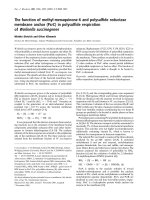
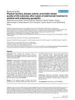
and other afflictions [2]. Arctiin is a lignan compound
isolated from Forsythiae Fructus (Figure 1A); it has been
found to significantly induce cell detachment and
decrease the number of PC-3 cells in human prostate
cancer [3]. Moreover, it has been demonstrated to pos-
sess many kinds o f bioactivities [4] and a number of
important pharmacological properties including being
demutagenic [5,6] cytotoxic, anti-proliferative [7,8], plate-
let activating factor (PAF) antagonistic [9], calcium
antagonistic [10], and anti-carcinogenetic. In animal stu-
dies, arctiin effectively inhibited the formation of 12-O-
tetradecanoylphorbol-13-acetate (TPA)-induced ear
edema in mice [11]. However, there has been a lack of
studies regarding the effects of arctiin on inflammation.
Chronic inflammation and infection has been demon-
strated to lead to an upregulation of a series of enzymes
and signaling proteins in affected tissues and cells. Inflam-
mation has been shown to be a multi-step process,
mediated by activated inflammatory and immune cells
including macrophages and monocytes [12]. Inflammatory
reactions, phagocytosis, natural cytotoxicity, cytokine pro-
duction, antibody response, and cellular imm unity are
defensive mechanisms that have been suggested to be
* Correspondence:
1
College of Pharmacy, SahmYook University, Seoul 139-742, Republic of
Korea
Full list of author information is available at the end of the article
Lee et al. Journal of Inflammation 2011, 8:16
/>© 2011 Lee et al; licensee BioMed Central Ltd. This is an Open Access article dist ributed under the terms of the Creative Commons
Attribution License (htt p://creativec ommons.org/licenses/by/2.0), which permits unr estricted use, distri bution, and reproduction in
any medium, provided the original work is properly cited.
modulated by therapeutic doses of antimicrobial agents
[13]. Activated macrophages include the inducible for ms
of nitric oxide synthase (iNOS) and cyclooxygenase-2
(COX-2), which have been reported to be responsible for
increasin g the levels of nitric oxide (NO) and prostaglan-
dins (PGs), the overproduction of proinflammatory cyto-
kines (e.g. TNF-a,IL-6,IL-1b)andinflammatory
mediators (e.g. PGE
2
, NO), and the mediation of many
inflammatory diseases [14].
Costimulatory molecules are one class of receptors,
which have recently been implicated as fulfilling this
role in the innate immune response. B7-1 and -2 repre-
sent one class of costimulatory receptors. They consist
of structurally related, cell-surface protein ligands, which
bind to receptors on lymphocytes that regulate immune
responses. In addition, theyarestimulatedviaCD28
while CTLA4 serves as both a stoichiometric inhibitor
of CD28-B7-1/-2 engagement as well as serving to
directly induce immunosuppressive signals within den-
dritic cells [15].
In th is study, we evaluated the potential of arctiin as a
therapeutic modality for inflammation in RAW264.7
mouse macrophage cells as well as in pri mary peritoneal
macrophages. Our results demonstrated that arctiin lar-
gely inhibited the excessi ve production of inflammatory
mediators such as NO, P GE
2
,TNF-a,IL-1b and IL-6 as
well as the suppression of COX-2 through the inhibition
of NF-kB translocation pathway.
Methods
Chemicals and reagents
Dul becco’s Modified Eagle Medium (DMEM)-1640, fetal
bovine serum (FBS), and penicillin/streptomycin were
purchased from Hyclone (Logan, USA). Escherichia coli
lipopolysaccharide (LPS) was purchased from sigma (St.
Louis, USA). b-actin,i-NOS,COX-2,p65,p-IBa, PE-
B7-1, and FITC-B7-2 anti-bodies were purchased from
BD Pharmingen™ (San Jose, USA). Enzyme immunoas-
say kits for the measurement of PGE
2
,IL-1b,IL-6,and
TNF-a were purchased from R&D system (Minneapolis,
USA).
Isolation of arctiin from Forsythiae Fructus
Forsythiae Fructus obtained from an herbal market in
Seoul, Korea, was extracted three times with hot MeOH
(3 hours) and then evaporated at 40°C under reduced
pressure to dryness. The MeOH ex tract was then resus-
pended in distilled water and successively partitioned
with CHCl
3
,EtOAc,andn-BuOH.TheBuOHfraction
was then loaded onto a silica gel column a nd eluted
with MeOH-CHCl
3
mixtures (1:5 to 1:1). The result was
white amorphous powders, which were identified as
authentic samples using spectrometric data of nuclear
magnetic resonance (
1
H-NMR), mass spectrometry
(MS).
1
H-NMR (300 MHz), DMSO-d
6
: δ 6.97(1H, d, J
= 8.3, H-5), 6.82 (1H, d,
J=8.0,H-5’ ), 6.77 (1H, d, J =
1.7, H-2), 6.65 (1H, dd, J = 8.3, 1.7, H-6), 6.65 (1H, s,
H-2’), 6.59 (1H, dd, J =8.0,1.8,H-6’), 4.83 (1H, d, J =
7.3, H-1’’), 4.00 (2H, m, H-9’), 3.70 (3H, s, OCH
3
), 3.69
(6H, s, OCH
3
), 2.76 (2H, m, H-7’), 2.54 (4H, m, H-7, 8,
8’ ). Positive FABMS (m/z): 557 (M + Na
+
), 372 (M +
gluco), 154, 1 36. The white amorphous p owder com-
pound analyzed by NMR and MS was identified to arc-
tiin (Figure 1A).
Animals
ICR mice (6-8 weeks old, specific pathogen-free) were
obtained from Orie nt-Bio Co. (Seongnam, Kore a). Ani-
mals were fed with standard laboratory chow (PMI Lab
Diet, Richmond, USA) and autoclaved distilled water
(DW). They were acclimatized in an animal facility (Sah-
myook University, Korea) and maintained at 22 ± 2°C in
50 ± 10% relative humidity and a light/dark ( 12 hrs/12
hrs) cycle for at least 7 days prior to the experiments.
Figure 1 Structure of arctiin (A); Cell viability (B). Cell viability was evaluated as described in ‘Materials and Methods’.
Lee et al. Journal of Inflammation 2011, 8:16
/>Page 2 of 9
Cell culture
Male ICR mice (6-8 weeks) were intraperitoneally injected
with 1.5 ml thioglycollate broth for recruitment of macro-
phages. RAW 264.7 cells were obtained from the A merican
Type Culture Collection (ATCC, Rockville, USA). These
cells were grown at 37°C in DMEM medium supplemented
with 10% FBS and 1% (v/v) penicillin (10,000 U/ml)/strep-
tomycin (10,000 U/ml) in a humidified 5% CO
2
-95% air
incubator u nder standard conditions.
Cell viability assay
A commercially available cell viability assay was employed
to evaluate the cytotoxic effect of arctiin using thiazolyl
blue tetrazolium bromide (SIGMA, USA). RAW264.7 cells
(2 × 10
5
cells/well) were plated with various concentra-
tions of arctiin in 96-well microtiter plates (Nunc,
Roskilde, Denmark) and were then cultured at 37°C in a
5% CO
2
incubat or. Subsequently, 50 μlofMTTsolution
was added to each well, and the cells were then cultured
for 4 hrs at 37°C in the same incubator. Following this,
100 μl of solubilized solution were added to each well and
the plate was allowed to stand overnight in the incubator.
The optical density (OD) was then measured at 560 nm by
a microplate reader (Molecular devices, USA).
Nitrite measurement
RAW 264.7 cells were added to each well (200 μl; 1 ×
10
6
cells/ml) of a flat-bottomed 96-well plate according
to the following treatment condition: LPS (100 ng/ml),
LPS/arctiin (12.5, 25, 50, 100 μg/ml), and media only
(DMEM-10). Nitric oxide was measured in culture
supernatants by reaction with the Griess reagent (1%
sulfanilamide and 0.1% N-[1-naphthy]-ethylenediamine
dihydrochloride in 5% phosphoric acid; Roche) to 100 μl
of culture supernatant for 15 m in at room temperature
in the dark. The absorbance at 540 nm was then d eter-
mined using a microplate reader (Molecular devices,
USA) and a standard curve was generated using NaNO
2
.
Determination of pro-inflammatory cytokines and PGE
2
RAW 264.7 cells and primary macrophages were cul-
tured in 12-well flat plates at a density of 5 × 10
6
cells/
well. The cells were then treated with various concentra-
tions of arctiin and subsequently stimulated with LPS
(100 ng/ml) at 37°C for 48 hrs in humidified air with 5%
CO
2
. The supernatants were then collected and m ea-
sured for TNF-a,IL-1b,IL-6,andPGE
2
by an enzyme-
linked immunosorbent assay (ELISA) according to the
manufacturer’s protocol.
RT-PCR (reverse transcription polymerase chain reaction)
Total RNA was extracted from macrophages using the
RNeasy Mini Kit (QIAGEN, USA) in an RNase-free envir-
onment. The reverse transcription of 1 μgRNAwas
carried out using M-MLV reverse transcriptase (Promega,
USA), oligo (dT) 16 primer, dNTP (0.5 mM) and 1 U
RNase inhibitor. After incubation at 65°C for 5 min and
37°C for 60 min, M-MLV reverse transcriptase was inacti-
vated by heating at 70°C for 15 min. The polymerase chain
reaction (PCR) was performed in 50 mM KCl, 10 mM
Tris-HCl (pH8.3), 1.5 mM MgCl
2
,and2.5mMdNTPs
with 5 units of Taq DNA polymerase and 10 pM of each
primer set for IL-1b, IL-6, TNF-a,iNOS,andCOX-2.The
cDNA was amplified by 35 cycles of denaturing at 94°C
for 45 s, annealing at 60°C for 45 s, and extension at 72°C
for 1 min. Final extension was performed at 72°C for
5 min. The PCR products wer e electrophoresed on 1.5%
agarose gels and stained with ethidium bromide. The pri-
mer sequences were as follows: 5’- AGC TCC TCC CAG
GAC CAC AC-3’ (forward), 5’ -ACG CTG AGT ACC
TCA TTG GC-3’ (reverse) for i-NOS, 5’-AAG AAG AAA
GTT CAT TCC TGA TCC C-3’ (forward), 5’-TGA CTG
TGG GAG GAT ACA TCT CTC-3’ (reverse) for COX-2,
and 5’-GTG GGC CGC CCT AGG ACC AG-3’ (forward),
5’- GGA GGA AGA GGA TGC GGC AG T-3’ (reverse)
for b-actin as a control for PCR. The band intensity was
quantified by densitometric analysis (Infinity 3026, Vilber
Lourmat, France).
Preparation of cytosolic and nuclear extracts
The cells were collected after culture and washed twice
with cold PBS, resuspended in hypotonic buffer (10 mM
HEPES, pH 7.9, 10 mM KCl, 1.5 mM MgCl
2
,0.2mM
PMSF, 0.5 mM DTT, 10 μg/ml aportinin). After 15 min
incubation on ice, the cells were lysed by the addition of
0.1% NP-40 and vigorous vortexing for 1 min. The nuclei
were pelleted by centrifugation at 12,000 × g for 1 min at
4°C and resuspended in high salt buffer (20 mM HEPES,
pH 7.9, 25% glycerol, 400 mM KCl, 1.5 mM MgCl
2
,
0.2 mM EDTA, 0.5 mM DTT, 1 mM NaF, 1 mM sodium
orthovanadate). The cytosol ic and nuclear extracts were
stored in aliquots at -70°C.
Western blot analysis
RAW264.7 cells were washed with phosphate-buffered
saline (PBS) and lysed using lysis buffer (1% SDS, 1.0
mM sodium vanadate, 10 mM T ris-Cl buffer, pH 7.4)
for 5 min. Further, 20 μg of protein from the cell lysates
were applied to 8-12% SDS-polyacrylamide gels and
then transferred to nitrocellulose membranes. The
membranes were blocked in 5% skim milk solution for 1
h. They were then incubated with anti-TNF-a,anti-IL-
1b, anti-IL-6, anti-iNOS, anti-COX-2, anti-p- IBa,or
anti-p65 monoclonal antibodies for 2 h and subse-
quently washed 3 time s with PBS. After incubation with
an AP-labeled secondary a ntibody for 2 h, the bands
were visualized using an alkaline phosphatase subs trate
(VECTOR, USA).
Lee et al. Journal of Inflammation 2011, 8:16
/>Page 3 of 9
Flow cytometry
RAW 264.7 cells (1 X 10
6
cells/ ml) were cultured in
Petri-dishes. The cells were treated with various concen-
trations (12.5, 25, 50, 100 μg/ ml) of arctiin in the pre-
sence of LPS (100 ng/ ml). The dishes were incubated at
37°C for overnight in humidifi ed 5% CO
2
incubator
under standard conditions. The cells were then washed
with PBS. The washed cel ls were blocked with staining
buffer containing 10% normal mouse serum (NMS) for
20 min on ice. The blocked cells were incubate d with
co-stimulatory molecules such as B7-1 and B7-2 anti-
body (BD Biosciences, San Jose, USA) for 20 min on ice.
The incubated cells were washed three times with stain-
ing buffer and then fixed by 1% paraformaldehyde i n
PBS. The fixed cells were measured by flow cytometry
(Beckman coulter, Brea, USA).
Statistical analysis
All data are presented as mean ± SEM values. Signifi-
cant differences (P < 0.05) between groups were evalu-
ated using a one-way analysis of variance with SPSS
(Chicago, IL, USA) for Windows and Duncan’s Multiple
Range Test where appropriate.
Results
Prior to e valuating whether arctiin showed anti-inflam-
matory activity, we examined its eff ect on cell growth in
RAW264.7 cells and found that arctiin did not affect
normal cell growth at concentrations up to 100 μg/ml
(Figure 1 B). Thus, in the following experiments, arctiin
was studied at concentrations up to 100 μg/ml in order
to exclude any effects on the normal growth status of
cells.
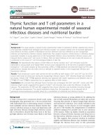
Effect of arctiin on PGE
2
and NO production in
macrophages
In the present study, we examined whether or not arc-
tiin suppress macrophages activation induced by LPS,
one of the most potent macrophages activation factors.
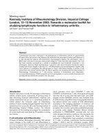
Arctiin significantly suppressed COX-2 protein expres-
sion (Figure 2A) in LPS-stimulated RAW 264.7 cells.
The pro-inflammatory mediator, PGE
2
is also generated
by the COX-2 enzyme in response to stimulation by
LPS. Results showed that arctiin significantly inhibited
PGE
2
production (Figure 2B) and mRNA of COX-2 in
RAW 264.7 cells (Figure 2C) and p rimary macrophages
(Figure 2D) by western blot analyses and subsequent
RT-PCR.
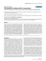
NO is known to be a pro-inflammatory mediator in
inflammatory diseases. Several studies have demonstrated
that overproduction of NO by iNOS was associated with
inflammatory responses and also with serious disorders,
including RA. Ther efore, we inves tigated whether arctiin
inhibits NO production in macrophages that were acti-
vated with LPS. Interestingly, in LPS (100 ng/ml) stimu-
lated RAW264.7 cells, when various concentrations of
Figure 2 Arctiin inhibits the production of COX-2 (A), PGE
2
(B) and mRNA (C, D) in LPS-stimulated macrophages. RAW 264.7 cells (A-C)
and primary macrophages (D) were treated with different concentrations of arctiin (12.5~100 μg/ml) in the presence of LPS (100 ng/ml) and
was monitored as described in ‘Materials and Methods’. Each bar represents the means ± S.D. from three separate experiments.
††
P< 0.01
compares to the control. *P< 0.05, **P< 0.01 compared to the LPS.
Lee et al. Journal of Inflammation 2011, 8:16
/>Page 4 of 9
arc tiin (12.5, 25, 50, 100 uGu/ml) were added to the cul-
ture media at the time of cell stimulation, LPS-induced
production of NO was significantly inhibited in a dose-
dependent manner (Figure 3A). Subsequent RT-PCR and
westernblotanalysesshowedthatarctiininhibited
proteinexpressionofi-NOS(Figure3B)andmRNA
(Figure 3C) in RAW 264.7 cells as well as the primary
macrophages (Figure 3D).
Arctiin down-regulates production of pro-inflammatory
cytokines
The in vitro anti-inflammatory activity of arctiin was
monitored by evaluating the gene and protein expression
levels of inflammation-related enzymes (iNOS and COX-
2) and several proinflammatory cytokines (IL-1b,IL-6,
and TNF-a) with ELISA and western blot analysis. As
shown in Figure 4A, arctiin significantly suppressed the
protein expression of pro-inflammatory cytokines in
LPS-stimulated macrophages. Moreover, productions of
cytokine were significantly attenuated by 100 μg/ml of
arctii n in the both RAW 264.7 cells (Figure 4B, D, and F)
and primary macrophages (Figure 4C, E, and D).
Effect of arctiin on the expression of co-stimulatory
molecules
Adhesion molecules play an important role in the
macrophage activation process. RAW264.7 cell surface
expression of B7-1 and B7-2 was examined using flow
cytometry. Results demonstrated that arctiin inhibited
cell surface molecules in a dose-dependent manner.
Further, LPS-stimulated RAW264.7 cells treated with a
high concentration of arctiin (100 μg/ml) had a greater
reduction than other concentrations (Figure 5).
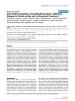
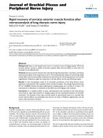
Effect of arctiin on the activation of NF-B
NF-кB proteins are present in the cytoplasm as inactive
heterodimers composed of two subunits, P50 and P65,
and are bound to the inhibitory protein IBa which pre-
vents it from the translocation into the nucleus of the
cell [16]. Upon stimulation, IBa is phosphorylat ed and
proteolytically degraded through a 26S proteasome-
mediated pathway which facilitates NF-кBtranslocation
into the nucleus and regulates gene transcription [17].
To investigate whether arctiin could affect nuclear trans-
location of NF-кB, western blot analysis of NF-кBp65
was carried out using RAW264.7 cell lysates. The
amount of NF-кB p65 was markedly increased upon
exposure to LPS alone, whereas arctiin inhibited it (Fig-
ure 6). Furthermore, we examined how arctiin modu-
lated translocation of NF- B, western blot analysis of
phosphorylation of I-Ba in cytopl asm. Arctiin signifi-
cantly attenuated IBa phosphorylation in RAW 264.7
cells (Figure 6). These results suggest that inhibition of
i-NOS, IL-1b, IL-6, TNF-a, and COX-2 gene ex pression
by arctiin may have been due to the down regulation of
NF-B activation.
Figure 3 Arctiin inhibits the production of NO (A), expression of iNOS protein (B), and mRNA (C, D) in LPS-stimulated macrophage s.
RAW 264.7 cells (A-C) and primary macrophages (D) were treated with different concentrations of arctiin (12.5 ~ 100 μg/ml) in the presence of
LPS (100 ng/ml) and was monitored as described in ‘Materials and Methods’. Each bar represents the means ± S.D. from three separate
experiments.
††
P< 0.01 compares to the control. **P< 0.01 compared to the LPS.
Lee et al. Journal of Inflammation 2011, 8:16
/>Page 5 of 9
Figure 4 Arctiin inhibits the production of pro-inflammatory cytokines in LPS-stimulated macrophages. RAW 264.7 cells (A:Western blot,
B, D, and F:ELISA) and primary macrophages (C, E, and G:ELISA) were treated with different concentrations of arctiin (12.5~100 μg/ml) in the
presence of LPS (100 ng/ml) and was monitored as described in ‘Materials and Methods’. Each bar represents the means ± S.D. from three
separate experiments.
†
P< 0.05,
††
P< 0.01 compares to the control. *P< 0.05, **P< 0.01 compared to the LPS.
Lee et al. Journal of Inflammation 2011, 8:16
/>Page 6 of 9
Discussion
Arctiin is lignin compound isolated from Forsythiae
Fructus. The anti-cancer [3,7,8] and platelet activat-
ing factor antagonistic effects [9] of arctiin have
been well documented but the mechanisms underly-
ing anti inflammatory effect are still not understood.
The present study found that arctiin significantly
inhibited the effects of LPS by suppressing a key
inflammatory pathway related to NF-B, PGE
2
and
NO production, and pro-inflammatory cytokines
expression.
Inf lammation is a reaction of t he body to injury or to
infectious, allergic, or chemical irritation. Leukocytes
destroy harmful microorganisms and dead cells, prevent-
ing the spread of the irritation and permitting the
injured tissue to repair itself. However, excessive or per-
sistent inflammation causes a variety of pathological
conditions, s uch as bacterial septic shock, and rheuma-
toid arthritis [18,19]. Inflammatory mediators (e.g. nitric
oxide and pro-inflammatory cytokines) have been
demonstrated to be critically involved in the develop-
ment of inflammatory diseases.
QXFOHDUS
S ,ț%Į
±
/3
6
QJ
PO
$UFWLLQȝJPO
±
±
ȕDFWLQ
Figure 6 Arctiin inhibits the IBa phosphorylation in cytoplasm and nuclear translocation of NF-B p65 in LPS-stimulated
macrophages. RAW 264.7 cells were treated with different concentrations of arctiin (12.5~100 μg/ml) in the presence of LPS (100 ng/ml) and
was monitored as described in ‘Materials and Methods’.
Figure 5 Arctiin inhibits the expression of co-stimulatory molecules in LPS-stimulated macrophages. RAW 264.7 cells (A-B) were treated
with different concentrations of arctiin (12.5~100 μg/ml) in the presence of LPS (100 ng/ml) and was monitored as described in ‘Materials and
Methods’. The surface B7-1 (A) and B7-2 (B) molecules were labeled with either anti-B7-1, B7-2 and the cell were stained using anti-Vb8.1+8.2-
FITC, anti-Vb2-PE, anti-Vb2-FITC (shaded histogram), which served as an isotype control for the nonspecific binding.
Lee et al. Journal of Inflammation 2011, 8:16
/>Page 7 of 9
Macrophages play importan t roles in regulating cell-
mediated immune responses, such as adaptive immune,
innate immune, and allergic reactions, as well as inflam-
mation in response to microbes and microbial products
such as LPS [20]. In addition to the well-known func-
tion of endocytosis, macrophages can be induced to
secrete nitric oxide and a series of cytokines including
TNF-a,IL-1b, IL-6, and IL-12 that express NF-B-
dependent i-NOS [21,22] and COX-2 [23]. Many dis-
eases, such as arteriosclerosis, chronic hepat itis and pul-
monary fibrosis, involve the overproduction of
inflammatory mediators [24-27] and t hus, inhibiting
their production may serve to prevent or suppress a
variety of inflammatory diseases, including rheumatoid
arthritis (RA), sepsis, and endotoxemia. Despite the
exact cause of autoimmune disease remaining obscure,
deregulated overproduction of pro-inflammatory cyto-
kines and a disruption in the regulation of cytokine sig-
nal transduction have been indicated as underlying
mechanisms of some autoimmune diseases such as RA
and Crohn’s disease [28,29].
Nitric oxide is synthesized via the oxidation of argi-
nine by a family of NOS, and it plays a vital role in reg-
ulating physiological processes, such as blood vessel
tone and neurotransmission, as well as in host defense
and immunity [30,31]. Pro-inflammatory cytokines, IL-
1b,IL-6,andTNF-a, have attracted more attention in
that they can be localized to the infe cted tissue, mani-
fested systemically throughout the body, and cause vaso-
dilation as well as symptoms of inflammation [32-34].
Our findings that arctiin inhibits the formation of NO
and pro-infl ammat ory cytokines showed the importance
of arctiin as an anti-inflammatory compound. The
reduced NO production by arctiin was a consequence of
an inhibition of iNOS, the key enzyme responsible for
NO production under pathological conditions.
PGE
2
plays major roles in the angiogenesis of syno-
vium through the expression of vascular endothelial
growth factors [35], s ynovial inflammation, and joint
erosion in RA [36]. Further, in the prostaglandin b io-
synthesis pathway, COX-2 is the key enzyme that cata-
lyzes the conversion of arachidonic acid to PGE
2
.Itis
generally accepted that PGE
2
is produced by COX-2 at
sites of inflammation, and that COX-1, another consti-
tutive isoform, is relevant in the production of prosta-
glandins that regulate normal cellular processes such as
vascular homeostasis regulation, gastric mucosal protec-
tion and renal integrity maintenance [37]. In the present
study, we found that arctiin suppressed PGE
2
produc-
tion via inhibition of COX-2 enzyme activity and this
may in part be responsible for some of anti-inflamma-
tory properties of this compound.
The transcriptio nal factor NF-B is import ant for the
expression of immune and inflammatory genes. The
activated NF-BthenbindsB motifs in the pro moters
via its p65 subunit, leading to expression of several
inflammatory genes. Because NF-B transcription fac-
torsareuniquelypositioned downstream of multiple
innate and adaptive signaling pathways, they seem ide-
ally placed to integrate and coordinate innate and adap-
tive signals required for formation of productive
immune responses. In the present study, we found that
arctiin could inhibit the NF-B nucleus translocation
induced by LPS through a reduction in IB phosphory-
lation status.
The B7 family is related immunoglobulin supergene
family members that are expressed by multiple cell
types involved in antigen presentation. Both B7-1 and
B7-2 are constitutively expressed on dendritic cells and
are regulated on monocytes, macrophages, B cells, and
T cells following activation . In agreement with our find-
ings, arctiin has also been shown to suppress co-stimu-
latory molecules such as B7-1 and B7-2 in macrophages.
In summary, these results suggest that arctiin has anti-
inflammatory effects on macrophages through the
reduced pro-inflammatory cytokines are associated with
NF-B inactiv ation and the suppression of NF-kB-regu -
lated proteins, and other bioactive substan ces as well as
through inhibition of the expression of co-stimulator y
molecules.
Acknowledgements
This study was supported by the Sahmyook University Research fund in
2010.
Author details
1
College of Pharmacy, SahmYook University, Seoul 139-742, Republic of
Korea.
2
School of Life Sciences, Handong Global University, Pohang 791-708,
Republic of Korea.
3
College of Pharmacy, Chungbuk National University,
Cheongju 361-763, Republic of Korea.
Authors’ contributions
SL participated in the design of this study and performed the statistical
analysis in whole research. SS carried out the Western blot assay of iNOS,
COX-2, and proinflammatory cytokines. HK carried out the RT-PCR assay. SH
carried out the FACS analysis in B7 family. KK carried out the preparation of
peritoneal macrophages, participated in the maintenance of SPF room and
care of animal. JK carried out the cytokine assay (ELISA). JHK participated in
the design of the study, and proofread a manuscript about in vitro
experiments design. CKL participated in the design of the study, and
proofread a manuscript about in vivo experiments design. NJH participated
in the test of toxicity of this compound, and proofread a manuscript about
cell toxicity. DY carried out the separation of arctiin from Forsythiae Fructus.
KK conceived of the study, and participated in its design and coordination.
All authors read and approved the final manuscript.
Competing interests
The authors declare that they have no competing interests.
Received: 17 June 2010 Accepted: 7 July 2011 Published: 7 July 2011
References
1. Lee GI, Ha JY, Min KR, Nakagawa H, Tsurufuji S, Chang IM: Inhibitory effects
of oriental herbal medicines on IL-8 induction in lipopolysaccharide-
activated rat macrophages. Planta Med 1995, 61(1):26-30.
Lee et al. Journal of Inflammation 2011, 8:16
/>Page 8 of 9
2. Kim TJ: Korean resources plants. Seoul;, first 1991.
3. Huang DM, Shen YC, Wu C, Huang YT, Kung FL, Teng CM, Guh JH:
Investigation of extrinsic and intrinsic apoptosis pathways of new
clerodane diterpenoids in human prostate cancer PC-3 cells. Eur J
Pharmacol 2004, 503(1-3):17-24.
4. Macrae WD, Tower GH: Biological activities of Lignans. Phytochemistry
1984, 23:1207-1220.
5. Morita K, Nishijima Y, Kada T: Chemical nature of a desmutagenic factor
from Burdock (Arctium lappa Linne). Agr Biol Chem 1985, 49:925-932.
6. Kazuki S, Seikichi K, Misao M, Zwe-Ling K, Hiroshi H: Antimutagenicity of
Dialyzates of Vegetables and Fruits (Food & Nutrition). Agr Biol Chem
1988, 52:1369-1375.
7. Ryu S, Ahn J, Kang Y, Han B: Antiproliferative effect of arctigenin and
arctiin. Arch Pharmacol Res 1995, 18:462-463.
8. Moritani S, Nomura M, Takeda Y, Miyamoto K: Cytotoxic components of
bardanae fructus (goboshi). Biol Pharm Bull 1996, 19:1515-1517.
9. Iwakami S, Wu JB, Ebizuka Y, Sankawa U: Platelet activating factor (PAF)
antagonists contained in medicinal plants: lignans and sesquiterpenes.
In Chem Pharm Bull. Volume 40. Tokyo; 1992:1196-1198.
10. Ichikawa K, Kinoshita T, Nishibe S, Sankawa U: The Ca2+ activity of lignans.
Chem Pharm Bull 1986, 34:3514-3517.
11. Liu S, Chen K, Schliemann W, Strack D: Isolation and identification of
arctiin and arctigenin in leaves of burdock (Arctium lappa L.) by
polyamide column chromatography in combination with HPLC-ESI [sol]
MS. Phytochem Anal 2005, 16:86-89.
12. Minagar A, Shapshak P, Fujimura R, Ownby R, Heyes M, Eisdorfer C: The
role of macrophage/microglia and astrocytes in the pathogenesis of
three neurologic disorders: HIV-associated dementia, Alzheimer disease,
and multiple sclerosis. J Neurol Sci 2002, 202:13-23.
13. Stevens DL: Immune modulatory effects of antibiotics. Curr Opin Infec Dis
1996, 9:165-169.
14. Nathan C: Nitric oxide as a secretory product of mammalian cells. FASEB
J 1992, 6:3051-3064.
15. Sharpe AH, Freeman GJ: The B7-CD28 superfamily. Nat Rev Immunol 2002,
2:116-126.
16. Gomez PF, Pillinger MH, Attur M, Marjanovic N, Dave M, Park J: Resolution
of inflammation: prostaglandin E2 dissociates nuclear trafficking of
individual NF-kappaB subunits (p65, p50) in stimulated rheumatoid
synovial fibroblasts. J Immunol 2005, 175:6924-6930.
17. Strayhorn WD, Wadzinski BE: A novel in vitro assay for deubiquitination of
I kappa B alpha. Arch Biochem Biophys 2002, 400:76-84.
18. Dinarello C: Proinflammatory and anti-inflammatory cytokines as
mediators in the pathogenesis of septic shock. Chest 1997,
112:321S-329S.
19. Palladino M, Bahjat F, Theodorakis E, Moldawer L: Anti-TNF-alpha
therapies: the next generation. Nat Rev Drug Discov 2003, 2:736-746.
20. Firestein GS: Invasive fibroblast-like synoviocytes in rheumatoid arthritis.
Passive responders or transformed aggressors? Arthritis Rheum 1996,
39:1781-1790.
21. Nathan C: Inducible nitric oxide synthase: what difference does it make?
J Clin Invest 1997, 100:2417-2423.
22. Bogdan C: Nitric oxide and the immune response. Nat Immunol 2001,
2:907-916.
23. Vila-del Sol V, Fresno M: Involvement of TNF and NF-kappa B in the
transcriptional control of cyclooxygenase-2 expression by IFN-gamma in
macrophages. J Immunol 2005, 174:2825-2833.
24. Isomaki P, Punnonen J: Pro- and anti-inflammatory cytokines in
rheumatoid arthritis. Ann Med 1997, 29:499-507.
25. Libby P, Ridker P, Maseri A: Inflammation and atherosclerosis. Am Heart
Assoc 2002, 150:1135-1143.
26. Tilg H, Wilmer A, Vogel W, Herold M, Nolchen B, Judmaier G: Serum levels
of cytokines in chronic liver diseases. Gastroenterology 1992, 103:264-274.
27. Coker RK, Laurent GJ: Pulmonary fibrosis: cytokines in the balance. Eur
Respir J 1998, 11:1218-1221.
28. Chenevier-Gobeaux C, Morin-Robinet S, Lemarechal H, Poiraudeau S,
Ekindjian JC, Borderie D: Effects of pro- and anti-inflammatory cytokines
and nitric oxide donors on hyaluronic acid synthesis by synovial cells
from patients with rheumatoid arthritis. Clin Sci (Lond) 2004, 107:291-296.
29. Nishimoto N: Cytokine signal regulation and autoimmune disorders.
Autoimmunity 2005, 38:359-367.
30. Furchgott R, Cherry P, Zawadzki J, Jothianandan D: Endothelial cells as
mediators of vasodilation of arteries. J Cardiovasc Pharmacol 1984, 6:
S336-343.
31. Wei X, Charles I, Smith A, Ure J, Feng G, Huang H: Altered immune
responses in mice lacking inducible nitric oxide synthase. Nature 1995,
375
:408-4011.
32. Haddad JJ, Safieh-Garabedian B, Saadé NE, Kanaan SA, Land SC:
Chemioxyexcitation (delta pO2/ROS)-dependent release of IL-1 beta, IL-6
and TNF-alpha: evidence of cytokines as oxygen-sensitive mediators in
the alveolar epithelium. Cytokine 2001, 13:138-147.
33. Moldoveanu A, Shephard R, Shek P: Exercise elevates plasma levels but
not gene expression of IL-1β, IL-6, and TNF-α in blood mononuclear
cells. J Appl Physiol 2000, 89:1499-1504.
34. Ben-Av P, Crofford L, Wilder R, Hla T: Induction of vascular endothelial
growth factor expression in synovial fibroblasts by prostaglandin E and
interleukin-1: a potential mechanism for inflammatory angiogenesis.
FEBS Lett 1995, 372:83-87.
35. Dayer J: Prostaglandin production by rheumatoid synovial cells:
stimulation by a factor from human mononuclear Cells. J Exp Med 1977,
145:1399-1404.
36. Lecomte M, Laneuville O, Ji C, DeWitt D, Smith W: Acetylation of human
prostaglandin endoperoxide synthase-2 (cyclooxygenase-2) by aspirin. J
Biol Chem 1994, 269:13207-13215.
37. Vane J, Bakhle Y, Botting R: Cyclooxygenases 1 and 2. Annu Rev Pharmacol
Toxicol 1998, 38:97-120.
doi:10.1186/1476-9255-8-16
Cite this article as: Lee et al.: Anti-inflammatory function of arctiin by
inhibiting COX-2 expression via NF-B pathways. Journal of Inflammation
2011 8:16.
Submit your next manuscript to BioMed Central
and take full advantage of:
• Convenient online submission
• Thorough peer review
• No space constraints or color figure charges
• Immediate publication on acceptance
• Inclusion in PubMed, CAS, Scopus and Google Scholar
• Research which is freely available for redistribution
Submit your manuscript at
www.biomedcentral.com/submit
Lee et al. Journal of Inflammation 2011, 8:16
/>Page 9 of 9