Báo cáo khoa hoc:" No evidence of enhanced oxidant production in blood obtained from patients with obstructive sleep apnea" potx
Bạn đang xem bản rút gọn của tài liệu. Xem và tải ngay bản đầy đủ của tài liệu tại đây (328.12 KB, 11 trang )
BioMed Central
Page 1 of 11
(page number not for citation purposes)
Journal of Negative Results in
BioMedicine
Open Access
Research
No evidence of enhanced oxidant production in blood obtained
from patients with obstructive sleep apnea
Izabela Grabska-Kobylecka
1
, Andrzej Kobylecki
1
, Piotr Bialasiewicz
1
,
Maciej Krol
1
, Golsa Ehteshamirad
1
, Marek Kasielski
2
and Dariusz Nowak*
1
Address:
1
Sleep and Respiratory Disorders Center of the Chair of Experimental and Clinical Physiology, Medical University of Lodz, 92-215 Lodz,
Mazowiecka St. 6/8, Poland and
2
Bases of Clinical Medicine Teaching Center, Medical University of Lodz, 90-153 Lodz, Kopcinskiego St. 20,
Poland
Email: Izabela Grabska-Kobylecka - ; Andrzej Kobylecki - ; Piotr Bialasiewicz - ;
Maciej Krol - ; Golsa Ehteshamirad - ; Marek Kasielski - ;
Dariusz Nowak* -
* Corresponding author
Abstract
Background: Obstructive sleep apnea syndrome (OSAS) is a recognized risk factor for
cardiovascular morbidity and mortality, perhaps due to causative exacerbations of systemic
oxidative stress. Putative oxidative stress related to numerous episodes of intermittent hypoxia,
may be an oxidants chief driving force in OSAS patients.
Methods: We assessed the resting and n-formyl-methionyl-leucyl-phenylalanine (fMLP)- induced
whole blood chemiluminescence (as a measure of oxidant production by polymorphonuclear
leukocytes and monocytes), ferric reducing ability of plasma (FRAP) and H
2
O
2
generation in the
whole blood of 27 untreated OSAS patients, 22 subjects after a night of CPAP therapy and 11
controls without OSAS. All of them were matched to age, BMI (body mass index) and smoking
habits. All parameters were measured before and after polysomnography-controlled sleep,
individual results were obtained as a mean from duplicated experiments.
Results: No significant differences were distinguished between evening and morning blood
chemiluminescence, H
2
O
2
activity and FRAP within and between all three study groups.
For instance patients with untreated OSAS had similar morning and evening resting whole blood
chemiluminescence (2.3 +/- 2.2 vs. 2.4 +/- 2.2 [aU·10
-4
phagocytes]), total light emission after
stimulation with fMLP (1790 +/- 1371 vs. 1939 +/- 1532 [aU·s·10
-4
phagocytes]), as well as FRAP
after 3 min. plasma incubation (602 +/- 202 vs. 671 +/- 221 [uM]). Although, in the subgroup of 11
patients with severe OSAS (apnea/hypopnea index 58 +/- 18/h and oxygen desaturation index 55
+/- 19/h), the morning vs. evening resting chemiluminescence and total light emission after
stimulation with fMLP observed a propensity to elevate 2.5 +/- 2.7 vs. 1.9 +/- 1.8 [aU·10
-4
phagocytes] and 1778 +/- 1442 vs. 1503 +/- 1391 [aU·s·10
-4
phagocytes], respectively, these did not
attain statistical significance (p > 0.05).
Conclusion: Our investigation exposed no evidence in the overproduction of oxidants via
circulating phagocytes, once considered a culprit in the oxidative stress of OSAS patients.
Published: 25 November 2008
Journal of Negative Results in BioMedicine 2008, 7:10 doi:10.1186/1477-5751-7-10
Received: 11 May 2008
Accepted: 25 November 2008
This article is available from: />© 2008 Grabska-Kobylecka et al; licensee BioMed Central Ltd.
This is an Open Access article distributed under the terms of the Creative Commons Attribution License ( />),
which permits unrestricted use, distribution, and reproduction in any medium, provided the original work is properly cited.
Journal of Negative Results in BioMedicine 2008, 7:10 />Page 2 of 11
(page number not for citation purposes)
Background
The presence of obstructive sleep apnea syndrome (OSAS)
is strongly associated with augmented morbidity and
mortality from cardiovascular diseases including arterial
hypertension, cardiac arrhythmias and ischemic heart dis-
ease [1,2]. OSAS is also considered to be a risk factor of
stroke and sudden cardiac death [3,4]. There appears to be
practicable links between OSAS and occurrences such as
apnea-induced intermittent hypoxia (IH) of tissues, sym-
pathetic over activities during sleep [5,6] as well as puta-
tive oxidative stress in relation to the systemic
inflammatory response [6,7]. Primed and or activated cir-
culating polymorphonuclear leukocytes (PMNs) and
monocytes can be a source of reactive oxygen species
(ROS) in OSAS patients [7]. Indeed, the in vitro incuba-
tion of whole blood under decreased partial oxygen pres-
sure (pO
2
≤ 46 mm Hg) resulted in the degranulation of
PMNs [8] and increased ROS production [9].
Moreover, healthy volunteers subjected to 20 min hypo-
baric hypoxia in a decompression chamber presented
with elevated production of ROS by PMNs [10].
Due to repeated apnea and/or hypopnea episodes, noctur-
nal PaO
2
can fall below 50 mmHg in OSAS patients
[11,12] and thus favor activation and enhance ROS pro-
duction by means of blood phagocytes. Scanty and con-
flicting data concerning ROS production by blood
phagocytes in the course of OSAS have been published so
far [13-16]. Müns et al. did not establish any alterations in
ROS production accompanying ingestion of Escherichia
coli by PMNs obtained from OSAS patients [13]. Other
researchers have illustrated that increasing the agonist n-
formyl-methionyl-leucyl-phenylalanine (fMLP), induced
production of superoxide radicals in isolated PMNs from
OSAS patients [14]. However, the limitation of that study
attributes to the control groups' significant difference in
respect to age, body mass index (BMI) as well as habitual
cigarette smoking. In addition, the control group included
cancer patients and healthy subjects not matched to OSAS
patients with comorbidity [14]. In another study Dyugov-
skaya et al. [15] identified subpopulations of monocytes
and PMNs in OSAS patients (using the flow cytometry
technique) producing more ROS than the cells from the
control group. Yet, in this study, the control group also
differed with regard to BMI, comorbidity and concomi-
tant pharmacological treatment. On the other hand, the
significant suppression of fMLP- induced respiratory burst
of isolated PMNs was reported in patients with severe
OSAS using the chemiluminescence technique [16-18].
Taking into account the divergences between these stud-
ies, we decided to monitor ROS production by PMNs and
monocytes in both untreated- and CPAP-treated OSAS
patients and age-, BMI- and cigarette-smoking habit-
matched controls (volunteers without OSAS). Two addi-
tional variables reflecting oxidant/antioxidant imbalances
were determined: the H
2
O
2
activity in the whole blood
[19] and the ferric reducing ability of plasma (FRAP) [20].
We found that the apnea/hypopnea episodes during sleep
did not change the morning and evening intensity of the
resting as well as fMLP-induced LBCL, FRAP and blood
H
2
O
2
activities in the OSAS patients. These suggest a lack
of apparent ROS overproduction via circulating PMNs in
OSAS patients.
Methods
Chemicals and solutions
Luminol, horseradish peroxidase (HRP, 222 I.U./mg),
2,4,6-tripyridyl-s-triazine (TPTZ), bovine catalase (2440
I.U./mg of solid substance), n-formyl-methionyl-leucyl-
phenylalanine (fMLP), FeCl
3
·6H
2
O and dimethyl sul-
phoxide (DMSO) were obtained from Sigma Chemical
Co. (St. Louis, MO, USA). Phenol red and phosphate buff-
ered saline (PBS, pH 7.4) came from ICN Biomedicals Inc
(Aurora, OH, USA) and Biomed (Lublin, Poland). All
other reagents were of analytical grade and were pur-
chased from the POCH (Gliwice, Poland). Stock solutions
of 1.4 M luminol in 0.1 M phosphate buffer (pH 7.4), 20
mM fMLP in DMSO, catalase in 0.9% NaCl (50 I.U/μl)
and HRP in 50 mM phosphate buffer (0.925 I.U/μl, pH
7.0), 10 mM TPTZ in 40 mM HCl, and 0.028 M phenol
red in water were stored in single use aliquots in the dark
at -70°C. fMLP was diluted just before use with 0.9%
NaCl to a final concentration 0.2 mM. Sterile, deionized,
pyrogen-free water for HPLC (resistance > 18 Ωcm, Water
Purification System USF ELGA, Buckinghamshire, UK)
was used throughout the study.
Study population and polysomnography
Sixty patients that underwent polysomnography at the
Sleep Laboratory were studied. Thirty-eight patients
underwent diagnostic polysomonography following a
preliminary diagnosis of OSAS. The remaining 22, previ-
ously diagnosed with OSAS met indication for continuous
positive airway pressure (CPAP) treatment.
The inclusion criteria incorporated an age span from 40 to
70 years along with a written informed consent. The
exclusion criteria involved pregnancy, presence of any
active infectious or inflammatory process, chronic
obstructive pulmonary disease (COPD), unstable angina,
uncontrolled hypertension or hypertension diagnosed
within the last three months, insulin-dependent diabetes,
and any surgery within the last three months, or treatment
with antibiotics, nonsteriodal anti-inflammatory drugs,
vitamins as well as any food supplements with antioxi-
dant potential within the preceding two weeks. All partic-
ipants, except for patients treated with CPAP, underwent
a standard overnight (7 hours from about 11:00 pm to
6:00 am) polysomnography (SleepLabPro, Jaeger, VIASYS
Journal of Negative Results in BioMedicine 2008, 7:10 />Page 3 of 11
(page number not for citation purposes)
Healthcare Hoechberg, Germany). This entailed an EEG,
electrooculography, chin muscles as well as an anterior
tibial electromyography (EMG), a unipolar EKG, snoring
detection, body position, measurement of oronasal air-
flow along with abdominal and chest respiratory move-
ments as previously described [21]. Participants
previously diagnosed with OSAS, underwent a similar
standard polysomnography trial CPAP treatment (CPAP
Respironics, RemStar Plus, Murrysville, Pennsylvania,
USA) along with a mask pressure monitoring as a substi-
tute in measurement of oronasal flow. The polysomnog-
raphy enabled differentiation in two groups of patients-
the OSAS group – 27 patients with OSAS (apnea/hypop-
nea index, AHI>5) including the subgroup of 11 patients
with severe OSAS (AHI ≥ 30), from patients without OSAS
(AHI<5) who served as a control group (n = 11). The third
group (CPAP-OSAS group) involved OSAS patients (n =
22), successful at the first attempt of CPAP treatment
(Table 1, 2, and 3).
Study Design
All participants were admitted to the Sleep Laboratory at
approximately 8:30 p.m., previously instructed to con-
sume a light final meal before 7:00 p.m. Venous blood
was collected twice for blood cell count and measurement
of oxidative stress markers – before and after polysomnog-
raphy-controlled sleep, at about 9:30 p.m. and 6:00 a.m.
(just after wakening up), respectively. Nine ml of venous
blood were drawn into sodium heparin vacuette tubes
(placed in ice-cold water) and EDTA-K
3
vacuette-tubes
(Greiner bio-one GmbH, Kremsmunster, Austria). The
heparinized blood was used for measuring whole blood
Table 1: Demographics of investigated groups
Subjects that underwent polysomnography
Parameter OSAS group, n = 27 Severe OSAS subgroup, n = 11 CPAP-OSAS group, n= 22 Controls, n = 11
Male/Female 25/2 10/1 17/5 9/3
Smokers 15 0 15 6
Age [yrs] 53 ± 13 55 ± 15 58 ± 8 50 ± 10
BMI [kg/m
2
] 31.1 ± 5.1 32.1 ± 6.5 34.3 ± 7.3 28.8 ± 5.5
NC [cm] 42.6 ± 2.6 43.7 ± 2.9 43.7 ± 4.1 41.5 ± 2.0
ESS 11 ± 1 11 ± 5 15 ± 1* 9 ± 1
TST [h] 4.8 ± 1.2 4.7 ± 1.3 5.2 ± 1.6 5.3 ± 1.0
AHI [n/h] 31 ± 5 58 ± 18 † 9 ± 2* 2 ± 1*
ODI [n/h] 31 ± 5 55 ± 19 † 9 ± 3* 2 ± 1*
Mean SaO
2
[%] 87 ± 1 84 ± 5 † 89 ± 1 91 ± 1*
Min SaO
2
[%] 74 ± 3 68 ± 15 † 81 ± 2 88 ± 1*
T
SaO2
<88% [min] 55 ± 12 99 ± 74 † 12 ± 4* 2 ± 1*
Snoring [%TST] 29 ± 24 28 ± 24 10 ± 14* 20 ± 25
OSAS – patients with untreated OSAS; Severe OSAS – patients with AHI ≥ 30; CPAP-OSAS – patients with OSAS treated successfully at a first
attempt with CPAP, controls – subjects without OSAS; BMI – body mass index; NC – neck circumference; ESS – Epworth sleepiness score; TST –
total sleep time; AHI (apnea/hypopnea index) – the number of apneas and hypopneas per hour of sleep; ODI (oxygen desaturation index) – the
number of desaturations ≥ 4% per hour of sleep; SaO
2
-nocturnal oxygen saturation; T
SaO2
– duration of nocturnal oxygen saturation < 88%.
Polysomnography data expressed as a mean ± SD. The daily cigarette consumption was 19 ± 12, 21 ± 13, and 17 ± 6 for current cigarette smokers
in OSAS, CPAP-OSAS and control groups respectively. Blood for chemiluminescence measurement and other determinations was taken before and
after polysomnographic controlled sleep.
* – p < 0.05 vs. OSAS group, † – p < 0.05 vs. CPAP-OSAS and controls groups.
Selected baseline characteristics of CPAP-OSAS patients before treatment initiation were: AHI 58 ± 29; ODI 56 ± 26; mean SaO
2
82 ± 7 %, min
SaO
2
75 ± 9 %, and T
SaO2
< 88% 105 ± 90 min. of 5.2 ± 1.7 h TST.
Journal of Negative Results in BioMedicine 2008, 7:10 />Page 4 of 11
(page number not for citation purposes)
H
2
O
2
activity, followed by its addition to other reagents
within 10 seconds from collection. Luminol enhanced
whole blood chemiluminescence (LBCL) measurement
procedure (EDTA-K
3
blood samples with determined
blood cell count) began no later than 30 min. following
blood collection. Plasma samples obtained from EDTA-K
3
blood (30 min incubation at 37°C, subsequent centrifu-
gation for 10 min at 1500·g and 4°C) were stored at -
70°C for no longer than 3 weeks until FRAP determina-
tion. Blood cell count was measured with Micros Analyzer
OT 45 (ABX, Montpellier, France). In the event of an at
night awakenings, only plain mineral water was allowed
to be drunk ad libitum. The Medical University of Lodz
Ethics Committee approved the study protocol and all
participants provided a written, informed consent.
Luminol enhanced whole blood chemiluminescence assay
A luminol enhanced whole blood chemiluminescence
(LBCL) technique was employed as a measure of resting
and agonist induced ROS production by circulating
phagocytes [17,18] in order to avoid any priming and/or
activation of oxidative cell response due to isolation pro-
cedures [18]. Moreover, to avoid any possible bias related
to patients' interindividual variability and differences in
comorbidity and pharmacological treatment, LBCL was
measured before (evening) and just after polysomnogra-
Table 2: Comorbidity in the investigated patient groups
Disease Number of patients with a given disease
OSAS n = 27 CPAP-OSAS n = 22 Controls n = 11
Arterial hypertension 16 16 4
Ischemic heart disease 7 7 2
Diabetes 2 4 0
Gout 0 4 0
OSAS – patients with untreated OSAS; CPAP-OSAS – patients with OSAS treated successfully after a first attempt with CPAP, controls – subjects
without OSAS.
Table 3: Ongoing pharmacological treatment in studied groups
Pharmacological treatment Number of patients receiving treatment
OSAS, n = 27 CPAP-OSAS, n = 22 Controls, n = 11
ACEI 12 10 2
Diuretics 11 6 1
Ca
2+
channel blocker 4 5 1
Beta-blockers 4 1 1
Nitrates 4 4 1
Digitalis 3 1 0
Statins 8 6 0
Allopurinol 0 4 0
Ticlopidine 2 0 1
Gliclazide 1 5 0
ACEI – angiotensin converting enzyme inhibitors; OSAS – patients with untreated OSAS; CPAP-OSAS – patients with OSAS treated successfully
after a first attempt with CPAP, controls – subjects without OSAS. Four, 2, and 6 subjects were free of any medication in OSAS, CPAP-OSAS and
controls group, respectively.
Journal of Negative Results in BioMedicine 2008, 7:10 />Page 5 of 11
(page number not for citation purposes)
phy-controlled sleep (morning) in a matched manner.
Therefore, evening results served as reference values
designed for morning data, presumably affected by
apnea/hypopnea episodes in addition to subsequent IH
during sleep.
LBCL as a measure of resting and fMLP-stimulated circu-
lating phagocytes (PMNs and monocytes) ability to pro-
duce ROS was determined according to Kukovetz et al.
[17] with some modifications [22]. Briefly, 3 μl of blood
sample was added to 947 μl of mixture solution (com-
posed of 1 ml sterile Ringer solution, 200 μl 5% D-glucose
solution, 3.6 ml deionized water and 5 ml 1.4 M luminol
solution) which was pre-warmed in darkness up to 37°C
for 60 min. The samples were placed in the 1251 lumi-
nometer (Bio-Orbit, Turku, Finland) and incubated for 30
min. at 37°C. Afterwards the resting chemiluminescence
was recorded continuously for 1 min. and then 50 μl fMLP
solution was added automatically to a final concentration
of 0.02 mM, with continuation of the light emission
measurement for an additional 7 min. All individual
results were obtained as a mean of four measurements
with LBCL parameters assessing resting chemilumines-
cence (rCl) – the average resting chemiluminescence prior
to the addition of fMLP; peak chemiluminescence (pCL)
– maximal chemiluminescence signal after the addition of
fMLP. Total light emission (tCL) – the area under the
chemiluminescence intensity curve after the addition of
fMLP until its return to baseline and peak time – the time
(seconds) from the addition of fMLP to the appearance of
pCL were also assessed. rCL and pCL were expressed in
arbitrary units (aU) per 10
4
phagocytes (PMNs and mono-
cytes) present in the assayed sample, while tCL in aU·s/
10
4
phagocytes. Preliminary experiments with platelet
rich plasma excluded the contribution of platelets to
fMLP-evoked LBCL (data not shown).
The H
2
O
2
activity in the whole blood
H
2
O
2
activity was measured in whole blood using the
phenol red oxidation method [19,23] with some modifi-
cations. Briefly, 600 μl of blood was either added to 60 μl
0.2 M NaN
3
solution or 60 μl of bovine catalase solution
(3000 I.U. per sample) in 0.9% NaCl. Both tubes were
subsequently incubated for 5 min at 37°C and then a 30
μl 0.028 M solution of phenol red in deionized water with
a 30 μl (27.75 I.U. per sample) of HRP solution in 50 mM
phosphate buffer (pH = 7.0) were then added to each test-
tube. Afterwards, the samples were incubated for 10 min
at 37°C and then centrifuged (10 min., 1500·g). One
hundred μl of supernatant was transferred into a cuvette
containing 900 μl PBS and 10 μl 1 M NaOH reading
(spectrophotometer Ultrospec III, LKB Biochrom Eng-
land) its absorbance at 610 nm (A
610
). The calibration
curve was made with 600 μl samples of standard H
2
O
2
solutions (increasing concentrations from 0.1 to 12 μM,
13 concentration points) in PBS added to 60 μl 0.2 M
NaN
3
solution and subsequently processed in the same
way as blood specimens. The concentration of H
2
O
2
(μM)
in the blood specimens was calculated according to the
regression equation: y = 33.12(x
1
-x
2
) – 0.23 (r = 0.97, p <
0.001) where y was the H
2
O
2
concentration and x
1
-x
2
was
the difference between the A
610
of a sample with NaN
3
(x
1
)
and A
610
of a sample with catalase (x
2
) being the blank (all
H
2
O
2
was decomposed by the enzyme). The method sen-
sitivity was 0.25 μM of H
2
O
2
. Individual results were
obtained as the mean of two measurements.
Ferric reducing ability of plasma
FRAP ascertainment was measured following the proce-
dure originally described by Benzie and Strain [20] with
some modifications [24], in which Fe
3+
to Fe
2+
ion reduc-
tion at low pH caused the formation of a coloured ferrous-
TPTZ complex, resulting in an increase in absorbance at
593 nm (A
593
). Briefly, 30 μl of plasma was mixed with 90
μl of deionized water and then added to 900 μl of a FRAP
reagent (pre-warmed to 37°C) while sample absorbance
at 593 nm was continuously measured over 8 min. at
37°C (Ultrospec III, with a Spectro-Kinetics software).
Control samples (blank) received 120 μl of water. FRAP
reagent was prepared just before the assay by adding in the
following order: 10 ml 300 mM acetate buffer (pH 3.6), 1
ml 10 mM TPTZ in 40 mM HCl, and 1 ml 20 mM aqueous
FeCl
3
solution. Calibration was performed with a FRAP
reagent containing the addition of FeCl
2
(total sample vol-
ume 1.02 ml, final concentrations from 20 to 2000 μM, 9
concentration points). Absorbance was linear (r = 0.98, p
< 0.001). Intra- and inter- assay coefficients of variations
tested on 10 aliquots of pooled plasma from 10 healthy
donors were less than 8%. Individual results were
obtained from duplicate experiments and were expressed
as a concentration of Fe
3+
ions reduced into Fe
2+
after 0, 1,
2, 3, 4, 5, 6, 7 and 8 min. incubation of plasma sample
with FRAP reagent. Calculations were done according to
the formula: Y [μM] = 1687.9 X – 3.3, where Y – concen-
tration of Fe
3+
reduced ions, X – difference between A
593
(assayed sample) and A
593
(blank).
In vitro effect of H
2
O
2
on FRAP
Nine hundred and fifty μl of pooled plasma samples
(obtained from 10 healthy donors) were mixed with 50 μl
of deionized water or 50 μl of various H
2
O
2
solutions and
were incubated for 5, 30 and 60 min at 37°C. The final
concentration of H
2
O
2
in plasma samples were 0 μM, 47
μM, 16.5 mM, and 82.5 mM, respectively. Then the sam-
ples were centrifuged (5 min., 1500·g, 4°C) and 30 μl of
plasma after mixing with 90 μl of water was added to 0.9
ml of FRAP reagent. A
593
was recorded over a 10 min.
incubation period at 37°C while the concentration of
reduced Fe
3+
ions was calculated as above.
Journal of Negative Results in BioMedicine 2008, 7:10 />Page 6 of 11
(page number not for citation purposes)
Statistical analysis
All data were expressed, dependent on the distribution, as
the mean and standard deviation (SD) and/or median
and quartile range. Shapiro-Wilk W test was used for nor-
mality testing. The differences between groups (normal
data) were assessed using an analysis of variance for inde-
pendent variables. Repeated measures ANOVA was
applied for dependent variables (evening vs. morning).
For data not normally distributed, the Kruskal-Wallis test
and Friedman test were used. In the case of significance,
appropriate post-hoc tests were implicated. An additional
comparison between two selected parameters with an
abnormal distribution was conducted using the Wilcoxon
test (for dependent variables) and the U Mann-Whitney
test (for independent variables). The t-student test for
dependent variables was used in comparison of two nor-
mally distributed parameters. A p value of < 0.05 was con-
sidered significant. For differences between means, the
95% confidence intervals were calculated.
Results
White blood cells, phagocytes and hematocrit
In all three investigated groups, morning white blood cell
counts were lower as compared to the evening counts (p <
0.05). Evening and morning quantities of PMNs collec-
tively with monocytes differed significantly (p < 0.05).
Indistinguishable manifestations arose in the subgroup of
11 patients with severe OSAS (data not shown). This
explains why the LBCL was expressed per 10
4
phagocytes
despite the relatively diminutive time-interval between
evening and morning blood collections, in addition to the
data analysis of LBCL as a dependent variable. Conversely,
hematocrit was significantly unchanged (p > 0.05) (Table
4). Therefore, correction of plasma volume changes in
FRAP and H
2
O
2
activities in the morning data was unnec-
essary.
Luminol enhanced whole blood chemiluminescence
No analyzed parameters of LBCL (rCL, pCL, tCL and peak-
time) altered significantly after sleep in patients with
OSAS and controls (Table 5). There were also no signifi-
cant differences between all three of the investigated
groups in respect to these parameters. Unpredictably, in
all groups, the tendency (although, insignificant) to
present a decreased LBCL (rCL, pCL and tCL) subsequent
to sleep was prominent.
Furthermore, patients with severe untreated OSAS (n =
11) did not reveal any differences between evening and
morning LBCL (p > 0.05). However, in the subgroup, the
observance of the reverse tendency to increase morning
LBCL parameters is notable (Table 5).
Ferric reducing ability of plasma
The 3 min. incubation of a plasma sample with a FRAP
reagent (containing Fe
3+
and TPTZ) is recommended in
receiving the most reliable results in plasma antioxidant
activity [20]. Table 6 shows this data after 3 min. incuba-
tion with no significant differences noted between
evening and morning plasma FRAP within all of the ana-
lyzed groups. There were also no significant differences
between the corresponding values found in OSAS, CPAP-
Table 4: White blood cells, phagocytes and hematocrit
OSAS CPAP-OSAS CONTROLS
Hematocrit [%] Evening 39.2 ± 3.8 40.8 ± 4.8 37.4 ± 5.5
Morning 38.6 ± 4.9 40.5 ± 6.1 38.6 ± 5.7
Diff & 95% CI 0.6 (-0.78–1.95) 0.3 (-0.74–1.38) 1.2 (0.42–1.99)
WBC [10
3
/μl] Evening 8.06 ± 2.00 8.01 ± 2.30 8.61 ± 2,00
Morning 7.20 ± 1.91 7.26 ± 2.23 7.67 ± 2.92
Diff & 95% CI 0.86 (0.86–1.18) 0.75 (0.75–1.29) 0.94 (0.46–1.65)
PMNs+monocytes [10
3
/μl] Evening 5.68 ± 1.67 5.75 ± 2.00 5.50 ± 2.26
Morning 5.13 ± 1.63 5.19 ± 1.82 4.67 ± 1.45
Diff & 95% CI 0.55 (0.32–0.80) 0.56 (0.15–0.98) 0.83 (0.31–1.36)
Data expressed as a mean ± SD (standard deviation)
WBC – white blood cells, PMNs – polymorphonuclear leukocytes, 95% CI – 95% confidence interval, Diff – difference of means with 95%
confidence interval
Journal of Negative Results in BioMedicine 2008, 7:10 />Page 7 of 11
(page number not for citation purposes)
OSAS, as well as the control groups. FRAP reflects the sum
of antioxidant activities in various compounds [20]. They
may encompass dissimilar potentials in reducing Fe
3+
ions, thereby varying along with the time of sample incu-
bation in their contribution to FRAP. Therefore, we addi-
tionally analyzed the effect of apneas-induced IH events
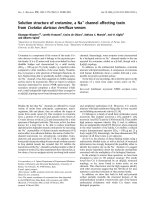
on FRAP obtained after various times of incubation. Fig-
ure 1 shows the mean evening and morning FRAP of
OSAS patients obtained after plasma sample incubation
from 0 till 8 min. No significant differences between the
evening and morning FRAP values were evident in each of
the times during incubation. Similar results were obtained
in CPAP-OSAS subjects, control group and the subgroup
of severe OSAS patients (data not shown). In vitro experi-
ments revealed that the incubation of plasma with H
2
O
2
at concentrations: 47 μM and 16.5 mM for 1 hour did not
suppress FRAP (data not shown). Even H
2
O
2
at a concen-
tration of 82.5 mM did not significantly decrease FRAP
(587 ± 12 μM vs. 603 ± 16 μM, n = 5, p > 0.05).
H
2
O
2
activity in the whole blood
Only eight OSAS patients had detectable H
2
O
2
levels in
the whole blood in the evening, while seven OSAS
patients observed detectable H
2
O
2
post awakening. In the
whole OSAS group, evening H
2
O
2
– 0.23 ± 0.44 μM (0;
0.30) did not differ (p > 0.05, n = 27) from the morning
– 0.22 ± 0.61 μM (0; 0.40) (95% CI -0.12 – 0.13). In the
subgroup of patients with severe OSAS, the ratio of posi-
tive H
2
O
2
readings were 4/11 and 2/11 for the evening
and morning measurements. For CPAP-OSAS and the
control group, the ratio of H
2
O
2
positive results were 4/22
and 3/11 for both evening and morning blood samples,
respectively. Similar to the whole group of OSAS patients,
no differences were found between the morning and
evening H
2
O
2
activities in the remaining two groups as
well as in the subgroup of patients with severe OSAS (data
not shown). Furthermore, no differences manifested
among all three groups (data not shown).
Discussion
Parameters of resting and fMLP-induced LBCL were the
co-primary variables in our study. The morning and
Table 5: Luminol enhanced whole blood chemiluminescence (LBCL) measured before and after polysomnography controlled sleep
Patients undergoing polysomnography
Chemiluminescence parameter Time of the day OSAS Severe OSAS CPAP-OSAS Controls
rCL [aU/10
4
p] Evening 2.4 ± 2.2 1.9 ± 1.8 2.2 ± 1.9 1.9 ± 1.8
Morning 2.3 ± 2.2 2.5 ± 2.8 1.5 ± 0.9 1.6 ± 1.5
Diff and 95% CI 0.1 (-0.7 – 0.8) 0.6 (0.15 – 1.02) 0.7 (0.1 – 1.2) 0.2 (-0.1 – 0.5)
pCL [aU/10
4
p] Evening 6.4 ± 5.1 4.9 ± 4.7 6.0 ± 4.4 5.5 ± 4.3
Morning 5.9 ± 4.6 5.9 ± 4.8 4.3 ± 2.9 4.7 ± 2.5
Diff and 95% CI 0.5 (-0.9 – 1.8) 0,9 (-0.4 – 2.2) 1.6 (0.4 – 2.9) 5.2 (-2,0 – 12.4)
tCL [aU·s/10
4
p] Evening 1939 ± 1532 1503 ± 1391 1805 ± 1278 1642 ± 1316
Morning 1790 ± 1371 1778 ± 1442 1313 ± 820 1416 ± 961
Diff and 95% CI 149 (-275 – 573) 275 (-117 – 668) 492 (132 – 852) 225 (22 – 430)
Peak time [s] Evening 281 ± 31 276 ± 37 293 ± 30 270 ± 24
Morning 275 ± 30 277 ± 37 284 ± 22 339 ± 23
Diff and 95% CI 6 (0.3 – 11.5) 1 (-5.3 – 6.5) 9 (0.1 – 16.3) 69 (-22 – 160)
rCl – the average resting chemiluminescence prior to addition of fMLP; pCL – maximal chemiluminescence signal after addition of fMLP; tCl – total
light emission after cell stimulation with fMLP; peak time – the time from fMLP addition to the appearance of pCL; OSAS – patients with the
untreated OSAS; Severe OSAS – patients with AHI ≥ 30; CPAP-OSAS – patients with OSAS treated successfully after a first attempt with CPAP,
controls – subjects without OSAS Results expressed as mean ± SD, difference of means with 95% confidence interval (Diff and 95% CI) No
significant differences (p > 0.05) were found for evening and morning LBCL parameters within and between the studied groups.
Journal of Negative Results in BioMedicine 2008, 7:10 />Page 8 of 11
(page number not for citation purposes)
evening chemiluminescence parameters like rCL, pCL and
tCL as well as FRAP were higher in OSAS patients in com-
parison with controls. Nevertheless, we did not find any
differences between morning and evening LBCL parame-
ters between and within the groups of OSAS, CPAP-OSAS
and the matched controls. Moreover, LBCL did not rise
significantly after polysomnography controlled sleep in
the subgroup of patients with severe untreated OSAS.
Therefore, our results suggest that OSAS related IH did not
enhance ROS production by blood PMNs and monocytes.
This finding is in contrast with the results of two previous
studies showing increased ROS production from isolated
PMNs (following stimulation with fMLP) and some sub-
populations of whole blood monocytes (resting and
phorbol myristate acetate – activated) of OSAS patients
[14,15].
Apart from the absence of cell isolation procedures and
the usage of monoclonal antibodies to surface cell mark-
ers possibly altering PMNs and monocytic responsiveness
to agonist stimulation, there were other significant differ-
ences between the protocols of our study and the afore-
mentioned. In previous studies, the control subjects were
not matched to OSAS patients in respect to age, BMI, cig-
arette smoking habits in addition to comorbidity [14,15].
Moreover, both investigations did not provide informa-
tion concerning concomitant pharmacologic treatment
[14,15]; furthermore, experiments on the effect of OSAS
related IH on monocytic activity were based on only 8 to
10 subjects out of the 18 enrolled [15] devoid of an unam-
biguous selective criterion.
In view of the fact that the majority of OSAS patients are
obese often developing a variety of cardiovascular and
metabolic diseases [1,2], there exists a challenge in find-
ing appropriate control subjects, chiefly in respects to
comorbidity and concomitant pharmacological treat-
ment. In our study, we overcome this impediment by
measuring LBCL prior to and following polysomno-
graphic controlled sleep, formulating comparisons within
and between study groups. These approaches act to possi-
bly eliminate biases related to patient medication, inten-
Ferric reducing ability of plasma (FRAP) before and after polisomnography controlled sleepFigure 1
Ferric reducing ability of plasma (FRAP) before and after polisomnography controlled sleep. Closed circles –
evening (before polisomnography controlled sleep). Open circles – morning (after polisomnography controlled sleep). Results
of FRAP in 27 subjects with OSAS expressed as mean and standard deviations represent the concentration of Fe
3+
reduced
ions after 0, 1, 2, 3, 4, 5, 6, 7 and 8 min incubation of patient plasma sample with the FRAP reagent. No significant differences (p
> 0.05) were found for evening and morning FRAP at all incubation time-points.
Journal of Negative Results in BioMedicine 2008, 7:10 />Page 9 of 11
(page number not for citation purposes)
sifying the quality of our results in showing no priming or
activation of blood phagocytes in untreated OSAS
patients.
The sympathetic over activity in OSAS patients resulting in
the rise of plasma norepinephrine and epinephrine levels
[25-27] may perhaps be responsible in the insignificant
changes between evening and morning blood LBCL in
OSAS patients. Reports have demonstrated a higher morn-
ing (following awakening) plasma norepinephrine con-
centration than those before sleep in OSAS patients by
24% [25]. Nonetheless, a night of successful CPAP ther-
apy resulted in a down-regulation of sympathetic activity
as well as a decrease in circulating catecholamines by 20%
[26]. Catecholamines in vitro, especially epinephrine at
physiologic levels revealed concentration dependent inhi-
bition of fMLP-induced degranulation and superoxide
radical production by human PMNs [28-30]; with a capa-
bility of suppressive monocytic activity as well [31,32].
Therefore, exposition of circulating phagocytes to
increased concentrations of catecholamines may result in
their insusceptibility to the priming effect of IH in respira-
tory burst, assuming responsibility for the negative results
in untreated OSAS patients. On the other hand, the obser-
vation of rapidly reversible sympathetic over-activation
due to successful treatment [26] elucidates the indiffer-
ences among morning and evening LBCL in CPAP-OSAS
group. This may result from the simultaneous reduction
of IH episodes along with plasma catecholamine suppres-
sion, together with a rapid turnover of circulating PMNs;
strongly supported by a recent study demonstrating the
inhibitory effect of exercise on hypobaric hypoxia-
induced enhancement of ROS production by PMNs in
healthy volunteers [33]. It cannot be excluded that our
OSAS patients encountered an inadequate amount of
apneas/hypopneas, consequently observing insufficient
blood desaturations to prime PMNs to fMLP stimulation.
In the abovementioned study, overnight hypobaric
hypoxia decreased the average SaO
2
from a baseline of
98% to 93% concluding a high altitude stay [33].
A decreased average SaO
2
(84%) along with minimal
SaO
2
(68%) was evident in our investigated patients, par-
ticularly those with severe OSAS, due to numerous tran-
sient desaturations. The activity of two secondary
variables (FRAP and H
2
O
2
activity in the whole blood) in
OSAS patients after sleep was compatible to the results of
LBCL. We did not observe sleep-induced ROS overproduc-
tion in the blood (LBCL, H
2
O
2
activity) of untreated OSAS
patients, therefore no suppression of morning FRAP was
noted. On the other hand, resistance of FRAP to 1 h in
vitro incubation with high concentrations of H
2
O
2
sug-
gests strong antioxidant plasma capacity. Therefore, FRAP
suppression related to significant expenditure of circulat-
ing antioxidants with high Fe
3+
reducing activity will occur
in vivo in the case of large and long (probably longer than
one night) systemic ROS overproduction.
The results of our study suggest that circulating phago-
cytes (PMNs and monocytes) are not the main culprit of
OSAS consequences in the human body. It does not
exclude augmented ROS production and activation of the
systemic ROS signaling [7,14,34]. Circulating phagocytes
probably do not take part in oxidative stress, which does
not synonymously reject the oxidative stress presence in
OSAS patients. It can take place near or exactly in blood
vessel endothelium, which can significantly accelerate
atherosclerosis.
Conclusion
In conclusion, untreated OSAS patients did not present
with elevated resting and fMLP induced LBCL when com-
pared with age-, BMI-, and smoking habits- matched con-
trols. Moreover, no significant alterations of evening vs.
morning LBCL, blood H
2
O
2
activities and FRAP were
noted in OSAS patients. These indicate that circulating
PMNs and monocytes did not produce increased amounts
of ROS and did not contribute to oxidative stress in OSAS
patients.
Competing interests
The authors declare that they have no competing interests.
Authors' contributions
IGK organized the whole study, prepared the solutions for
laboratorial experiments, carried out morphology, FRAP,
LBCL and H
2
O
2
activity in the whole blood measurement,
analyzed all patients histories and all experiments results,
Table 6: Ferric reducing ability of plasma (FRAP) before
(evening) and after (morning) polysomnography controlled
sleep
Patients group FRAP [μM] measured at Diff, 95% CI
Evening Morning
OSAS, n = 27 671 ± 221 602 ± 202 70 (7–133)
Severe OSAS, n = 11 729 ± 156 650 ± 170 79 (37–122)
CPAP-OSAS, n = 22 597 ± 199 591 ± 242 7 (-44–57)
Controls, n = 11 634 ± 229 597 ± 201 37 (-3–77)
OSAS – patients with the untreated OSAS; CPAP-OSAS – patients
with OSAS treated successfully after a first attempt with CPAP,
controls – subjects without OSAS. Results expressed as a mean ± SD
represent concentration of Fe
3+
reduced ions after 3 min. incubation
of plasma sample with the FRAP reagent. Diff, 95% CI – difference of
means with 95% confidence interval
No significant differences (p > 0.05) were found for evening and
morning FRAP within and between the studied groups.
Journal of Negative Results in BioMedicine 2008, 7:10 />Page 10 of 11
(page number not for citation purposes)
prepared the manuscript. AK presented the idea and the
aim of the study to our patients, carried out all polysom-
nographies, collected blood samples and participated in
LBCL and H
2
O
2
activity in the whole blood measurement.
PB took patients histories and described about 50 % poly-
somnographies. Based on them he diagnosed patients
and, in case of need, chose CPAP with adequate pressure.
MKr took patients histories, described about 50 %
remaining polysomnographies, based on them diagnosed
patients and also, in case, chose CPAP with adequate pres-
sure. GE carried out the in vitro experiment verifying the
effect of H
2
O
2
on FRAP. MKa performed the statistical
analysis. DN conceived of the study, and participated in
its design and coordination. He also drafted the manu-
script and had decisive influence on the discussion course.
All authors read and approved the final manuscript.
Acknowledgements
The authors would like to thank Jeffrey de Graft-Johnson, M.D., M.P.A.,
M.S., for his assistance in preparing the manuscript.
This study was supported by the Medical University of Lodz Insti-
tutional Grant No 503-0079-1
References
1. Shamsuzzaman ASM, Gersh BJ, Somers VK: Obstructive sleep
apnea. Implications for cardiac and vascular disease. JAMA
2003, 290:1906-1914.
2. Nieto FJ, Young TB, Lind BK, Shahar E, Samet JM, Redline S, D'Ago-
stino RB, Newman AB, Lebowitz MD, Pickering TG: Association of
sleep-disordered breathing, sleep apnea, and hypertension in
a large community-based study. JAMA 2000, 283:1829-1836.
3. Shahar E, Whitney CW, Redline S, Lee ET, Newman AB, Javier Nieto
F, O'Connor GT, Boland LL, Schwartz JE, Samet JM: Sleep-disor-
dered breathing and cardiovascular disease. Cross-sectional
results of the Sleep Heart Health Study. Am J Respir Crit Care
Med 2001, 163:19-25.
4. Peker Y, Hedner J, Kraiczi H, Löth S: Respiratory disturbance
index an independent predictor of mortality in coronary
artery disease. Am J Respir Crit Care Med 2000, 162:81-86.
5. Fletcher EC: Cardiovascular consequences of obstructive
sleep apnea: experimental hypoxia and sympathetic activity.
Sleep 2000, 23(Suppl 4):S127-S131.
6. Phillips CL, Yang Q, Williams A, Roth M, Yee BJ, Hedner JA, Berend
N, Grunstein RR: The effect of short-term withdrawal from
continuous positive airway pressure therapy on sympathetic
activity and markers of vascular inflammation in subjects
with obstructive sleep apnea. J Sleep Res 2007, 16:217-225.
7. Yamauchi M, Nakano H, Maekawa J, Okamoto Y, Ohnishi Y, Suzuki T,
Kimura H: Oxidative stress in obstructive sleep apnea. Chest
2005, 127:1674-1679.
8. Sanidas D, Garnham A, Mian R: Activation of human leukocytes
by acute hypoxia. Exp Physiol 2000, 85:263-266.
9. Sanidas D, Garnham A, Mian R: Hypoxia-induced chemilumines-
cence in human leukocytes: the role of Ca
2+
. Eur J Pharmacol
2002, 453:183-187.
10. Klokker M, Kharazmi A, Galbo H, Bygbjerg I, Pedersen BK: Influence
of in vivo hypobaric hypoxia on function of lymphocytes, neu-
trocytes, natural killer cells, and cytokines. J Appl Physiol 1993,
74:1100-1106.
11. Bradley TD, Martinez D, Rutherford R, Lue F, Grossman RF, Moldof-
sky H, Zamel N, Phillipson EA: Physiological determinants of
nocturnal arterial oxygenation in patients with obstructive
sleep apnea. J Appl Physiol 1985, 59:1364-1368.
12. Chaouat A, Weitzenblum E, Krieger J, Oswald M, Kessler R: Pulmo-
nary hemodynamics in the obstructive sleep apnea syn-
drome. Results in 220 consecutive patients. Chest 1996,
109:380-386.
13. Müns G, Rubinstein I, Singer P: Phagocytosis and oxidative burst
of granulocytes in the upper respiratory tract in chronic and
acute inflammation. J Otolaryngol 1995, 24:105-110.
14. Schulz R, Mahmoudi S, Hattar K, Sibelius U, Olschewski H, Mayer K,
Seeger W, Grimminger F: Enhanced release of superoxide from
polymorphonuclear neutrophils in obstructive sleep apnea.
Impact of continuous positive airway pressure therapy. Am J
Respir Crit Care Med 2000, 162:566-570.
15. Dyugovskaya L, Lavie P, Lavie L: Increased adhesion molecules
expression and production of reactive oxygen species in leu-
kocytes of sleep apnea patients. Am J Respir Crit Care Med 2002,
165:934-939.
16. Sokolnicka I, Zieliński J, Skopińska-Rózewska E, Barcz E, Pływacze-
wski R, Zabuska-Jabłońska K: Malfunction of some immunologic
functions in patients with obstructive sleep apnea. Pneumonol
Alergol Pol 2000, 68:247-254.
17. Kukovetz EM, Bratschitsch G, Hofer HP, Egger G, Schaur RJ: Influ-
ence of age on the release of reactive oxygen species by
phagocytes as measured by a whole blood chemilumines-
cence assay. Free Radical Biol Med 1997, 22:433-438.
18. Kopprasch S, Graessler J, Kohl M, Bergmann S, Schröder HE: Com-
parison of circulating phagocyte oxidative activity measured
by chemiluminescence in whole blood and isolated polymor-
phonuclear leukocytes. Clin Chim Acta 1996, 253:145-157.
19. Lacy F, O'Connor DT, Schmid-Schonbein GW: Plasma hydrogen
peroxide production in hypertensives and normotensive
subjects at genetic risk of hypertension. J Hypertens 1998,
16:291-303.
20. Benzie IFF, Strain JJ: The Ferric reducing ability of plasma
(FRAP) as a measure of "antioxidant power": the FRAP
assay. Analytical Biochemistry 1996, 239:70-76.
21. Czupryniak L, Loba J, Pawlowski M, Nowak D, Bialasiewicz P: Treat-
ment with continuous positive airway pressure may affect
blood glucose levels in nondiabetic patients with obstructive
sleep apnea syndrome. Sleep 2005, 28:601-603.
22. Szkudlarek U, Luczynska M, Kasielski M, Kaucka S, Nowak D:
Exhaled hydrogen peroxide correlates with the release of
reactive oxygen species by blood phagocytes in healthy sub-
jects. Respiratory Medicine 2003, 97:718-725.
23. Pick E, Keysari Y: A simple colorimetric method for the meas-
urement of hydrogen peroxide produced by cells in culture.
J Immunol Methods 1980, 38:161-170.
24. de Graft-Johnson J, Kolodziejczyk K, Krol M, Nowak P, Krol B,
Nowak D: Ferric-reducing ability power of selected plant
polyphenols and their metabolites: implications for clinical
studies on the antioxidant effects of fruits and vegetable con-
sumption. Basic Clin Pharmacol Toxicol 2007, 100:345-352.
25. Noda A, Okada T, Yasuma F, Nakashima N, Yokota M: Cardiac
hypertrophy in obstructive sleep apnea syndrome. Chest
1995, 107:1538-1544.
26. Baruzzi A, Riva R, Cirignotta F, Zucconi M, Cappelli M, Lugaresi E:
Atrial natriuretic peptide and catecholamines in obstructive
sleep apnea syndrome. Sleep 1991, 14:83-86.
27. Coy TV, Dimsdale JE, Ancoli-Israel S, Clausen J: Sleep apnea and
sympathetic nervous system activity: a review. J Sleep Res
1996, 5:42-50.
28. O'Dowd YM, El-Benna J, Perianin A, Newsholme P: Inhibition of
formyl-methionyl-leucyl-phenylalanine-stimulated respira-
tory burst in human neutrophils by adrenaline: inhibition of
Phospholipase A2 activity but not p47phox phosphorylation
and translocation. Biochem Pharmacol 2004, 67:183-190.
29. Tintinger GR, Theron AJ, Anderson R, Ker JA: The anti-inflamma-
tory interactions of epinephrine with human neutrophils in
vitro are achieved by cyclic AMP-mediated accelerated rese-
questration of cytosolic calcium. Biochem Pharmacol 2001,
61:1319-1328.
30. Trabold B, Gruber M, Fröhlich D: Functional and phenotypic
changes in polymorphonuclear neutrophils induced by cate-
cholamines. Scand Cardiovasc J 2007, 41:59-64.
31. Lünemann JD, Buttgereit F, Tripmacher R, Baerwald CG, Burmester
GR, Krause A: Norepinephrine inhibits energy metabolism of
human peripheral blood mononuclear cells via adrenergic
receptors. Biosci Rep 2001, 21:627-635.
32. Farmer P, Pugin J: Beta-adrenergic agonists exert their "anti-
inflammatory" effects in monocytic cells through the Ikap-
Publish with Bio Med Central and every
scientist can read your work free of charge
"BioMed Central will be the most significant development for
disseminating the results of biomedical research in our lifetime."
Sir Paul Nurse, Cancer Research UK
Your research papers will be:
available free of charge to the entire biomedical community
peer reviewed and published immediately upon acceptance
cited in PubMed and archived on PubMed Central
yours — you keep the copyright
Submit your manuscript here:
/>BioMedcentral
Journal of Negative Results in BioMedicine 2008, 7:10 />Page 11 of 11
(page number not for citation purposes)
paB/NF-kappaB pathway. Am J Physiol Lung Cell Mol Physiol 2000,
279:L675-682.
33. Choukèr A, Demetz F, Martignoni A, Smith L, Setzer F, Bauer A, Hölzl
J, Peter K, Christ F, Thiel M: Strenuous physical exercise inhibits
granulocyte activation induced by high altitude. J Appl Physiol
2005, 98:640-647.
34. Prabhakar NR, Kumar GK, Nanduri J, Semenza GL: ROS signaling
in systemic and cellular responses to chronic intermittent
hypoxia. Antioxid Redox Signal 2007, 9:1397-1403.