Báo cáo khoa hoc:" Comparison between the HCV IRES domain IV RNA structure and the Iron Responsive Element" pps
Bạn đang xem bản rút gọn của tài liệu. Xem và tải ngay bản đầy đủ của tài liệu tại đây (391.64 KB, 8 trang )
BioMed Central
Page 1 of 8
(page number not for citation purposes)
Journal of Negative Results in
BioMedicine
Open Access
Research
Comparison between the HCV IRES domain IV RNA structure and
the Iron Responsive Element
Ebenezer Tumban
1,2
, Jenna M Painter
2
and William B Lott*
1,2,3
Address:
1
Molecular Biology Program, New Mexico State University, Las Cruces, NM 88003-8001, USA,
2
Department of Chemistry and
Biochemistry, New Mexico State University, Las Cruces, NM 88003-8001, USA and
3
Institute for Health and Biomedical Innovation, School of Life
Sciences, Queensland University of Technology, Brisbane, QLD 4001, Australia
Email: Ebenezer Tumban - ; Jenna M Painter - ; William B Lott* -
* Corresponding author
Abstract
Background: Serum ferritin and hepatic iron concentrations are frequently elevated in patients
who are chronically infected with the hepatitis C virus (HCV), and hepatic iron concentration has
been used to predict response to interferon therapy, but these correlations are not well
understood. The HCV genome contains an RNA structure resembling an iron responsive element
(IRE) in its internal ribosome entry site (IRES) structural domain IV (dIV). An IRE is a stem loop
structure used to control the expression of eukaryotic proteins involved in iron homeostasis by
either inhibiting ribosomal binding or protecting the mRNA from nuclease degradation. The HCV
structure, located within the binding site of the 40S ribosomal subunit, might function as an
authentic IRE or by an IRE-like mechanism.
Results: Electrophoretic mobility shift assays showed that the HCV IRES domain IV structure does
not interact with the iron regulatory protein 1 (IRP1) in vitro. Systematic HCV IRES RNA
mutagenesis suggested that IRP1 cannot accommodate the shape of the wild type HCV IRES dIV
RNA structure.
Conclusion: The HCV IRES dIV RNA structure is not an authentic IRE. The possibility that this
RNA structure is responsible for the observed correlations between intracellular iron
concentration and HCV infection parameters through an IRE-like mechanism in response to some
other cellular signal remains to be tested.
Background
Hepatitis C virus (HCV) is a positive-sense single-stranded
RNA virus that infects about 1% of the world's population
[1]. Fifty percent of acute infections progress to chronic
liver infection and can lead to cirrhosis of the liver and
hepatocellular carcinoma [2,3]. A correlation between
chronic HCV infection and intracellular iron homeostasis
has been empirically established, but the mechanism of
this relationship is not understood [4,5]. Increased intrac-
ellular iron concentration has been shown to enhance
HCV IRES-dependent translation, and two cellular factors,
p85 and p110, bind to both the HCV internal ribosome
entry site (IRES) and the iron responsive element (IRE) in
an iron-dependent manner [6]. Serum ferritin and hepatic
iron concentrations are frequently elevated in chronically
infected patients [7], and hepatic iron concentration has
been used as a predictor of response to interferon therapy
[8].
Published: 18 February 2009
Journal of Negative Results in BioMedicine 2009, 8:4 doi:10.1186/1477-5751-8-4
Received: 13 November 2007
Accepted: 18 February 2009
This article is available from: />© 2009 Tumban et al; licensee BioMed Central Ltd.
This is an Open Access article distributed under the terms of the Creative Commons Attribution License ( />),
which permits unrestricted use, distribution, and reproduction in any medium, provided the original work is properly cited.
Journal of Negative Results in BioMedicine 2009, 8:4 />Page 2 of 8
(page number not for citation purposes)
An RNA structural element located at the junction
between the open reading frame (ORF) and the 5'
untranslated region (UTR) of the HCV genome bears strik-
ing structural and sequential similarities to the iron
responsive element (IRE), an RNA structure found in
some eukaryotic mRNA that controls gene expression in
response to intracellular iron concentration [9]. If this
HCV RNA element functions as an authentic IRE, then
iron depletion in an HCV infected cell would be expected
to specifically inhibit viral protein expression and could
potentially decrease subsequent viral replication. A possi-
ble relationship between this HCV RNA structure and the
IRE was investigated.
HCV belongs to the Hepacivirus genus of the Flaviviridae
family [10]. The RNA genome is approximately 9.6 kb and
serves as a template for both translation and replication.
The single long ORF is flanked at the 5' and 3' ends by
highly structured UTRs, which are essential for initiation
of translation and replication, respectively [11]. The hepa-
civirus and pestivirus genera of Flaviviridae initiate transla-
tion via virtually identical non-scanning cap-independent
mechanisms that utilize an internal ribosome entry site
(IRES) to recruit and assemble the ribosome directly at the
initiation site [12-15]. The HCV IRES (figure 1) is a com-
plex and highly conserved RNA structure that is located
predominantly within 5' UTR, but is believed to extend
into the 5' proximal region of the ORF [16]. It is canoni-
cally divided into four structural domains. Domain I is
required for efficient viral replication and is not required
for viral translation. Domains II and III are necessary and
sufficient to recruit and assemble the ribosome at the start
site [13]. The domain IV (dIV) RNA structure, which is not
essential for efficient translation [17] and is unique to
hepaciviruses, has no known function. The pestivirus IRES
contains structural domains that are analogous to HCV
IRES domains I-III, but lacks domain IV. The border
between the 5' UTR and the ORF of the pestivirus genome
is believed to be unstructured.
The authentic HCV initiation codon resides in the termi-
nal loop of the RNA hairpin structure in the HCV IRES
dIV. This structure is unlikely to be tolerated in the RNA
binding cleft of the ribosome while the initiation codon
occupies the ribosomal P site [17-19], and the ribosomal
toeprint on the HCV genome at +15 from the start codon
is characteristic of a eukaryotic ribosome bound to
unstructured mRNA [20]. Consequently, increased stabil-
ity of the HCV IRES dIV structure predictably decreases the
efficiency of HCV IRES-dependent translation [17]. In
addition, the HCV IRES dIV structure must presumably
melt to allow the N-terminus of the HCV polyprotein to
be translated from the codons in the 5' region of the ORF
that are involved in the HCV IRES dIV RNA structure. The
apparent requirement that the HCV IRES dIV structure
unwinds to allow translation initiation suggests that its
presumed viral function occurs either before the ribosome
has been recruited to the mRNA or after the translating
ribosome has exited the start site, which is consistent with
a regulatory RNA element.
The IRE is an example of a translation regulatory RNA ele-
ment that does not otherwise actively participate in the
translation initiation mechanism [9]. The interaction
between the IRE and its cognate binding partner, an iron
regulatory protein (IRP1 or IRP2), either inhibits gene
expression by inhibiting 40S ribosomal subunit binding
or enhances gene expression by protecting the mRNA
from nuclease degradation [21], depending on where the
IRE is located in the mRNA. To inhibit ribosomal binding,
the IRE must reside within the 40S ribosomal subunit
binding site near the 5' cap structure. The IRE cannot bind
to the 40S ribosomal subunit while it is bound to an IRP,
but it has little effect on the translation initiation effi-
ciency when it is not bound by an IRP. The active IRP con-
centration is controlled by the intracellular iron
concentration. IRP1 is an aconitase enzyme conformer
that is formed when the intracellular iron concentration is
insufficient to form the characteristic aconitase ironsulfur
cluster. IRP2 is homologous to IRP1 but does not form the
aconitase iron-sulfur cluster [22]. Like the HCV IRES dIV
structure, the IRE must melt to allow the mRNA to occupy
the ribosomal RNA binding cleft.
The HCV IRES dIV RNA structure shares many of the IRE
consensus structural characteristics[23,24], most closely
resembling the human erythroidspecific δ-aminolevulinic
acid synthase (eALAS) IRE [25] (figure 2, boxed inset).
The eALAS IRE stem contains an unpaired C residue in the
5' arm (C-bulge) that is separated from the terminal loop
by 5 base pairs. The C-bulge is implicated in a sequence-
specific binding interaction with the IRP [26]. A promi-
nent feature of the IRE terminal loop is an intraloop C-G
base pair, which is required for its interaction with an IRP.
The C-G intraloop base pair solvent-exposes the guanine
base at the center of the resultant terminal tri-loop, which
is also required for sequence-specific high-affinity binding
to the IRP [26]. The consensus sequence of the IRE termi-
nal loop consists of six bases, the first five of which are
usually CAGUG. The sixth base can be any nucleotide, so
long as it cannot base pair with C. The HCV IRES dIV RNA
hairpin incorporates similar features, including a C-con-
taining bulge in 5' arm of the stem that is separated from
the terminal loop by five base pairs and the potential to
form an intraloop C-G pair. In fact, the HCV IRES dIV
stem differs from the consensus C-bulge IRE in only three
significant ways (figure 2, boxed inset). It has an extra ade-
nosine residue in the terminal loop, two extra adenosine
residues in the 3' arm of the stem directly across from the
unpaired cytosine residue, and an adenosine residue
Journal of Negative Results in BioMedicine 2009, 8:4 />Page 3 of 8
(page number not for citation purposes)
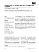
The HCV 5'UTR secondary structureFigure 1
The HCV 5'UTR secondary structure. The base pairing convention described by Honda et al [17] is used to depict the
predicted base pairing in the HCV IRES RNA structure for genotype 1b. The structural domains are labelled I-IV. The authentic
HCV start codon at HCV nt 342–344 is boxed. The wild type HCV IRES dIV RNA sequence used in this study represents HCV
nt 331–354.
AU
CG
CG
GU
GC
AU
AA
GC CU
UGGG
G
A
U
A
CG
GC
U
G
A
GC
G
U
U
U
C
U
C
G
U
A
GC
AU
CG
GC
UA
GC
CG
C
A
A
A
C
C
A
GC
GC
CG
GC
UA
CG
UA
GC
GU
UG
GC
AU
UA
GCCG UA GUG
CGGA A GCGC
AG
A
U
U
G
G
G
U
C
U
U
G
A
GGG
CCC
CA CCGG
GUGGCC
C
G
U
G
A
U
G
AA
A
GC
GC
AU
CG
CG
GA
UG
UA
AU
AU
U
UA
GC
GC
GC
CG
AU
GC
A
A
U
G
C
A
CC
C
C
U
U
U
C
UU
G
G
A
U
A
A
CUCACUAAAC
A
U
G
A
CUCCCGGG
GA GGGCCC
GC
UA
CG
CG
GC
GC
AU
GC
U
C
U
A
C
A
G
AU
UG
C
GC
UG
U
U
C
U
U
AU
CG
GA
CG
AU
CG
A
A
G
A
G
U
A
CG
UG
GC
CG
GU
AU
U
A
G
C
C
A
U
ACCCCCC
G
GCCA GA CA CUC CA CCA UGA A U CA CUCC
GC
CG
CG
CG
CG
CG
UU
GA
I
II
III
IV
Journal of Negative Results in BioMedicine 2009, 8:4 />Page 4 of 8
(page number not for citation purposes)
instead of a guanosine residue at the apex of the putative
tri-loop formed by the intraloop C-G base pair. A mutant
HCV IRES dIV structure in which all three differences are
reconciled would fit the consensus definition of an
authentic IRE [23,24] and would be expected to bind to
an IRP with wild type affinity.
If HCV were to utilize an IRE-like mechanism to control
viral expression, the RNA structure used for this regulation
must be positioned within the 40S ribosomal subunit
binding site. In sharp contrast to normal eukaryotic cap-
dependent translation, the HCV IRES-dependent transla-
tion initiation mechanism requires that the 40S ribos-
omal subunit binds directly to the HCV genomic RNA
sequence flanking the 5' UTR-ORF boundary to allow the
start codon to occupy the ribosomal P site [20]. This
mechanism would force at least part of any putative IRE-
like RNA element into the ORF. Thus the HCV IRES dIV
structure is properly positioned in the HCV mRNA to
function as an IRE-like RNA element.
Results
RNA binding to hIRP1
Since the HCV IRES dIV structure differs from the consen-
sus IRE at only three characteristics, a mutant RNA panel
was constructed to evaluate the relative binding contribu-
tion of each characteristic. The panel systematically
mutated the wild type HCV IRES dIV structure to a con-
sensus IRE structure (figure 2).
Electrophoretic mobility shift assays (EMSA) were used to
detect binding interactions between hIRP1 and the RNA
species shown. Although a 100 ng concentration of hIRP1
was reported to bind to the wild type IRE [22], only the
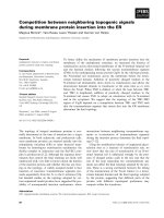
RNA sequences and secondary structuresFigure 2
RNA sequences and secondary structures. Sequences and predicted secondary structures of the RNA panel evaluated in
this study. The nucleotides in bold in each construct represent deviations from the consensus C-bulge IRE. Inset: the eALAS
IRE and wild type HCV IRES dIV RNA are boxed for comparison.
&*
*&
8$
*&
&*
$8
*&
*&
&
$
8
&
*
&*
*&
8$
*&
&*
$8
*&
*&
&
$
8
&
&*
*&
8$
*&
&*
$8
*&
*&
&
$
*
8
&
&*
*&
8$
*&
&*
$8
*&
*
&
&$
8
&
&*
*&
8$
*&
&*
$8
*&
*&
&$
8
&
&*
*&
8$
*&
&*
$8
*&
*&
&$
*8
&
&
&*
*&
8$
*&
&*
$8
*&
*&
$
*8
&
8$
&*
&*
8*
*&
8$
8$
*&
*
$
&&
*8
&
&*
*&
8$
*&
&*
$8
*&
*&
&
$
8
&
8$
&*
&*
8*
*&
8$
8$
*&
&
&
**
8&
&
Journal of Negative Results in BioMedicine 2009, 8:4 />Page 5 of 8
(page number not for citation purposes)
eALAS IRE and ET1+2+3 bound to hIRP1 at this concen-
tration in our experiments (data not shown). Six of the
mutant RNA sequences (ET1+2, ET3, ET2, ET1+3, ET2+3,
ET1+2+3) and the eALAS IRE detectably bound to hIRP1
at an hIRP1 concentration of 240 ng (figure 3). No detect-
able binding to hIRP1 was observed for the wild type HCV
IRES dIV, ET1, ET2, or the negative control IRE. These
experiments yielded the following binding trend:
(eALAS IRE ≈ ET1+2+3) > ET2+3 > ET1+3 > ET3 > (ET2 ≈
ET1+2) > (ET1 ≈ HCV IRES dIV ≈ IRE (-) control)
Estimate of the dissociation constant, K
d
EMSA was used quantitatively to estimate the dissociation
constants (K
d
) of the hIRP1-ET2+3 and hIRP1-ET1+2+3
complexes. Unsurprisingly, ET1+2+3, which adheres to
the consensus definition of an IRE [23,24], bound to
hIRP1 with approximately wild type affinity (K
d
~ 50 pM,
data not shown). ET2+3 bound to hIRP1 with a K
d
= 54 ±
13 nM (figure 4B), which is about three orders of magni-
tude weaker than the consensus IRE.
Discussion
The HCV IRES has been described as an RNA structural
element that regulates viral translation. Strictly speaking,
however, the HCV IRES mechanism describes the initia-
tion of translation, not necessarily its regulation. The HCV
IRES domains I-III have been well characterized in recent
years, and much is known about the roles of these struc-
tures in the recruitment and assembly of the eukaryotic
ribosome on the HCV RNA genome. The role of domain
IV, however, remains enigmatic. It is not required for effi-
cient initiation and is not present in the closely related
pestivirus IRES, yet it is conserved across all HCV geno-
types. Mutational analysis of this structure demonstrated
only a modest relationship between HCV IRES-dependent
translational efficiency and structural stability [17]. This
observation might simply reflect the effect of RNA struc-
ture near the start codon, and does not necessarily imply
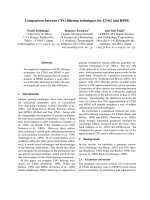
Relative RNA – hIRP1 binding interactionsFigure 3
Relative RNA – hIRP1 binding interactions. EMSA showing the relative binding ability of hIRP1 to the RNA panel shown
in figure 2.
eALAS
IRE
WT dIV
ET1
ET2
ET3
ET1+2
ET1+3
ET2+3
IRE
(-) CNTRL
ET1+2+3
Free RNA
RNA-IRP1
Complex
Journal of Negative Results in BioMedicine 2009, 8:4 />Page 6 of 8
(page number not for citation purposes)
a role in translation initiation. These observations are con-
sistent with a regulatory RNA element that modulates
translation efficiency in response to an appropriate bio-
logical signal but does not otherwise participate directly in
the recruitment and assembly of the ribosome. The IRE-
IRP mechanism represents a useful model for this type of
interaction.
A cursory comparison of the structure and genomic loca-
tion of the HCV IRES dIV RNA structure to the C-bulge
eALAS IRE is provocative and suggests a correlated similar-
ity of function. The wild type HCV IRES dIV RNA does not
measurably bind to hIRP1, however, demonstrating that it
is not an authentic IRE. The ET1+2+3 RNA, which adheres
to the consensus definition of an IRE, bound to IRP1 with
wild type efficiency as expected. Deleting the two A resi-
dues in the bulge of the wild type HCV IRES dIV RNA (the
ET3 effect) contributed the greatest relative effect on bind-
ing affinity, followed by deletion of an A residue in the ter-
minal loop (the ET2 effect), suggesting the importance of
the correct three dimensional RNA structure on hIRP1
binding. The G residue at the apex of the terminal tri-loop
and C-bulge residue are conserved in the consensus IRE
and are presumably required for sequence-specific inter-
actions between the IRE and recognition motifs on the
IRP [9]. These residues are displayed in three dimensions
relative to each other by the structural features of the stem
and the terminal loop. Altering the IRE structure likely
misaligns the recognition nucleotides with their respec-
tive recognition motifs on hIRP1. A well-defined and rigid
IRE binding pocket would not accept a stem of the wrong
shape, regardless of the sequential positions of the recog-
nition nucleotides. The ET2 and ET3 effects are consistent
with the reported IRE characteristics required for efficient
IRE-IRP interaction [27]. Mutating the A residue of the
start codon to a G residue (the ET1 effect), a sequence-
only mutation, showed the least effect on hIRP1 binding.
The ET1 effect was unexpected, since the literature reports
that the G residue in the terminal loop is essential [28].
Either purine residue in this position yielded significant
IRP1 binding in our hands, with only a slight preference
for G (compare ET2+3 to ET1+2+3 in figure 3).
Conclusion
The primary conclusion from this work is that the wild
type HCV IRES dIV RNA structure is not an authentic IRE,
as it does not bind appreciably to the recombinant IRP1
protein. The hypothesis that HCV utilizes this structure to
control viral expression by an IRE-like mechanism
remains viable, although a putative cellular or viral factor
that fulfils the analogous IRP function must be identified
to properly evaluate this hypothesis. The p85 and p100
proteins that have been recently shown to bind to both
the HCV IRES and the iron responsive element with high
affinity [6] could serve this purpose. For now, the role of
the highly conserved HCV IRES dIV structure remains
unresolved.
ET2+3 – hIRP1 binding interaction concentration dependenceFigure 4
ET2+3 – hIRP1 binding interaction concentration dependence. (A) EMSA showing the hIRP1 concentration depend-
ence on binding to ET2+3. Lanes showing hIRP1 concentrations of 74 pM-5.1 nM were omitted for clarity. (B) Plotted data
used to determine the K
d
for the IRP1-ET2+3 interaction.
9.1 nM
7.1 nM
11 nM
31 nM
51 nM
71 nM
91 nM
110 nM
310 nM
510 nM
710 nM
910 nM
1.1
M
(-) CNTRL
Free RNA
RNA-IRP1
Complex
$ %
.
G
s Q0
Journal of Negative Results in BioMedicine 2009, 8:4 />Page 7 of 8
(page number not for citation purposes)
Methods
Expression and purification of human IRP1
Human IRP1 (hIRP1) fused to glutathione S-transferase
(GST) was expressed and purified as previously described
[22] with some modifications. The pGEX-2T plasmid (Dr.
Lukas Kuhn, ISREC, Epalinges, Switzerland) was trans-
formed into HB101 cells and hIRP1 expression was
induced with 0.1 or 0.5 mM IPTG (Sigma-Aldrich, St
Louis, MO) overnight at room temperature. Each lysis
reaction contained 2 mL (approximately 0.2 g) of cell
lysate, 10 μL of protease cocktail inhibitor (Sigma-
Aldrich, St Louis, MO) and 15,000 U of lysozyme (Nova-
gen, San Diego, CA). The cells were lysed by sonication in
PBS buffer (150 mM NaCl, 16 mM Na
2
HPO
4
, 4 mM
NaH
2
PO
4
, pH 7.3) containing 1% triton-X-100 (Sigma-
Aldrich, St Louis, MO). The cell lysate was then incubated
at 4°C for 30 minutes and spun at 10,000 rpm for 30 min-
utes. hIRP1 was purified on a 50% glutathione sepharose
resin column (Amersham, Piscataway, NJ). The protein
was eluted with 50 mM Tris and 10 mM reduced glutath-
ione (pH 8.0), concentrated using microcon YM 50 (Mil-
lipore, Jaffrey, NH) and its concentration, 1.2 μg/μL, was
determined using the Micro Lowry method (Sigma-
Aldrich, St Louis, MO). Purified and unpurified hIRP1
was visualized on an SDS-PAGE and confirmed by West-
ern blot using rabbit anti-rat IRP1 polyclonal antibodies
(Alpha Diagnostic, San Antonio, TX).
RNA synthesis
RNA was synthesized by in vitro transcription from dou-
ble-stranded DNA oligonucleotides[29,30]. Seven pairs of
DNA mutant oligonucleotides corresponding to RNA
sequences (ET1, ET2, ET3, ET1+2, ET1+3, ET2+3, and
ET1+2+3) were derived from wild type genotype 1b HCV
dIV RNA (figure 2). The sequences of the cDNA oligonu-
cleotides representing the wild type HCV dIV RNA (HCV
nt 331–354), the seven RNA mutants, a positive control
IRE (erythroid δ-aminolevulinate synthase IRE (eALAS
IRE)), and a negative control IRE[31], all fused down-
stream of a T7 bacteriophage promoter were synthesized
and purified by PAGE (IDT, Coralville, IA). Complemen-
tary oligonucleotides (76 μM each) were annealed in
annealing buffer (10 mM MgCl
2
, 200 mM Tris-HCl, pH 8)
at 95°C for 3 minutes, and cooled to room temperature.
RNA was transcribed in vitro from 456 nM of each
annealed DNA using T7 MAXIscipt (Ambion Inc. Austin,
Texas) following the manufacturer's instructions except
that α-
32
P CTP (800 Ci/mmole) (MP Biologicals, Irvine,
CA) and 12.5 U of RNase Inhibitor (Ambion Inc. Austin,
Texas) were added. The reactions were incubated at 37°C
for 1 hour, treated with DNase for 25 minutes, quenched
by the addition of 25 nM EDTA (Ambion Inc. Austin,
Texas), and the RNA transcripts were purified on a 20%
denaturing PAGE gel.
Electrophoretic mobility shift assays
EMSAs were used to detect and visualize binding interac-
tions between hIRP1 and the oligoribonucleotides in vitro.
Each RNA transcript (0.3 ng, approximately 3.0 × 10
5
cpm) was folded at 75°C in 20 mM MgCl
2
for 2 minutes
and was cooled to room temperature. hIRP1 (with GST-
tag) was activated with 2% β-mercaptoethanol prior to
use. Binding reactions consisted of 0.3 ng of each folded
RNA transcript and 240 ng of hIRP1 in binding buffer (10
mM HEPES pH 7.6, 2 mM MgCl
2
, 40 mM KCl, 5% glyc-
erol, and 1 mM DTT) to a 20 μL total volume. Reaction
mixtures were incubated at room temperature for 30 min-
utes. Heparin (0.63 μg/μL) was then added, and the mix-
ture was incubated at room temperature for 10 additional
minutes. RNA-protein complexes were resolved on a dis-
continuous native polyacrylamide gel (7% top and 14%
bottom) in 0.5× TBE buffer to allow both the free and
IRP1-bound RNA to be visualized on the same gel. The gel
was dried, exposed to a phosphor-imager plate (Molecular
Dynamics) overnight. The bands were visualized on a
Storm phosphorimager (Molecular Dynamics) and quan-
tified using ImageQuaNT software (Molecular Dynam-
ics).
EMSA was used quantitatively to estimate the dissociation
constants (K
d
) of the hIRP1-ET2+3 and hIRP1-ET1+2+3
complexes. Activated hIRP1 was serially diluted in bind-
ing buffer (10 mM HEPES pH 7.6, 2 mM MgCl
2
, 40 mM
KCl, 5% glycerol, and 1 mM DTT) to give a final concen-
tration range of 74 pM to 1.1 μM. Diluted hIRP1 was incu-
bated with 5.2 nM α
32
P-CTP labeled RNA. Complexed
and free RNA species were resolved on a discontinuous
native gel (figure 4A), visualized and quantified as previ-
ously described. The reported K
d
is an average of three
independent experiments and was calculated by nonlin-
ear curve fit using the Origin program (MicroCal)[32,33]
(figure 4B). Calculations assumed that the plateau in the
curve represents complete RNA binding and that there
was only one hIRP1 binding site on the RNA. To allow for
the possibility that the hIRP1-RNA complexes might have
dissociated during resolution on the native gel, the com-
plex bands were quantified to include all radioactivity that
ran ahead of these complexes with respect to the control
lane lacking hIRP1.
Competing interests
The authors declare that they have no competing interests.
Authors' contributions
ET synthesized and purified the RNA and hIRP1, carried
out the EMSA experiments, and helped draft the manu-
script. JMP reproduced and verified the results. WBL con-
ceived of the study, participated in its design and
coordination, and drafted the manuscript. All authors
read and approved the final manuscript.
Publish with BioMed Central and every
scientist can read your work free of charge
"BioMed Central will be the most significant development for
disseminating the results of biomedical research in our lifetime."
Sir Paul Nurse, Cancer Research UK
Your research papers will be:
available free of charge to the entire biomedical community
peer reviewed and published immediately upon acceptance
cited in PubMed and archived on PubMed Central
yours — you keep the copyright
Submit your manuscript here:
/>BioMedcentral
Journal of Negative Results in BioMedicine 2009, 8:4 />Page 8 of 8
(page number not for citation purposes)
Acknowledgements
This work was supported by NIH grant number RR-16480 from the New
Mexico IDeA Networks of Biomedical Research Excellence (NM-INBRE) of
the National Center for Research Resources, NIH grant number S06 GM
08136-31 from the Support of Continuous Research Excellence (SCORE)
program. We thank Dr. William Severson and Professor Michael Johnson
for their helpful discussions, and Dr. Lukas Kuhn (ISREC, Epalinges, Swit-
zerland) for his kind gift of the hIRP1 expression construct.
References
1. Cuthbert JA: Hepatitis C: progress and problems. Clin Microbiol
Rev 1994, 7(4):505-532.
2. Saito I, Miyamura T, Ohbayashi A, Harada H, Katayama T, Kikuchi S,
Watanabe Y, Koi S, Onji M, Ohta Y, et al.: Hepatitis C virus infec-
tion is associated with the development of hepatocellular
carcinoma. Proc Natl Acad Sci USA 1990, 87(17):6547-6549.
3. Kiyosawa K, Sodeyama T, Tanaka E, Gibo Y, Yoshizawa K, Nakano Y,
Furuta S, Akahane Y, Nishioka K, Purcell RH, et al.: Interrelation-
ship of blood transfusion, non-A, non-B hepatitis and hepato-
cellular carcinoma: analysis by detection of antibody to
hepatitis C virus. Hepatology 1990, 12(4 Pt 1):671-675.
4. Di Bisceglie AM, Axiotis CA, Hoofnagle JH, Bacon BR: Measure-
ments of iron status in patients with chronic hepatitis. Gas-
troenterology 1992, 102(6):2108-2113.
5. Fillebeen C, Muckenthaler M, Andriopoulos B, Bisaillon M, Mounir Z,
Hentze MW, Koromilas AE, Pantopoulos K: Expression of the sub-
genomic hepatitis C virus replicon alters iron homeostasis in
Huh7 cells. J Hepatol 2007, 47(1):12-22.
6. Cho H, Lee HC, Jang SK, Kim YK: Iron increases translation ini-
tiation directed by internal ribosome entry site of hepatitis
C virus. Virus Genes 2008, 37(2):154-160.
7. Piperno A, D'Alba R, Fargion S, Roffi L, Sampietro M, Parma S, Arosio
V, Fare M, Fiorelli G: Liver iron concentration in chronic viral
hepatitis: a study of 98 patients. Eur J Gastroenterol Hepatol 1995,
7(12):1203-1208.
8. Olynyk JK, Reddy KR, Di Bisceglie AM, Jeffers LJ, Parker TI, Radick JL,
Schiff ER, Bacon BR: Hepatic iron concentration as a predictor
of response to interferon alfa therapy in chronic hepatitis C.
Gastroenterology 1995, 108(4):1104-1109.
9. Rouault TA: The role of iron regulatory proteins in mamma-
lian iron homeostasis and disease. Nat Chem Biol 2006,
2(8):406-414.
10. Lindenbach BD, Rice CM:
Molecular biology of flaviviruses. Adv
Virus Res 2003, 59:23-61.
11. Takamizawa A, Mori C, Fuke I, Manabe S, Murakami S, Fujita J, Onishi
E, Andoh T, Yoshida I, Okayama H: Structure and organization of
the hepatitis C virus genome isolated from human carriers.
J Virol 1991, 65(3):1105-1113.
12. Poole TL, Wang C, Popp RA, Potgieter LN, Siddiqui A, Collett MS:
Pestivirus translation initiation occurs by internal ribosome
entry. Virology 1995, 206(1):750-754.
13. Pisarev AV, Shirokikh NE, Hellen CU: Translation initiation by
factorindependent binding of eukaryotic ribosomes to inter-
nal ribosomal entry sites. C R Biol 2005, 328(7):589-605.
14. Wang C, Sarnow P, Siddiqui A: Translation of human hepatitis C
virus RNA in cultured cells is mediated by an internal ribos-
ome-binding mechanism. J Virol 1993, 67(6):3338-3344.
15. Tsukiyama-Kohara K, Iizuka N, Kohara M, Nomoto A: Internal
ribosome entry site within hepatitis C virus RNA. J Virol 1992,
66(3):1476-1483.
16. Hwang LH, Hsieh CL, Yen A, Chung YL, Chen DS: Involvement of
the 5' proximal coding sequences of hepatitis C virus with
internal initiation of viral translation. Biochem Biophys Res Com-
mun 1998, 252(2):455-460.
17. Honda M, Brown EA, Lemon SM: Stability of a stem-loop involv-
ing the initiator AUG controls the efficiency of internal initi-
ation of translation on hepatitis C virus RNA. Rna 1996,
2(10):955-968.
18. Rijnbrand R, Bredenbeek PJ, Haasnoot PC, Kieft JS, Spaan WJ, Lemon
SM: The influence of downstream protein-coding sequence
on internal ribosome entry on hepatitis C virus and other fla-
vivirus RNAs. Rna 2001, 7(4):585-597.
19. Fletcher SR, Ali IK, Kaminski A, Digard P, Jackson RJ: The influence
of viral coding sequences on pestivirus IRES activity reveals
further parallels with translation initiation in prokaryotes.
Rna 2002, 8(12):1558-1571.
20. Pestova TV, Shatsky IN, Fletcher SP, Jackson RJ, Hellen CU:
A
prokaryoticlike mode of cytoplasmic eukaryotic ribosome
binding to the initiation codon during internal translation ini-
tiation of hepatitis C and classical swine fever virus RNAs.
Genes Dev 1998, 12(1):67-83.
21. Ke Y, Wu J, Leibold EA, Walden WE, Theil EC: Loops and bulge/
loops in iron-responsive element isoforms influence iron reg-
ulatory protein binding. Fine-tuning of mRNA regulation? J
Biol Chem 1998, 273(37):23637-23640.
22. Kaldy P, Menotti E, Moret R, Kuhn LC: Identification of RNA-
binding surfaces in iron regulatory protein-1. Embo J 1999,
18(21):6073-6083.
23. Barton HA, Eisenstein RS, Bomford A, Munro HN: Determinants
of the interaction between the iron-responsive element-
binding protein and its binding site in rat L-ferritin mRNA. J
Biol Chem 1990, 265(12):7000-7008.
24. Allerson CR, Cazzola M, Rouault TA: Clinical severity and ther-
modynamic effects of iron-responsive element mutations in
hereditary hyperferritinemia-cataract syndrome. J Biol Chem
1999, 274(37):26439-26447.
25. Dandekar T, Stripecke R, Gray NK, Goossen B, Constable A, Johans-
son HE, Hentze MW: Identification of a novel iron-responsive
element in murine and human erythroid delta-aminole-
vulinic acid synthase mRNA. Embo J 1991, 10(7):1903-1909.
26. Addess KJ, Basilion JP, Klausner RD, Rouault TA, Pardi A: Structure
and dynamics of the iron responsive element RNA: implica-
tions for binding of the RNA by iron regulatory binding pro-
teins. J Mol Biol 1997, 274(1):72-83.
27. Jaffrey SR, Haile DJ, Klausner RD, Harford JB: The interaction
between the iron-responsive element binding protein and its
cognate RNA is highly dependent upon both RNA sequence
and structure. Nucleic Acids Res 1993, 21(19):4627-4631.
28. Mikulits W, Schranzhofer M, Beug H, Mullner EW: Post-transcrip-
tional control via iron-responsive elements: the impact of
aberrations in hereditary disease. Mutat Res 1999,
437(3):219-230.
29. Milligan JF, Groebe DR, Witherell GW, Uhlenbeck OC: Oligoribo-
nucleotide synthesis using T7 RNA polymerase and synthetic
DNA templates. Nucleic Acids Res 1987, 15(21):8783-8798.
30. Milligan JF, Uhlenbeck OC: Synthesis of small RNAs using T7
RNA polymerase. Methods Enzymol 1989, 180:51-62.
31. Henderson BR, Menotti E, Kuhn LC: Iron regulatory proteins 1
and 2 bind distinct sets of RNA target sequences. J Biol Chem
1996, 271(9):4900-4908.
32. Haynes SR: RNA-Protein Interaction Protocols. In Methods in
Molecular Biology Volume 118. Humana Press; 1999:105-128.
33. Severson W, Partin L, Schmaljohn CS, Jonsson CB: Characteriza-
tion of the Hantaan nucleocapsid protein-ribonucleic acid
interaction. J Biol Chem 1999, 274(47):33732-33739.