Báo cáo khoa hoc:" Effect of venlafaxine on bone loss associated with ligature-induced periodontitis in Wistar rats" pptx
Bạn đang xem bản rút gọn của tài liệu. Xem và tải ngay bản đầy đủ của tài liệu tại đây (1001.31 KB, 8 trang )
Carvalho et al. Journal of Negative Results in BioMedicine 2010, 9:3
/>Open Access
RESEARCH
© 2010 Carvalho et al; licensee BioMed Central Ltd. This is an Open Access article distributed under the terms of the Creative Commons
Attribution License ( which permits unrestricted use, distribution, and reproduction in
any medium, provided the original work is properly cited.
Research
Effect of venlafaxine on bone loss associated with
ligature-induced periodontitis in Wistar rats
Rosimary S Carvalho*
1,2
, Carolina M de Souza
1
, Julliana CS Neves
1
, Sergio A Holanda-Pinto
2
, Lívia MS Pinto
2
,
Gerly AC Brito
3
and Geanne M de Andrade
1
Abstract
Background: The present study investigated the effects of venlafaxine, an antidepressant drug with
immunoregulatory properties on the inflammatory response and bone loss associated with experimental periodontal
disease (EPD).
Materials and Methods: Wistar rats were subjected to a ligature placement around the second upper left molar. The
treated groups received orally venlafaxine (10 or 50 mg/kg) one hour before the experimental periodontal disease
induction and daily for 10 days. Vehicle-treated experimental periodontal disease and a sham-operated (SO) controls
were included. Bone loss was analyzed morphometrically and histopathological analysis was based on cell influx,
alveolar bone, and cementum integrity. Lipid peroxidation quantification and immunohistochemistry to TNF-α and
iNOS were performed.
Results: Experimental periodontal disease rats showed an intense bone loss compared to SO ones (SO = 1.61 ± 1.36;
EPD = 4.47 ± 1.98 mm, p < 0.001) and evidenced increased cellular infiltration and immunoreactivity for TNF-α and
iNOS. Venlafaxine treatment while at low dose (10 mg/kg) afforded no significant protection against bone loss (3.25 ±
1.26 mm), a high dose (50 mg/kg) caused significantly enhanced bone loss (6.81 ± 3.31 mm, p < 0.05). Venlafaxine
effectively decreased the lipid peroxidation but showed no significant change in TNF-α or iNOS immunoreactivity.
Conclusion: The increased bone loss associated with high dose venlafaxine may possibly be a result of synaptic
inhibition of serotonin uptake.
Introduction
Although depression and periodontitis are common con-
ditions in older adults, past studies could not establish
that these two conditions are related [1]. However, recent
studies evidence that stress and depression may affect the
onset and progression of periodontal disease through
behavioral and physiologic mechanisms [2,3]. Depression
may dysregulate regulatory mechanisms within the brain
involved in immune regulation, and thereby alter
immune responses and influence the development and
progression of infections and inflammatory diseases,
including periodontitis [4,5]. In this context, using liga-
ture-induced model of experimental periodontitis Breivik
et al [6], have shown an enhanced susceptibility to perio-
dontitis in the animal model of depression, which could
be reversed by an antidepressant drug, tianeptine. In a
clinical situation, depression may thus have a negative
effect on periodontal treatment outcome that warrants
an antidepressant therapy [7].
Antidepressant treatment contributes to immune regu-
lation in patients with major depressive disorder [8]. Ven-
lafaxine and fluoxetine exert negative immunoregulatory
effects by a change in lymphocyte subsets and by sup-
pressing the interferon-γ and interleukin-10 production
ratio in whole-blood cells [9,10]. Studies have reported an
association between depression and low bone mineral
density. Depression may induce bone loss and osteoporo-
tic fractures, primarily via specific immune and endo-
crine mechanisms, while use of specific antidepressants
such as the selective serotonin reuptake inhibitors (SSRIs)
are potential contributory factors [11]. Also, there has
been a growing body of evidence indicating that inhibi-
* Correspondence:
1
Laboratory of Neurosciences and Behavior, Department of Physiology and
Pharmacology, Federal University of Ceará, Rua Coronel Nunes de Melo, 1127,
CEP 60430-270, Fortaleza, CE, Brazil
Full list of author information is available at the end of the article
Carvalho et al. Journal of Negative Results in BioMedicine 2010, 9:3
/>Page 2 of 8
tion of serotonin uptake has negative effects on the skele-
ton [12,13]. Venlafaxine is a well-known antidepressant
that acts by inhibiting primarily the reuptake of serotonin
and noradrenaline [14] and only partially the dopaminer-
gic uptake [15]. Animal studies indicated that it could
enhance serotonin and noradrenaline concentrations in
hippocampus [16] attenuate anxiety and depression
behaviors in REM deprived animals [17] and suppress the
central nervous system and peripheral inflammation
[18,19].
In the light of these literature findings, the present
study was aimed to verify the possible effects of venlafax-
ine on the inflammatory response and in relation to bone
loss associated with ligature-induced experimental peri-
odontal disease in Wistar rats.
Materials and methods
Animals
Experiments were performed on male Wistar rats (180-
220 g), housed in standard conditions (12-h light/dark
cycle and 22 ± 2°C), with free access to food and water
except during the test period. The experimental protocol
for surgical procedures and animal treatments was duly
approved by Institutional Animal Ethics Committee of
the Federal University of Ceará in accordance with the
guidelines of the National Institute of Health, Bethesda.
Induction of experimental periodontitis (EPD)
Experimental periodontitis was induced in rats under
Ketamine (5%, Vetanarcol
®
, König, Argentina, 60 mg/kg,
i.p) - Xylazine (2%, Kensol
®
, König, Argentina, 10 mg/kg, i.
p) anesthesia by placement of a sterile nylon (3-0) thread
ligature around the cervix of the maxillary left second
molar. The ligature was knotted on the buccal side of the
tooth, resulting in a subgingival position palatal and a
supragingival position buccally, as described elsewhere
[20]. The animals were euthanized by cervical dislocation
on day 11. The sham group was submitted to the place-
ment and immediate withdrawal of the nylon ligature
around the cervix of second upper molar.
Drug treatments
For treatments, venlafaxine (EFEXOR XR, Wyeth-White-
hall, Brazil) was solubulized in distilled water (vehicle).
All treatments (venlafaxine or vehicle) were given orally 1
hr before the induction of EPD, and once daily for 10
days. Animals were assigned randomly to the following
six groups. Group 1: sham-operated (SO), Group 2: vehi-
cle-treated experimental periodontitis (EPD); Groups 3
and 4: rats without EPD treated with 10 or 50 mg/kg ven-
lafaxine; Groups 5 and 6: EPD rats treated orally with
venlafaxine 10 or 50 mg/kg.
Measurement of alveolar bone loss
The excised maxillae were fixed in 10% neutral formalin
for 24 hours. Both maxillary halves were then defleshed
and stained with aqueous methylene blue (1%) in order to
differentiate bone from teeth. Measurements of bone loss
were made along the axis of each root surfaces of all
molar teeth. Three recordings for the first (three roots)
and two recordings for the second and third molar teeth
(two roots each) were made. The total alveolar bone loss
was obtained by taking the sum of the recordings from
buccal tooth surface and subtracting the values of the
right maxilla (unligated control) from the left one, in mil-
limeters (mm) [21]. Morphometric analysis of the alveo-
lar bone was performed with standardized digital
photographed (× 1.5) and the distance was measured
with software Image Tool 1.37.
Histopathological analysis
The alveolar bone specimens were fixed in 10% neutral
buffered formalin and demineralized in 5% nitric acid.
Following this, these specimens were then dehydrated,
embedded in paraffin, and sectioned along the molars in
a mesio-distal plane for hematoxylin-eosin. Sections of 6
μm thickness, corresponding, the area between the first
and second molars where a ligature had been placed,
were evaluated by light microscopy (× 40). Parameters
such as inflammatory cell influx, alveolar bone and
cementum integrity were analyzed by a histologist in a
single-blind fashion and graded as follows: Score 0:
absence of or only discrete cellular infiltration (inflamma-
tory cell infiltration is sparse and restricted to the region
of the marginal gingival), preserved alveolar process and
cementum. Score 1: moderate cellular infiltration
(inflammatory cellular infiltration present all over the
insert gingival), some but minor alveolar process resorp-
tion and intact cementum. Score 2: accentuated cellular
infiltration (inflammatory cellular infiltration present in
both gingival and periodontal ligament), accentuated
degradation of the alveolar process, and partial destruc-
tion of cementum. Score 3: accentuated cellular infiltrate,
complete resorption of the alveolar process and severe
destruction of cementum [22].
TNF-α and iNOS immunohistochemistry
Thin sections of periodontal tissue (5 μm) were obtained
by using a microtome and transferred to a gelatin coated
slide. The tissue section was first deparaffinized and then
rehydrated. The gingival and periodontal tissues slices
after washing with 0.3% Triton X- 100 in phosphate buf-
fer, and quenching of endogenous peroxidase (3% hydro-
gen peroxide), were incubated with primary antibody
(tumor necrosis factor-α (TNF-α), 1:250 or inducible
nitric oxide synthase (iNOS), 1:250, Sigma-USA), for
overnight at 4°C. After washing with phosphate buffer,
the slices were then incubated with secondary antibody
for 1 hour, the immunoreactivity to TNF-α was visualized
using a colorimetric-based detection kit following the
manufacturer protocol (Dako LSAB + Kit, peroxidase,
Carvalho et al. Journal of Negative Results in BioMedicine 2010, 9:3
/>Page 3 of 8
DAKO, USA), and to iNOS using the alkaline phos-
phatase detection kit (EnVision TM/AP K1396, Dako
Cytomation kit).
Thiobarbituric Acid Reactive Substances (TBARS)
TBARS levels in the gingivomucosal tissue were deter-
mined as an indicator of lipid peroxidation according to a
previously described method [23]. Gingival tissues were
cut into small pieces and then homogenized in ice-cold
phosphate buffer (50 mM pH 7.4) to give a 10% homoge-
nate. 250 μL of homogenates were transferred to test
tubes and incubated in a water bath at 37°C for 60 min.
After this period, 400 μL of 35% perchloric acid was
added and centrifuged at 10 500 g for 10 min. To the
supernatant solution, 400 μL of 0.6% thiobarbituric acid
solution was added and the mixtures were then placed in
a water bath and heated for 30 min at 95-100°C. After
cooling, the absorbance was measured with a microplate
reader at a wavelength of 532 nm. The standard curve
was prepared with several concentrations of malondial-
dehyde (MDA) under the same conditions.
Statistical Analysis
Data on alveolar bone loss are expressed as mean ± SD.
All other data are presented as mean ± S.E.M. Results
were analyzed using one-way analysis of variance
(ANOVA), followed by Tukey's multiple comparison test.
The Kruskal- Wallis and Dunn`s tests were used for his-
topathological analysis. A significance level of 0.05 was
applied.
Results
Effect of venlafaxine treatment in EPD
Periodontal disease induction by ligature placement
caused a significant alveolar bone loss, observed at 11
th
day (SO = 1.61 ± 1.36 mm; EPD = 4.47 ± 1.98 mm, p <
0.001). Venlafaxine treatment, while at small dose (10
mg/kg) (EPD + Venla 10 = 3.25 ± 1.26 mm) produced no
change it caused a significant increase in bone loss at
higher dose (50 mg/kg) (EPD + Venla 50 = 6.81 ± 3.31
mm, p < 0.05) (Figure 1). These data can be clearly seen in
Figure 2A that shows the macroscopic aspects of the
sham group with no resorption of the alveolar bone when
compared to the untreated group (EPD), where severe
bone resorption with root exposure is observed (Figure
2B). Figures 2C and 2D show the macroscopic appear-
ance of periodontium subjected to experimental perio-
dontitis and treated with venlafaxine 10 or 50 mg/kg,
respectively, where severe bone loss is observed.
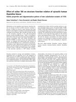
The histological analysis of the region between the first
and second molars of sham operated, shows the structure
of the normal periodontium, where gingival (g), peri-
odontal ligament (pl), alveolar bone (ab), cementum (c),
can be observed (Figure 3A; Table 1). The histopathology
of the periodontium of the animals subjected to experi-
mental periodontitis that received no treatment (EPD)
revealed inflammatory cell infiltration coupled with
severe cementum destruction and alveolar process
Figure 1 Effect of venlafaxine treatment on the alveolar bone
loss in experimental periodontitis in rats (EPD). Measurements
were made along the axis of each root of the first molar (three roots)
and two recordings for the second and third molar teeth (two roots
each). The total alveolar bone loss was obtained by taking the sum of
recordings from the buccal tooth and subtracting the value of the right
maxilla (unligated control) from the left (mm). Venlafaxine (10 or 50
mg/kg, orally) was administered 1 hour before ligature placement and
daily for 10 days. Control groups, sham (SO), and EPD were treated with
saline. Values represent mean ± S.D., * vs SO; ** vs EPD (p < 0.05, ANO-
VA and Tukey's test).
SO
EPD
Venla 10 + EPD
Venla 50 + EPD
0
2
4
6
8
10
*
Alveolar Bone Loss
(mm)
**
Figure 2 Macroscopic aspects of periodontium of rats submitted
to EPD and treated with venlafaxine. A sham operated (SO) with no
resorption of the alveolar bone when compared to the untreated
group (EPD), where severe bone resorption with root exposure is ob-
served B. Figure 2C and 2D shows the periodontium subjected to EPD
and treated with venlafaxine (10 or 50 mg/kg, administered orally 1
hour before ligature placement and daily for 10 days) where severe
bone loss is observed. Original magnification × 1.5.
Carvalho et al. Journal of Negative Results in BioMedicine 2010, 9:3
/>Page 4 of 8
destruction (Figure 3B; Table 1), receiving median score 2
(range 2 to 3). The venlafaxine (10 mg/kg) treatment was
not able to prevent the inflammatory parameters induced
by experimental periodontitis (Figures 3C), receiving
median scores 2(1-3) (Table 1). This value was not statis-
tically different when compared to the EPD group. Rats
treated with venlafaxine (10 or 50 mg/kg) alone did not
manifest bone loss or inflammatory changes in periodon-
tium (not shown in figure 2 or 3).
Immunohistochemical reaction for TNF- α and iNOS
The periodontium of rats submitted to experimental
periodontitis and received no treatment (EPD) showed
marked immune-staining for both TNF- α (Figure 4B, 4E)
and iNOS (Figure 4H, 4K) when compared to the perio-
dontium of the sham group (Figures 4A, 4D, and 4G, 4J,
respectively). Venlafaxine (10 mg/kg) failed to reduce the
TNF- α as well as the iNOS immune-staining in the peri-
odontium of rats submitted to experimental periodontitis
(Figures 4C, 4F and 4I, 4L, respectively).
TBARS production in EPD rats and the effect of venlafaxine
treatment
The extent of lipid peroxidation was analyzed in terms of
thiobarbituric acid reactive substances (TBARS),
expressed as malondialdehyde (MDA) in gingival tissue.
Rats submitted to experimental periodontitis (EPD)
showed an increase in lipid peroxidation compared with
sham group. Venlafaxine (10 mg/kg) prevented (p < 0.05)
the malondialdehyde formation (Figure 5).
Discussion
In the present study, we examined the effects of sero-
tonin-norepinephrine reuptake inhibitor, venlafaxine, on
the inflammatory events and bone loss associated with
EPD, for three reasons. The first reason is that venlafax-
ine has been described to possess anti-inflammatory and
immunoregulatory effects, and chronic periodontitis is
an inflammatory disorder and an immunologically com-
promised disease [18,19,24]. Secondly, there were few
controversial reports on state of depression in patients
with periodontitis and depression itself might contribute
to bone loss [1,3,25]. A third reason is that there have
been reports suggesting that selective serotonin reuptake
inhibitors can promote bone loss [12,13]. We used the lig-
ature-induced periodontitis in rats as a model system for
the study, since it is a highly reproducible experimental
model wherein ligation acts as a mechanical trauma on
the dentogingival area, thereby reducing tissue integrity
and allowing for intense host-plaque interaction, and
finally a bacterial plaque-formation. Lima et al have
shown that placement of a nylon thread around the sec-
ond upper molars induced significant alveolar bone loss
commencing day-3 of periodontitis induction, reaching a
maximum between days 7 and 11, and declining at the
14
th
day [21]. These data are in accordance with another
study that demonstrated a maximum bone loss 9
th
day
after ligature placement, and when sacrificed on day-14,
these animals showed shorter but thicker buccal alveolar
bone covered by the new bone, and a new dento-epithe-
lial junction formed subjacent to the ligature [20]. The
nylon thread functions as a bacterial plaque retentive fac-
tor that contributes to periodontitis [26], thus bacterial
stimulation induces a host response that leads to inflam-
matory cell infiltration, osteoclast formation, bone loss,
and the loss of tooth attachment [27]. The role of bacteria
and the host response in periodontitis has long been rec-
ognized. Together, these two factors lead to release of
inflammatory mediators and ultimately to alveolar bone
loss [28]. Among the mediators, the prostaglandins (PG),
mainly PGE2, and the cytokines, interleukin-1 (IL-1) and
tumor necrosis factor (TNF-α) play an active role in the
development of periodontal inflammation [29]. These
cytokines may in turn stimulate nitric oxide (NO) pro-
Table 1: Histological analysis of rat maxillae with EPD and
treated with venlafaxine.
GROUP SCORE
SO 0 (0-0)
EPD 2 (2-3) *
EPD + Venlafaxine (10 mg/kg) 2 (1-3) *
Data are reported as medians with range within parenthesis, n =
5. * vs SO (p < 0.004 (Kruskal-Wallis and Dunn' tests).
Figure 3 Histopathology of the periodontium of rats submitted
to EPD and treated with venlafaxine. Venlafaxine (10 mg/kg) was
administered orally 1 hour before ligature placement and daily for 10
days). Photomicrographs show the region between the first and sec-
ond molars of rats: A) sham operated (SO) normal periodontium,
where gingival (g), periodontal ligament (pl), alveolar bone (ab), ce-
mentum (c), B) periodontium of rat subjected to EPD showing inflam-
matory cell infiltration with severe cementum destruction and alveolar
process, C) periodontium of rat with EPD and treated with venlafaxine
showing no evidence of prevention of inflammation or bone resorp-
tion (arrow). H&E stain; original magnification × 40. Scale bars = 100
μm.
g
pl
ab
c
A C B
100 µm
Carvalho et al. Journal of Negative Results in BioMedicine 2010, 9:3
/>Page 5 of 8
duction, which has a role to play in periodontal disease
progression and in bone resorption [30,31].
In this study, the periodontium of rats submitted to
experimental periodontitis showed marked immunoreac-
tivity to both TNF-α and the iNOS isoenzyme that syn-
thesize NO from L-arginine as compared to
periodontium of the SO group, reinforcing the participa-
tion of TNF-α and NO in the development of EPD and
bone loss. Venlafaxine treatment didn't suppress effec-
tively the TNF-α nor the iNOS immunoreactivities. Voll-
mar et al have shown its immunomodulatory properties,
in murine experimental autoimmune encephalomyelitis
(EAE), a T-cell-mediated central nervous system demyeli-
nating disease model of multiple sclerosis. Venlafaxine
ameliorated the clinical symptoms of the disease possibly
by suppressed production of pro-inflammatory cytokines
Figure 4 Photomicrographs of periodontal tissue of rats on EPD and treated with venlafaxine showing the immunoreactivity to TNF-α (A-
F) and iNOS (G-L). (A,D,G,J) SO: sham operated rats; (B,E,H,K) rats subjected to EPD; (C,F,I,L) rats subjected to EPD and treated with venlafaxine (10 mg/
kg). A,B,C,G,H,I -100 ×, bar scale = 100 μm); D,E,F,J,K,L - 400 ×, bar scale = 50 μm). polpa (p) gingival (g), periodontal ligament (pl) and dentina (d).
A
B C
D
E
F
G
H I
J
K
L
100
µ
m
100
µ
m
50
µ
m
50
µ
m
lp
g
d
Carvalho et al. Journal of Negative Results in BioMedicine 2010, 9:3
/>Page 6 of 8
interleukin-12 (IL-12), p40, TNF-α and interferon-γ
(IFN-γ). These findings differ from ours, largely due to
the reason of differences in the model and treatment pro-
tocol, in which venlafaxine was administered at a higher
dose (60 mg/kg) and over a longer period (14 days) [32].
Venlafaxine treatment not only failed in preventing the
bone loss, but, at a high dose, also significantly enhanced
the bone loss. Evidence regarding a functional serotonin
(5-hydroxytryptamine) signaling system in bone has gen-
erated considerable recent interest. The specific bio-
chemical nature of serotoninergic pathways and their
direct and/or indirect effects on bone metabolism are still
unclear. Serotonin is involved in the pathophysiology of
depression, and therefore studies of depression and anti-
depressant treatments (as modulators of the serotonin
system) are relevant with regard to bone outcomes. SSRIs
have been associated with lower bone mineral density
(BMD) and increased rates of bone loss, as well as
increased rates of fracture after accounting for falls [33].
Selective serotonin-reuptake inhibitors (SSRIs) antago-
nize the serotonin (5-hydroxytryptamine) transporter (5-
HTT), and are frequently prescribed to children and ado-
lescents to treat depression. However, recent findings of
functional serotonergic pathways in bone cells and pre-
liminary clinical evidence demonstrating detrimental
effects of SSRIs on bone growth have raised questions
regarding the effects of these drugs on the growing skele-
ton. 5-HTT null mutant mice had a consistent skeletal
phenotype of reduced mass, altered architecture, and
inferior mechanical properties, whereas bone mineral
accrual was impaired in growing mice treated with a SSRI
[34]. These findings indicate that SSRIs do negatively
impact the skeleton and that further research is required
to decipher their precise influence.
Our observations on venlafaxine differ from the results
obtained in studies of Breivik et al [6], in which the use of
an antidepressant, tianeptine significantly inhibited the
alveolar bone loss in rats on ligature-induced periodonti-
tis. This discrepancy can be clarified the following way.
While venlafaxine is a member of SNRIs (serotonin-nora-
drenalin reuptake inhibitors), tianeptine is an atypical
antidepressant drug. In contrast to tricycle antidepres-
sants and selective serotonin reuptake inhibitors (SSRIs),
it has been suggested that tianeptine decreases sero-
tonin's bioactivity and its accumulation in serotonergic
synapses of the central nervous system by promoting
serotonin reuptake, and normalizing serotonergic neu-
rotransmission [35,36]. Venlafaxine, which has a mecha-
nism of action opposite to that of tianeptine (i.e.
inhibition of serotonin uptake) clearly explains its dose-
related effect on bone loss, observed in the present exper-
iment. Thus we show for the first time that SNRIs such as
venlafaxine are likely to worsen the bone loss in peri-
odontal disease.
Oxidative stress has been documented in periodontal
disease [37,38]. Patients with periodontitis have a signifi-
cantly higher level of TBARS than healthy people and this
suggests that TBARS of gingival tissue are closely associ-
ated with periodontal status and its measurements can
help in the treatment and monitoring of progression of
periodontal disease [37]. In this study we found that ani-
mals submitted to experimental periodontitis had high
levels of lipid peroxidation, the finding consistent with
earlier observations and the treatment with venlafaxine
(10 mg/kg) reduced significantly the lipid peroxidation in
these animals. Studies also showed an antioxidant effect
of venlafaxine, in rats rendered depressive [39,40]. How-
ever, these effects of venlafaxine do not seem to be favor-
able influenced in this study on the periodontitis
outcome (data not shown).
Several lines of evidence suggest that nitric oxide over-
production is associated with periodontal disease, the
presence of inducible nitric oxide synthase (iNOS) activ-
ity in inflamed gingival tissue of young patients has been
demonstrated [41]. Increased iNOS activity has also been
reported in rat experimental model of periodontitis, sug-
gesting that the gingivomucosal immune and epithelial
cells are able to induce this enzyme [22]. In periodontitis,
inducible nitric oxide synthase expression may have ben-
eficial as well as detrimental roles. Beneficial effects may
include antimicrobial activity, immune modulation, and
inhibition of microvascular thrombosis, as well as
increased tissue perfusion. On the other hand, detrimen-
tal effects may include a cytotoxic action toward the host
tissues, including alveolar bone resorption due to the
Figure 5 Malondialdehyde (MDA) levels in gingival tissue of rats
submitted to EPD and treated with venlafaxine. Animals were
treated with venlafaxine (10 mg/kg, orally) during 10 days. On day 11,
the gingival tissue was removed and analyzed for lipid peroxidation.
Values represent mean ± S.E.M. * vs SO, ** vs EPD (p < 0.05, ANOVA and
Tukey"s test).
SO
EPD
SO + Venla 10
EPD + Venla 10
0.0
0.5
1.0
1.5
2.0
2.5
3.0
3.5
*
**
MDA (
P
M)
Carvalho et al. Journal of Negative Results in BioMedicine 2010, 9:3
/>Page 7 of 8
stimulating effect of nitric oxide on the activity of the
osteoclasts [42]. In this study we observed an increase on
iNOS immunoreactivity in ligature-induced periodonti-
tis, a finding that corroborates with the study of Lohinai
et al [30]. It implies that venlafaxine lacks efficacy in sup-
pressing EPD-associated increase in iNOS expression.
Venlafaxine also failed to modify the cellular infiltration
response in the gingivomucosal tissues, in our experi-
mental conditions.
In conclusion, our results show that the tissue damage
induced by ligature is associated with bone loss, inflam-
matory response and increased immunoreactivity to
TNF-α and iNOS. We speculate that the venlafaxine
treated rats were not protected against bone loss possibly
for the reason that its antidepressant action involves syn-
aptic inhibition of serotonin uptake. Future studies
should address on other more selective reuptake inhibi-
tors (SSRIs) to know whether they also behave in same
fashion in EPD. Possibly, atypical antidepressants like
tianeptine that increase/favour synaptic uptake of sero-
tonin may be more useful to combat periodontitis-associ-
ated alveolar bone loss.
Competing interests
The authors declare that they have no competing interest.
Authors' contributions
RSC and GMA contributed equally in realizing experiments, data collection and
analysis. CMS and JCSN collaborated in immuno-histochemical studies (TNF-α
e iNOS) and in the evaluation of oxidant stress. SAHP and LMSP helped in
inducing experimental periodontitis in rats. GACB performed the histopatho-
logic analysis. The authors declare that they read and approved the final manu-
script.
Acknowledgements
The authors are grateful to Rao Satyanarayana for insightful discussions of
manuscript (Federal University of Ceará), Maria Vilani Bastos and Ivan Rodrigues
de Sousa (Federal University of Ceará) for technical assistance. The financial
support from the Ceará State Research Foundation (FUNCAP) and the Brazilian
National Research Council (CNPq) thanks.
Author Details
1
Laboratory of Neurosciences and Behavior, Department of Physiology and
Pharmacology, Federal University of Ceará, Rua Coronel Nunes de Melo, 1127,
CEP 60430-270, Fortaleza, CE, Brazil,
2
Department of Clinical Odontology,
Faculty of Pharmacy, Odontology and Nursing, Federal University of Ceará, Rua
Monsenhor Furtado, s/n, CEP 60441-750, Fortaleza, CE, Brazil and
3
Department
of Morphology, Faculty of Medicine, Federal University of Ceará, Rua Delmiro
de Farias, s/n, CEP 60416-030, Fortaleza, CE, Brazil
References
1. Persson GR, Persson RE, MacEntee CI, Wyatt CC, Hollender LG, Kiyak HA:
Periodontitis and perceived risk for periodontitis in elders with
evidence of depression. J Clin Periodontol 2003, 30:691-696.
2. Peruzzo DC, Benatti BB, Ambrosano GMB, Nogueira-Filho GR, Sallum EA,
Casati MZ, Nociti H Jr: A Systematic review of stress and psychological
factors as possible risk factors for periodontal disease. J Periodontol
2007, 78:1491-1504.
3. Rosanja AE, Low KG, McCormick CM, Rosanja DA: Stress, depression,
cortisol, and periodontal disease. J Periodontol 2009, 80:260-266.
4. Pavlov VA, Tracey KJ: Neural regulators of innate immune responses and
inflammation. Cell Mol Life Sci 2004, 61:2322-2331.
5. Behl Y, Siqueira M, Ortiz J, Li J, Desta T, Faibish D, Graves DT: Activation of
the acquired immune response reduces coupled bone formation in
response to a periodontal pathogen. J Immunol 2008, 81:8711-8718.
6. Breivik T, Gundersen Y, Myhrer T, Fonnum F, Osmundsen H, Murison R,
Gjermo P, Von Hörsten S, Opstad PK: Enhanced susceptibility to
periodontitis in an animal model of depression: reversed by chronic
treatment with the anti-depressant tianeptine. J Clin Periodontol 2006,
33:469-477.
7. Elter JR, White BA, Gaynes BN, Bader JD: Relationship of clinical
depression to periodontal treatment outcome. J Periodontol 2002,
73:441-449.
8. Maes M: The immunoregulatory effects of antidepressants. Hum
Psychopharmacol 2001, 16:95-103.
9. Kubera M, Lin AH, Kenis G, Bosmans E, van Bockstaele D, Maes M: Anti-
inflammatory effects of antidepressants through suppression of the
interferon-gamma/interleukin-10 production ratio. J Clin
Psychopharmacol 2001, 21:199-206.
10. Bas¸terzi AD, Yazici K, Buturak V, Cimen B, Yazici A, Eskandari G, Tot Acar S,
Tasdelen B: Effects of venlafaxine and fluoxetine on lymphocyte
subsets in patients with major depressive disorder: A flow cytometric
analysis. Prog Neuropsychopharmacol Biol Psychiatry 2009. doi: 10.1016/
j.jpnpbp.2009.09.025
11. Cizza G, Primma S, Csako G: Depression as a risk factor for osteoporosis.
Trends Endocrinol Metab 2009, 20:367-373.
12. Warden SJ, Haney EM: Skeletal effects of serotonin (5-
hydroxytryptamine) transporter inhibition: evidence from in vitro and
animal-based studies. J Musculoskelet Neuronal Interact 2008, 8:121-132.
13. Haney EM, Warden SJ, Bliziotes MM: Effects of selective serotonin
reuptake inhibitors on bone health in adults: time for
recommendations about screening, prevention and management.
Bone 2009. doi: 10.1016/j.bone.07.083
14. Feighner JP, Entsuah AR, McPherson MK: Efficacy of once-daily
venlafaxine extended release (XR) for symptoms of anxiety in
depressed outpatients. J Affect Disord 1998, 47:55-62.
15. Lemke MR: Antidepressant effects of dopamine agonists: Experimental
and clinical findings. Nervenarzt 2007, 78:31-38.
16. Piacentini MF, Clinckers R, Meeusen R, Sarre S, Ebinger G, Michotte Y:
Effects of venlafaxine on extracellular 5-HT, dopamine and
noradrenaline in the hippocampus and on peripheral hormone
concentrations in the rat in vivo. Life Sci 2003, 73:2433-2442.
17. De Oliveira RA, Cunha GMA, Borges KDM, Bruin EASF, Viana GSB, Bruin
VMS: The effect of venlafaxine on behaviour, body weight and striatal
monoamine levels on sleep-deprived female rats. Pharmacol Biochem
Behav 2004, 79:499-506.
18. Aricioğlu F, Buldanlioğlu U, Salanturoğlu G, Ozyalçin NS: Evaluation of
antinociceptive and anti-inflammatory effects of venlafaxine in the rat.
Agri 2005, 17:41-46.
19. Vollmar P, Haghikia A, Dermietzel R, Faustmann PM: Venlafaxine exhibits
an anti-inflammatory effect in an inflammatory co-culture model. Int J
Neuropsychopharmacol 2008, 11:111-117.
20. Sallay K, Sanavi F, Ring I, Pham P, Behling UH, Nowotny A: Alveolar bone
destruction in the immunosuppressed rat. J Periodontal Res 1982,
17:263-274.
21. Lima V, Bezerra MM, De Menezes Alencar VB, Vidal FD, Da Rocha FA, De
Castro Brito GA, De Albuquerque Ribeiro Lima V, Bezerra MM, Alencar
VBM: Effects of chlorpromazine on alveolar bone loss in experimental
periodontal disease in rats. Eur J Oral Sci 2000, 108:123-129.
22. Leitão RFC, Ribeiro RA, Chaves HV, Rocha FAC, Lima V, Brito GAC: Nitric
oxide synthase inhibition prevents alveolar bone resorption in
experimental periodontitis in rats. J Periodontol 2005, 76:956-963.
23. Draper HH, Hadely M: Malondialdehyde determination as an índex of
lipid peroxidation. Methods Enzymol 1990, 186:421-431.
24. Van Dyke TE: The management of inflammation in periodontal disease.
J Periodontol 2008, 79:1601-1608.
25. Ababneh KT, Al Shaar MBA, Taani DQ: Depressive symptoms in relation
to periodontal health in a Jordanian sample. Int J Dent Hyg 2009. Online
publication date: 1-Jul
26. Williams RC: Periodontal disease. N Engl J Med 1990, 8:373-382.
Received: 7 April 2010 Accepted: 14 June 2010
Published: 14 June 2010
This article is available from: 2010 Carvalho et al; licensee BioMed Central Ltd. This is an Open Access article distributed under the terms of the Creative Commons Attribution License ( ), which permits unrestricted use, distribution, and reproduction in any medium, provided the original work is properly cited.Journal of Negative Results in BioMedicine 2010, 9:3
Carvalho et al. Journal of Negative Results in BioMedicine 2010, 9:3
/>Page 8 of 8
27. Wahl SM, Costa GL, Mizel DE, Allen JB, Skalericu U, Mangan DF: Role of
transforming growth factor beta in the pathophysiology of chronic
inflammation. J Periodontol 1993, 64:407-415.
28. Bascones-Martínez A, Muñoz-Corcuera M, Noronha S, Mota P, Bascones-
Ilundain C, Campo-Trapero J: Host defence mechanisms against
bacterial aggression in periodontal disease: Basic mechanisms. Med
Oral Patol Oral Cir Bucal 2009, 14:680-685.
29. Lima V, Vidal FDP, Da Rocha FA, De Castro Brito GA, De Albuquerque
Ribeiro R: Effects of TNF-α inibitors pentoxifylline and thalidomide on
alveolar bone loss in short-term experimental periodontal disease in
rats. J Periodontol 2004, 75:156-162.
30. Lohinai Z, Benedek P, Fehér E, Gyorfi A, Rosivall L, Fazekas A: Protective
effects of mercaptoethylguanidine, a select inhibitor of inducible nitric
oxide synthase in ligature inducible periodontitis in rats. Br J Pharmacol
1998, 123:353-360.
31. Batista AC, Silva TA, Chun JH, Lara VS: Nitric oxide synthesis and severity
of human periodontal disease. Oral Diseases 2002, 8:254-260.
32. Vollmar P, Nessler S, Kalluri SR, Hartung HP, Hemmer B: The
antidepressant venlafaxine ameliorates murine experimental
autoimmune encephalomyelitis by suppression of pro-inflammatory
cytokines. Int J Neuropsychopharmacology 2009, 12:525-536.
33. Haney EM, Warden SJ, Bliziotes MM: Effects of selective serotonin
reuptake inhibitors on bone health in adults: time for
recommendations about screening, prevention and management?
Bone 2010, 46:13-17.
34. Warden SJ, Robling AG, Sanders MS, Bliziotes MM, Turner CH: Inhibition of
the serotonin (5-hydroxytryptamine) transporter reduces bone accrual
during growth. Endocrinology 2005, 146:685-693.
35. Uzbay TI: Tianeptine: potential influences on neuroplasticity and novel
pharmacological effects. Prog Neuropsychopharmacol Biol Psychiatry
2008, 32:915-924.
36. Uzbekov MG: Antidepressant action of tianeptine is connected with
acceleration of serotonin turnover in the synapse: a hypothesis.
Neuropsychopharmacol Hung 2009, 11:83-87.
37. Panjamurthy K, Manoharan S, Ramachandran CR: Lipid peroxidation and
antioxidant status in patients with periodontitis. Cell Mol Biol Lett 2005,
10:255-264.
38. Giannopoulou C, Krause KH, Müller F: The NADPH oxidase NOX2 plays a
role in periodontal pathologies. Semin Immunopathol 2008, 30:273-278.
39. Eren I, Naziroğlu M, Demirdas¸ A, Celik O, Uğuz AC, Altunbas¸ak A, Ozmen I,
Uz E: Venlafaxine modulates depression-induced oxidative stress in
brain and medulla of rat. Neurochem Res 2007, 32:497-505.
40. Kumar A, Garg R: A role of nitric oxide mechanism involved in the
protective effects of venlafaxine in sleep deprivation. Behav Brain Res
2008, 194:169-173.
41. Gaspirc B, Masera A, Skaleric U: Immunolocalization of inducible nitric
oxide synthase in localized juvenile periodontitis patients. Connect
Tissue Res 2002, 43:413-418.
42. Hukkanen M, Hughes FJ, Buttery LD, Gross SS, Evans TJ, Seddon S, Riveros-
Moreno V, Macintyre I, Polak JM: Cytokine-stimulated expression of
inducible nitric oxide synthase by mouse, rat, and human osteoblast-
like cells and its functional role in osteoblast metabolic activity.
Endocrinology 1995, 136:5445-5453.
doi: 10.1186/1477-5751-9-3
Cite this article as: Carvalho et al., Effect of venlafaxine on bone loss associ-
ated with ligature-induced periodontitis in Wistar rats Journal of Negative
Results in BioMedicine 2010, 9:3