Báo cáo khoa hoc:" VEGF receptors on PC12 cells mediate transient activation of ERK1/2 and Akt: comparison of nerve growth factor and vascular endothelial growth factor" doc
Bạn đang xem bản rút gọn của tài liệu. Xem và tải ngay bản đầy đủ của tài liệu tại đây (564.03 KB, 6 trang )
BioMed Central
Page 1 of 6
(page number not for citation purposes)
Journal of Negative Results in
BioMedicine
Open Access
Research
VEGF receptors on PC12 cells mediate transient activation of
ERK1/2 and Akt: comparison of nerve growth factor and vascular
endothelial growth factor
Ingrid Berger, Sonja Stahl, Natalia Rychkova and Ute Felbor*
Address: Department of Human Genetics, University of Würzburg, Germany
Email: Ingrid Berger - ; Sonja Stahl - ; Natalia Rychkova - ;
Ute Felbor* -
* Corresponding author
Abstract
Vascular endothelial growth factor (VEGF) and endostatin are angiogenic and anti-angiogenic
molecules, respectively, that have been implicated in neurogenesis and neuronal survival. Using
alkaline phosphatase fusion proteins, we show that the PC12 neuronal cell line contains cell
membrane receptors for VEGF but not for endostatin and the collagen XV endostatin homologue.
Immunocytochemistry confirmed that proliferating and differentiated PC12 cells express VEGF
receptors 1, 2 and neuropilin-1. While no functional effects of VEGF on PC12 cell proliferation and
differentiation could be observed, a slight VEGF-induced reduction of caspase-3 activity in
differentiated apoptotic PC12 cells was paralleled by transient activation of ERK1/2 and Akt. In
direct comparison, nerve growth factor proved to be a strikingly more potent neuroprotective
agent than VEGF.
Background
VEGF, VEGF receptor antagonists, and the C-terminal col-
lagen XVIII fragment endostatin, an inhibitor of angio-
genesis and tumor growth [1], have been tested for use in
long-term therapies to enhance or reduce vascularisation
[2]. Therefore, knowledge of VEGF and endostatin recep-
tor expression patterns as well as of their non-endothelial
cell functions is important. VEGF was originally identified
as a vascular permeability factor [3] which turned out to
be crucial for vasculo- and angiogenesis [4]. Later, non-
endothelial VEGF target cells have been described in a
variety of organs [5]. More recently, autocrine and para-
crine functions have been observed in neurogenesis and
neuronal survival in vitro and in vivo, both in the central
nervous system and the peripheral nervous system [6].
Endostatin was implicated in neuronal cell migration and
axon guidance in Caenorhabditis elegans [7]. Fc-endostatin
dimers were also reported to have motogenic activity on
rat pheochromocytoma PC12 cells cultured on Matrigel
[8], an extracellular matrix preparation used for differenti-
ation of endothelial cells into tube-like structures. NGF-
treated PC12 cells are an established model for analysis of
neuronal differentiation, neuronal survival and neuro-
trophin signal transduction [9]. Finally, increased neuro-
nal and paracellular endostatin deposits were found in
patients with Alzheimer's disease [10].
VEGF exerts its anti-apoptotic effect on hypoxic neurons
via VEGF receptor 2 (VEGFR-2), neuropilin-1 (NRP1), the
Ras/mitogen-activated protein kinase (MAPK) and the
phosphatidylinositol 3-kinase (PI3K)/Akt kinase path-
ways [11-13] as in VEGFR-2-dependent endothelial sur-
Published: 01 June 2006
Journal of Negative Results in BioMedicine 2006, 5:8 doi:10.1186/1477-5751-5-8
Received: 04 February 2006
Accepted: 01 June 2006
This article is available from: />© 2006 Berger et al; licensee BioMed Central Ltd.
This is an Open Access article distributed under the terms of the Creative Commons Attribution License ( />),
which permits unrestricted use, distribution, and reproduction in any medium, provided the original work is properly cited.
Journal of Negative Results in BioMedicine 2006, 5:8 />Page 2 of 6
(page number not for citation purposes)
vival [14]. Ras/MAPK and PI3K/Akt are also involved in
PC12 cell survival signaling stimulated by nerve growth
factor (NGF) [15,16]. Since VEGF has also been suggested
to act as a neurotrophin in motoneuron degeneration
[17], we intended to evaluate the effects of VEGF and
endostatins on neuronal differentiation and survival in
direct comparison with the prototypic neurotrophin NGF.
PC12 cells were first probed with dimeric fusion proteins
composed of the human placental isozyme of alkaline
phosphatase (AP) at the N-terminus and murine (m)
VEGF
164
or endostatins at the C-terminus. While the
endostatin affinity probes did not react with PC12 cells,
AP-mVEGF
164
strongly bound to proliferating and differ-
entiated PC12 cells. Although PC12 cells were subse-
quently shown to express VEGF receptors 1, 2 and
neuropilin-1, only a minor neuroprotective effect was
observed for VEGF when compared to NGF.
Materials and methods
Cell culture
PC12 cells were a gift from Drs. M. Sendtner and S. Wiese
(Department of Neurology, University of Wuerzburg, Ger-
many). Cow pulmonary artery endothelial (CPAE) cells
were purchased from ATCC (CCL-209). PC12 cells were
cultured in DMEM with glutamax-I (Gibco) supple-
mented with 10% horse serum, 5% fetal bovine serum,
100 U/ml penicillin G, and 100 µg/ml streptomycin
(Gibco) in 5% CO
2
at 37°C. For differentiation experi-
ments, PC12 cells were plated on poly-L-ornithine coated
tissue culture dishes and allowed to adhere over night (o/
n). After one wash with serum-free DMEM, the cells were
differentiated in serum-free DMEM containing 50 ng/ml
human recombinant NGF (PAN Biotech) for 3 days [18].
Although Fc-endostatin dimer application induced the
formation of multicellular PC12 aggregates on Matrigel
[8], Matrigel was not chosen for the current study since it
is an extracellular matrix preparation generally used for
endothelial tube formation assays.
Alkaline phosphatase staining of PC12 cells
For construction and expression of AP fusion proteins see
[19]. PC12 cells were either grown to 80% confluence or
differentiated in 6-well plates, and AP staining was per-
formed as described in [20]. Staining was monitored with
a Nikon Eclipse TE2000-U inverted microscope and doc-
umented using the Spot Insight QE Color imaging soft-
ware (Visitron). Quantitative measurement of AP fusion
protein binding to proliferating PC12 cells was carried out
as described previously [21].
Immunocytochemistry
PC12 cells plated on poly-L-ornithine coated glass cover-
slips were fixed in phosphate-buffered saline (PBS) con-
taining 4% paraformaldehyde at room temperature for 20
min, washed three times in prewarmed tris-buffered
saline (TBS) for 5 min, and incubated in blocking buffer
(TBS with 10% goat serum) at room temperature for 1 h.
After washing once with prewarmed TBS, the cells were
incubated with anti-Flt-1 (sc-316), anti-Flk-1 (sc-504) or
anti-Neuropilin (sc-5541) antibodies (2 µg/ml, Santa
Cruz Biotechnology) in blocking buffer at 4°C o/n.
Unbound primary antibodies were removed by washing,
and the cells were incubated in 20 mM ammonium chlo-
ride solution for 30 min to reduce autofluorescence. Cells
were stained for 1 h at 37°C with a secondary Cy3-conju-
gated goat anti-rabbit antibody (1:500, Dianova) in
blocking buffer. After washing, the coverslips were
mounted in Kaiser's glycerol gelatine (Merck). Fluorescent
preparations of proliferating PC12 cells were documented
at 600-fold magnification (Nikon Eclipse TE2000-U).
Image acquisition of differentiated PC12 cells was per-
formed with a Zeiss Axiophot at 1000-fold magnification.
Cell proliferation and cell death analyses
PC12 cell proliferation upon stimulation with 50 and 100
ng/ml VEGF
165
(R&D Systems) was assayed after 24 h, 48
h, and 72 h using the CellTiter 96
®
AQueous One Solution
Cell Proliferation Assay (Promega). These assays were per-
formed using two different cell densities (2 × 10
4
cells/cm
2
and 6.7 × 10
4
cells/cm
2
) and full serum as well as serum-
deprived conditions (0.1% and 0.4% horse serum). In
addition, increasing VEGF
165
concentrations from 0.2 to
400 ng/ml were added to PC12 cells cultured in 5% fetal
bovine serum, and [
3
H]thymidine incorporation was
measured 72 hours after onset of stimulation. The fluoro-
metric CaspACE™ Assay System and western blot analyses
with an anti-cleaved caspase-3 antibody (1:1000, Cell Sig-
naling, #9664) were used to monitor apoptosis of differ-
entiated PC12 cells (see below). All experiments were
performed in triplicate.
SDS-PAGE/Western blot analyses of differentiated
apoptotic PC12 cell lysates
PC12 cells were grown to 50% confluence in 10 cm tissue
culture dishes and NGF-differentiated for 72 h in serum-
free DMEM. For induction of apoptosis, cells were washed
three times with serum-free DMEM and incubated for 7.5
h under serum-deprived conditions in NGF-free DMEM.
After addition of exogenous recombinant human VEGF
165
(R&D Systems) for the indicated time frames, PC12 cells
were washed once with ice-cold PBS containing 100 µM
sodium orthovanadate, followed by centrifugation at
5000 rpm for 5 min at 4°C. The cells were lysed in 200 µl
of ice-cold lysis-buffer (HEPES, pH 7.8, 150 mM KOAc, 50
mM ß-glycerolphosphate, 25 mM NaF, 10 mM MgCl
2
, 5
mM EGTA, 1 mM EDTA, 10% glycerol, 1% Triton X-100,
0,05% (v/v) ß-mercaptoethanol, 1 µg/ml aprotinin, 6 µg/
ml chymostatin, 1 µg/ml leupeptin, 1 µg/ml pepstatin A,
1 mM PMSF, 1 mM sodium orthovanadate). Supernatants
were collected after centrifugation at 14 000 rpm for 10
Journal of Negative Results in BioMedicine 2006, 5:8 />Page 3 of 6
(page number not for citation purposes)
min at 4°C. Standardized samples containing 50 µg of
whole protein (Bradford assay) were separated using 10–
20% gradient gels. Proteins were wetblotted onto nitrocel-
lulose and probed with anti-phospho-ERK1/2 (1:2000,
Sigma, M8159) or anti-phospho-Akt-Ser473 (1:1000, Cell
Signaling, #9271) antibodies. The blots were stripped and
reprobed with antibodies detecting the respective non-
phosphorylated proteins (anti-ERK1, 1:1000, Santa Cruz
Biotechnology, sc-94; anti-Akt, 1:1000, Cell Signaling,
#9272). Horseradish peroxidase-conjugated secondary
antibodies (1:2000, Dianova) were visualized by
enhanced chemiluminescence detection (Western Light-
ning™ Chemiluminescence Reagent System, Perk-
inElmer). Experiments were performed in triplicate.
Results
Differential cell binding of VEGF and endostatins
To determine the expression profile of binding partners
for murine VEGF
164
, endostatin and the collagen XV
endostatin homologue on neuronal and endothelial cell
lines, PC12 and CPAE cells were incubated with AP fusion
proteins. The AP-mVEGF
164
affinity probe strongly stained
proliferating and differentiated PC12 cells (Fig. 1a, b). In
contrast, AP-mVEGF
110
which lacks the C-terminal
heparan sulfate binding domain, the endostatin domain
of collagen XVIII (AP-mESXVIII), and the collagen XV
endostatin homologue (AP-mESXV) did not bind to PC12
cells. Both AP-mVEGF
164
and AP-mESXVIII labelled CPAE
cells while AP-mVEGF
110
, AP-mESXV and control AP did
not (Fig. 1c). Quantitative measurement of AP fusion pro-
tein binding to proliferating PC12 cells confirmed the
above results (Fig. 1d). In agreement with lack of AP-
mESXVIII and AP-mESXV binding to PC12 cells, recom-
binant human endostatins [22] did neither promote nor
inhibit PC12 cell differentiation (data not shown). Thus,
PC12 cells are not a useful model for understanding
endostatin effects on cells.
PC12 cells express high-affinity VEGF receptors
Consistent with AP staining, it was shown by immunoflu-
orescence that PC12 cells express the high-affinity recep-
tor tyrosine kinases VEGF receptor 1 and 2 (VEGFR-1,
VEGFR-2) as well as the low-affinity receptor neuropilin-
1 (Fig. 2). These receptors are expressed on the cell surface
of proliferating (Fig. 2a–c) and differentiated (Fig. 2e)
PC12 cells and seem to possess a clustered morphology
reminiscent of activated tyrosine kinases. Comparable
results were obtained with antibodies from Santa Cruz
Biotechnology and Dianova (data not shown).
VEGF induces transient activation of ERK1/2 and Akt
kinase in differentiated apoptotic PC12 cells
While the addition of VEGF
165
had no effect on PC12 cell
proliferation and neurite formation, a consistent but non-
significant reduction of caspase-3 activity became appar-
ent when VEGF
165
was administered to differentiated
apoptotic PC12 cells (data not shown). To analyze VEGF
signaling in PC12 cell survival, the activation of extracel-
lular signal-regulated kinases ERK1/2 and Akt was exam-
ined by Western blot analyses using phospho-specific
antibodies and their respective non phosphorylated coun-
terparts. Control cultures demonstrated that ERK1/2
(p44/p42 MAPK) and Akt activation are sustained for 79.5
h in the presence of NGF (Fig. 3, lane 1). Removal of NGF
for 7.5 h after 72 hours of differentiation led to a signifi-
cant reduction of ERK1/2 and Akt phosphorylation (Fig.
3, lane 2). Exogenous addition of 100 ng/ml VEGF
165
to
NGF-deprived PC12 cells resulted in transient ERK1/2
activity within 7–10 min which decreased almost to con-
trol levels 20 min after stimulation (Fig. 3, lanes 3, 4, and
data not shown). Higher concentrations of recombinant
VEGF (200 ng/ml) did not increase or prolong ERK1/2
activity (data not shown). Similar activation kinetics were
observed for phosphorylation of Akt at serine 473 (Fig. 3).
For comparison of signaling mechanisms, apoptotic PC12
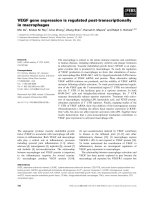
Alkaline phosphatase (AP) staining of (a, d) proliferating and (b) differentiated PC12 cells revealed binding of the AP-mVEGF
164
affinity probe while AP-murine endostatin (AP-mESXVIII) only stained (c) endothelial cellsFigure 1
Alkaline phosphatase (AP) staining of (a, d) proliferating and
(b) differentiated PC12 cells revealed binding of the AP-
mVEGF
164
affinity probe while AP-murine endostatin (AP-
mESXVIII) only stained (c) endothelial cells.
Journal of Negative Results in BioMedicine 2006, 5:8 />Page 4 of 6
(page number not for citation purposes)
cells were also NGF-stimulated. NGF-stimulation induced
a much more pronounced and sustained activation of
ERK1/2 and Akt which was even more prominent than in
NGF treated non-apoptotic control cells (Fig. 3, lanes 5,
6).
Discussion
We here report that endostatin affinity probes derived
from collagens XVIII and XV do not bind to PC12 cells
indicating that these cells do not express endostatin cell
membrane receptors. This observation is consistent with
absent effects of recombinant endostatins on neurite out-
growth (data not shown) and lack of binding of AP-
mESXVIII and AP-mESXV to murine embryonal nerve tis-
sues [19]. AP-mESXVIII predominantly labelled blood
vessels while AP-mESXV binding was restricted to the
lense capsule [19] which correlates with the current results
that only AP-mESXVIII, but not AP-mESXV, strongly
stained CPAE cells. As opposed to the endostatin affinity
probes, AP-mVEGF
164
showed strong binding to PC12
cells. Undifferentiated PC12 cells were known to express
VEGF to stimulate angiogenesis [23]. We now demon-
strate that proliferating and differentiated PC12 cells also
express VEGFR-1 and -2 and NRP1. NRP1 acts as an iso-
form-specific VEGF co-receptor which only binds VEGF
165
[24]. Since C-terminally deleted AP-mVEGF
110
did not
bind to PC12 cells, NRP1 appears to be required for the
interaction of VEGF
165
with PC12 cells.
Despite prominent expression of VEGF receptors on PC12
cells, exogenous VEGF
165
had no effect on PC12 cell pro-
liferation and neurite formation. One reason might be
endogenous VEGF-expression of proliferating PC12 cells
which is downregulated only 48 h after induction of dif-
ferentiation with NGF [23]. This would also explain the
slight anti-apoptotic effect of VEGF
165
on PC12 cells that
had been differentiated for three days prior to VEGF
165
stimulation. However, only an insignificant decrease of
cell proliferation could be observed upon treatment with
an antibody against rat VEGF
164
(data not shown). Thus,
our data are in line with the observation that VEGFR-1-
expressing cells show a poor mitogenic response to VEGF
stimulation [5]. Analysis of VEGF
165
-induced signal trans-
duction in differentiated apoptotic PC12 cells demon-
strated activation of ERK1/2 and Akt. The transient nature
of VEGF
165
-triggered ERK1/2 phosphorylation in PC12
cells provides a further explanation for the observation
that VEGF
165
was not able to induce PC12 cell differentia-
tion which is known to require sustained activation of the
MAPK cascade [25]. The inefficient rescue of PC12 cells
from apoptosis through VEGF
165
is likely also due to its
short-lived and relatively small effect on ERK1/2 and Akt.
VEGF-induced neuroprotective signaling via VEGFR-2,
NRP1 and the two above-mentioned signaling cascades
was shown in hypoxic and glucose-deprived hippocampal
neuron × neuroblastoma (HN33) hybrid cells [11], in rat
primary hippocampal neurons that had been exposed to
glutamate [12], and in hypoxic murine primary cortical
neurons [13]. In these in vitro model systems of cerebral
ischemia, no comparison of VEGF and NGF activation
kinetics was performed. Our results on growth factor stim-
ulated PC12 cells show that both the anti-apoptotic effect
and the activation of ERK1/2 and Akt were transient and
minor when compared to NGF. It remains to be clarified
whether this is a consequence of experimental conditions,
cell line-specific or a general feature of VEGF-induced neu-
roprotection.
Conclusion
Based on experiments using growth factor deprivation,
the present study suggests that NGF protects neuronal
cells from cell death much more efficiently than VEGF
165
.
The significant NGF-induced reduction of caspase-3 activ-
ity in differentiated apoptotic PC12 cells correlates with a
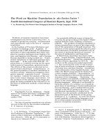
Immunodetection of VEGF receptors expressed on the cell surface of (a-d) proliferating and (e) differentiated PC12 cellsFigure 2
Immunodetection of VEGF receptors expressed on the cell
surface of (a-d) proliferating and (e) differentiated PC12
cells. PC12 cells were stained with polyclonal antibodies
against (a) VEGFR-1, (b, e) VEGFR-2, and (c) neuropilin-1.
The primary antibody was omitted in (d) controls. Differenti-
ated PC12 cells were also immunoreactive for VEGFR-1 and
neuropilin-1 (data not shown).
Publish with BioMed Central and every
scientist can read your work free of charge
"BioMed Central will be the most significant development for
disseminating the results of biomedical research in our lifetime."
Sir Paul Nurse, Cancer Research UK
Your research papers will be:
available free of charge to the entire biomedical community
peer reviewed and published immediately upon acceptance
cited in PubMed and archived on PubMed Central
yours — you keep the copyright
Submit your manuscript here:
/>BioMedcentral
Journal of Negative Results in BioMedicine 2006, 5:8 />Page 5 of 6
(page number not for citation purposes)
much more pronounced and prolonged activation of
downstream effectors when compared to VEGF
165
. Thus,
the angiogenic compound VEGF
165
may only be a minor
player in neurogenesis and neuronal survival and may
only have little therapeutic and side effects on neuronal
cells.
Acknowledgements
This work was supported by an Emmy Noether-grant from the Deutsche
Forschungsgemeinschaft (Fe 432/6-4). Sonja Stahl receives a stipend from
the Graduiertenkolleg 1048.
References
1. O'Reilly MS, Boehm T, Shing Y, Fukai N, Vasios G, Lane WS, Flynn E,
Birkhead JR, Olsen BR, Folkman J: Endostatin: an endogenous
inhibitor of angiogenesis and tumor growth. Cell 1997,
88:277-285.
2. Cross MJ, Dixelius J, Matsumoto T, Claesson-Welsh L: VEGF-
receptor signal transduction. Trends Biochem Sci 2003,
28:488-494.
3. Senger DR, Galli SJ, Dvorak AM, Perruzzi CA, Harvey VS, Dvorak HF:
Tumor cells secrete a vascular permeability factor that pro-
motes accumulation of ascites fluid. Science 1983, 219:983-985.
4. Ferrara N, Gerber HP, LeCouter J: The biology of VEGF and its
receptors. Nat Med 2003, 9:669-676.
5. Matsumoto T, Claesson-Welsh L: VEGF receptor signal trans-
duction. Sci STKE 2001, 2001(112):RE21.
6. Storkebaum E, Lambrechts D, Carmeliet P: VEGF: once regarded
as a specific angiogenic factor, now implicated in neuropro-
tection. Bioessays 2004, 26:943-954.
7. Ackley BD, Crew JR, Elamaa H, Pihlajaniemi T, Kuo CJ, Kramer JM:
The NC1/endostatin domain of Caenorhabditis elegans type
XVIII collagen affects cell migration and axon guidance. J Cell
Biol 2001, 152:1219-1232.
8. Kuo CJ, LaMontagne KRJ, Garcia-Cardena G, Ackley BD, Kalman D,
Park S, Christofferson R, Kamihara J, Ding YH, Lo KM, Gillies S, Folk-
man J, Mulligan RC, Javaherian K: Oligomerization-dependent
regulation of motility and morphogenesis by the collagen
XVIII NC1/endostatin domain. J Cell Biol 2001, 152:1233-1246.
9. Kaplan DR, Miller FD: Signal transduction by the neurotrophin
receptors. Curr Opin Cell Biol 1997, 9:213-221.
10. Deininger MH, Fimmen BA, Thal DR, Schluesener HJ, Meyermann R:
Aberrant neuronal and paracellular deposition of endostatin
in brains of patients with Alzheimer's disease. J Neurosci 2002,
22:10621-10626.
11. Jin KL, Mao XO, Greenberg DA: Vascular endothelial growth
factor: direct neuroprotective effect in in vitro ischemia. Proc
Natl Acad Sci U S A 2000, 97:10242-10247.
12. Matsuzaki H, Tamatani M, Yamaguchi A, Namikawa K, Kiyama H,
Vitek MP, Mitsuda N, Tohyama M: Vascular endothelial growth
factor rescues hippocampal neurons from glutamate-
induced toxicity: signal transduction cascades. FASEB J 2001,
15:1218-1220.
13. Ogunshola OO, Antic A, Donoghue MJ, Fan SY, Kim H, Stewart WB,
Madri JA, Ment LR: Paracrine and autocrine functions of neuro-
nal vascular endothelial growth factor (VEGF) in the central
nervous system. J Biol Chem 2002, 277:11410-11415.
14. Gerber HP, McMurtrey A, Kowalski J, Yan M, Keyt BA, Dixit V, Fer-
rara N: Vascular endothelial growth factor regulates
endothelial cell survival through the phosphatidylinositol 3'-
kinase/Akt signal transduction pathway. Requirement for
Flk-1/KDR activation. J Biol Chem 1998, 273:30336-30343.
15. Xia Z, Dickens M, Raingeaud J, Davis RJ, Greenberg ME: Opposing
effects of ERK and JNK-p38 MAP kinases on apoptosis. Sci-
ence 1995, 270:1326-1331.
16. Wert MM, Palfrey HC: Divergence in the anti-apoptotic signal-
ling pathways used by nerve growth factor and basic fibrob-
last growth factor (bFGF) in PC12 cells: rescue by bFGF
involves protein kinase C delta. Biochem J 2000, 352 Pt
1:175-182.
17. Sopher BL, Thomas PSJ, LaFevre-Bernt MA, Holm IE, Wilke SA, Ware
CB, Jin LW, Libby RT, Ellerby LM, La Spada AR: Androgen receptor
YAC transgenic mice recapitulate SBMA motor neuronopa-
thy and implicate VEGF164 in the motor neuron degenera-
tion. Neuron 2004, 41:687-699.
18. Gotz R, Karch C, Digby MR, Troppmair J, Rapp UR, Sendtner M: The
neuronal apoptosis inhibitory protein suppresses neuronal
differentiation and apoptosis in PC12 cells. Hum Mol Genet
2000, 9:2479-2489.
19. Rychkova N, Stahl S, Gaetzner S, Felbor U: Non-heparan sulfate-
binding interactions of endostatin/collagen XVIII in murine
development. Dev Dyn 2005, 232:399-407.
20. Flanagan JG, Cheng HJ, Feldheim DA, Hattori M, Lu Q, Vander-
haeghen P: Alkaline phosphatase fusions of ligands or recep-
tors as in situ probes for staining of cells, tissues, and
embryos. Methods Enzymol 2000, 327:19-35.
21. Stahl S, Gaetzner S, Mueller TD, Felbor U: Endostatin phenyla-
lanines 31 and 34 define a receptor binding site. Genes to Cells
2005, 10(9):929-39.
22. Gaetzner S, Deckers MM, Stahl S, Lowik C, Olsen BR, Felbor U:
Endostatin's heparan sulfate-binding site is essential for inhi-
bition of angiogenesis and enhances in situ binding to capil-
lary-like structures in bone explants. Matrix Biol 2005,
23:557-561.
23. Claffey KP, Wilkison WO, Spiegelman BM: Vascular endothelial
growth factor. Regulation by cell differentiation and acti-
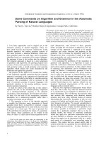
Western blot analyses of VEGF-induced signal transduction in differentiated PC12 cells after NGF withdrawalFigure 3
Western blot analyses of VEGF-induced signal transduction
in differentiated PC12 cells after NGF withdrawal. Lysates of
control cells maintained in the presence of NGF were loaded
in lane 1. NGF-deprived PC12 cells (lane 2) treated with
VEGF
165
(lanes 3, 4) or NGF (lanes 5, 6) demonstrated that
VEGF
165
induced transient activation of ERK1/2 and Akt after
7 min. In contrast, NGF produced a stronger and persistent
phosphorylation of ERK1/2 and Akt than VEGF
165
.
Publish with BioMed Central and every
scientist can read your work free of charge
"BioMed Central will be the most significant development for
disseminating the results of biomedical research in our lifetime."
Sir Paul Nurse, Cancer Research UK
Your research papers will be:
available free of charge to the entire biomedical community
peer reviewed and published immediately upon acceptance
cited in PubMed and archived on PubMed Central
yours — you keep the copyright
Submit your manuscript here:
/>BioMedcentral
Journal of Negative Results in BioMedicine 2006, 5:8 />Page 6 of 6
(page number not for citation purposes)
vated second messenger pathways. J Biol Chem 1992,
267:16317-16322.
24. Soker S, Fidder H, Neufeld G, Klagsbrun M: Characterization of
novel vascular endothelial growth factor (VEGF) receptors
on tumor cells that bind VEGF165 via its exon 7-encoded
domain. J Biol Chem 1996, 271:5761-5767.
25. Traverse S, Gomez N, Paterson H, Marshall C, Cohen P: Sustained
activation of the mitogen-activated protein (MAP) kinase
cascade may be required for differentiation of PC12 cells.
Comparison of the effects of nerve growth factor and epider-
mal growth factor. Biochem J 1992, 288 ( Pt 2):351-355.