Báo cáo y học: "Early Differential signaling mechanisms regulate expression of CC chemokine receptor-2 during monocyte maturatio" ppsx
Bạn đang xem bản rút gọn của tài liệu. Xem và tải ngay bản đầy đủ của tài liệu tại đây (765.1 KB, 14 trang )
BioMed Central
Page 1 of 14
(page number not for citation purposes)
Journal of Inflammation
Open Access
Research
Differential signaling mechanisms regulate expression of CC
chemokine receptor-2 during monocyte maturation
Roderick J Phillips*
1,3
, Marin Lutz
1
and Brett Premack
1,2,4
Address:
1
Department of Physiology David Geffen School of Medicine at University of California, Los Angeles, Los Angeles, California, 90095 USA,
2
Jonsson Comprehensive Cancer Center, David Geffen School of Medicine at University of California, Los Angeles, Los Angeles, California, 90095
USA,
3
Department of Discovery Research, Intermune, 3280 Bayshore Blvd, Brisbane, California, 94005 USA and
4
Department of Technology
Development, ChemoCentryx Inc., 1539 Industrial Road, San Carlos, California USA
Email: Roderick J Phillips* - ; Marin Lutz - ; Brett Premack -
* Corresponding author
HumanCellular DifferentiationCell Surface MoleculesGene Regulation
Abstract
Background: Peripheral blood monocytes and monocyte-derived macrophages are key
regulatory components in many chronic inflammatory pathologies of the vasculature including the
formation of atherosclerotic lesions. However, the molecular and biochemical events underlying
monocyte maturation are not fully understood.
Methods: We have used freshly isolated human monocytes and the model human monocyte cell
line, THP-1, to investigate changes in the expression of a panel of monocyte and macrophage
markers during monocyte differentiation. We have examined these changes by RT-PCR and FACS
analysis. Furthermore, we cloned the CCR2 promoter and analyzed specific changes in
transcriptional activation of CCR2 during monocyte maturation.
Results: The CC chemokine receptor 2 (CCR2) is rapidly downregulated as monocytes move
down the macrophage differentiation pathway while other related chemokine receptors are not.
Using a variety of biochemical and transcriptional analyses in the human THP-1 monocyte model
system, we show that both monocytes and THP-1 cells express high levels of CCR2, whereas THP-
1 derived macrophages fail to express detectable CCR2 mRNA or protein. We further
demonstrate that multiple signaling pathways activated by IFN-γ and M-CSF, or by protein kinase
C and cytoplasmic calcium can mediate the downregulation of CCR2 but not CCR1.
Conclusion: During monocyte-to-macrophage differentiation CCR2, but not CCR1, is
downregulated and this regulation occurs at the level of transcription through upstream 5'
regulatory elements.
Background
Chemokines are a superfamily of small (8–10 kDa) pro-
teins, which coordinate cellular responses to inflamma-
tion, insult or injury [1-4]. They also play a pivotal role in
the regulation of leukocyte trafficking and extravasation
through the luminal surface of endothelial cells into sites
Published: 31 October 2005
Journal of Inflammation 2005, 2:14 doi:10.1186/1476-9255-2-14
Received: 15 December 2004
Accepted: 31 October 2005
This article is available from: />© 2005 Phillips et al; licensee BioMed Central Ltd.
This is an Open Access article distributed under the terms of the Creative Commons Attribution License ( />),
which permits unrestricted use, distribution, and reproduction in any medium, provided the original work is properly cited.
Journal of Inflammation 2005, 2:14 />Page 2 of 14
(page number not for citation purposes)
of tissue inflammation. The chemokine superfamily
includes at least 20 receptors and more than 50 ligands [1-
5]. The chemokine ligands can be separated into two
major categories depending on whether they express a CC
or CXC amino acid motif in their N-termini. This dichot-
omy appears to be functionally important since many CC
chemokines preferentially target monocytes and T cells,
while CXC chemokines such as IL-8 (CXCL8) tend to
attract neutrophils. The CC chemokines bind to a family
of G-protein coupled serpentine (seven transmembrane
spanning) receptors, which are termed CC chemokine
receptors (CCRs; [1,3,6]). Currently ten of the CC recep-
tors have been identified and monocytes predominantly
express three of them: CCR1, CCR2 and CCR5 [2,7,8].
These receptors can bind and signal to different CC chem-
okines including MCP-1 (CCL2), MIP-1α (CCL3) and
RANTES (CCL3) [3,4,9] and these same chemokines are
secreted by endothelial cells when activated by LDL or
inflammatory cytokines [10-13] or when the endothelium
is damaged [14,15].
Indeed, the recruitment of peripheral blood monocytes to
the site of injured endothelium by pro-inflammatory
chemokines is a key regulatory component in the forma-
tion of an atherosclerotic lesion [16,17]. The monocytes
subsequently adhere to the endothelium and eventually
migrate into the sub-intima [18,19]. Here, they receive a
series of differentiation signals including macrophage-col-
ony stimulating factor (M-CSF) and minimally oxidized
LDL that enables them to mature into macrophages. These
macrophages then engulf large quantities of cholesterol to
become lipid-laden foam cells. And it is the accumulation
of these foam cells that eventually leads to the formation
of characteristic fatty streaks, intermediate lesions and
fibrous plaques [20,21].
To date, though, the actual role of chemokines and their
receptors in atherosclerosis has not been clearly estab-
lished. However, recent studies using transgenic mouse
models of atherosclerosis have provided convincing evi-
dence that CCR2 is required for disease progression in
apolipoprotein E-null mice [22,23]. In these animals, dis-
ruption of the CCR2 gene greatly decreases lesion forma-
tion without affecting plasma lipid or lipoprotein
concentrations. Using a slightly different approach Roll-
ins and colleagues have demonstrated that CCL2, the lig-
and for CCR2, plays an equally important role in the
development of atherosclerosis in low-density lipoprotein
receptor deficient mice [24,25]. Here, deletion of CCL2
leads to a significant reduction in lipid deposition within
the aorta.
Despite the promising experimental results from these
systems, relatively little is known about how the expres-
sion of chemokine receptor genes is regulated in normal
or diseased human tissues. A recent paper by Yamamoto
and colleagues [26] examined the basal regulatory mech-
anisms underlying expression of the CCR2 gene in the
human monocyte cell line, THP-1. Indeed, this group
characterized two key elements that seemed to be neces-
sary and sufficient for the basal regulation of CCR2
expression: an Oct-1 binding sequence located 36 bp
upstream of the TATA box and a tandem CAAT/enhancer-
binding protein (C/EBP) binding sequence located, unu-
sually, in the 5' UTR (at +50 to +77 bp). However, studies
have not directly examined the molecular mechanisms by
which basal expression of CCR2 is rapidly downregulated
during the differentiation of monocytes into macro-
phages.
In an effort to address this issue, we have further devel-
oped a model of monocyte differentiation using THP-1
cells, which can be induced to mature into macrophages
using either phorbol esters and ionomycin or a physiolog-
ical combination of interferon-γ (IFN-γ) and M-CSF. In
common with other studies, we report here that THP-1
cell maturation mediated by either high concentrations of
PMA (50 nM) alone, or very low concentrations of PMA
(1 nM) plus ionomycin (1 µM) is characterized by an
increase in size, the development of an adherent pheno-
type and the up-regulation of a panel of differentiation
markers [27-30]; in addition, CCR2, but not CCR1, was
specifically down-regulated during differentiation. Modu-
lation of CCR2 by PMA (50 nM), but not PMA (1 nM)
plus ionomycin (1 µM), was found to be sensitive to inhi-
bition by the broad-spectrum protein kinase inhibitor
staurosporine. Furthermore, transient transfection of
THP-1 cells with a CCR2-specific reporter construct indi-
cated that PMA (50 nM) and PMA (1 nM) plus ionomycin
(1 µM) mediated the downregulation of CCR2 through
inhibition of CCR2-specific gene transcription. Moreover,
physiological treatment of THP-1 monocytes with two
known differentiation factors, IFN-γ and M-CSF, also pro-
moted a differentiation phenotype essentially identical to
that observed using pharmacologic stimuli. These data
indicate that the activation of several intracellular signal-
ing pathways selectively regulate the expression of CCR2
during monocyte maturation into macrophages.
Materials and methods
Cell lines
The THP-1 human monocytic cell line (ATCC) was grown
in RPMI 1640 medium (GibcoBRL) containing 10 % fetal
calf serum (FCS; GibcoBRL), 100 U/ml penicillin and 100
µg/ml streptomycin (GibcoBRL). The cells were main-
tained in culture at 37°C and 5% C0
2
. Typically, cells (7 ×
10
6
per point) were stimulated with 50 nM phorbol myr-
istate acetate (PMA; Sigma) or 1 nM PMA plus 1 µM ion-
omycin (Calbiochem) in the presence or absence of the
PKC inhibitor staurosporine (Calbiochem).
Journal of Inflammation 2005, 2:14 />Page 3 of 14
(page number not for citation purposes)
Isolation and culture of human peripheral blood
monocytes
Peripheral blood mononuclear cells (PBMCs) were iso-
lated from freshly prepared leukopacks (buffy coats) that
were between 2–4 hours old. Briefly, 20 ml of blood from
leukopacks were diluted using PBS (1:1) and layered over
15 ml of Ficoll-Paque PLUS (Amersham Pharmacia Bio-
tech). Cells were then centrifuged at 400 × g for 20 min-
utes at room temperature. After this time, PBMCs were
collected from the interphase and washed (× 2) with PBS
and centrifuged at 150 × g for 10 minutes. Monocytes
were further isolated from PBMCs using Percoll (Amer-
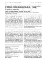
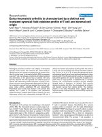
Macrophage-derived monocytes selectively downregulate CCR2, but not CCR1, during differentiationFigure 1
Macrophage-derived monocytes selectively downregulate CCR2, but not CCR1, during differentiation. (a).
Changes in morphology between freshly isolated monocytes (left panel) and cells cultured for 5 days (right panel) were deter-
mined using a Nikon Diaphot Camera set up and Axon Imaging Workbench software. Magnification is at 60 ×. (b). Freshly iso-
lated monocytes were either cultured for 5 days (broken line) or immediately stained (solid line) for a panel of macrophage
markers: CD36 (left panel), CD11b (middle panel) or CD68 (right panel). Dotted histograms represent the isotype controls.
(c). Panel I. Genomic DNA was prepared from freshly isolated monocytes and assayed for germ line expression of chemokine
receptors CCR1-CCR9 and CXCR1-CXCR5 by PCR using primers designed in-house. Note each primer pair amplified a single
product only, thus confirming that the primers are functional and specific. Panel II. Messenger RNA was prepared from freshly
isolated monocytes (upper panel) and cells that had been cultured for either 2 days (middle panel) or 5 days (lower panel). Sub-
sequently, RT-PCR was performed using primers for chemokine receptors CCR1-CCR9, CXCR1-CXCR5 and GAPDH.
Marker is a 100 bp DNA ladder. Similar results were obtained in three other experiments. (d). Freshly isolated monocytes
(upper panel plots 1, 4, 7, 10, 13 and 16) and cells that had been cultured for either 2 days (middle panel plots 2, 5, 8, 11, 14
and 17) or 5 days (lower panel plots 3, 6, 9, 12, 15 and 18) were stained for CCR1, CCR2, CCR5, CCR7, CXCR2 and CXCR4.
Cells were then analyzed for changes in chemokine receptor expression by flow cytometry. Similar results were obtained in
three other experiments.
A
Day 0
Day 5
B
CD36 CD11b CD68
Day 2
Day 0
Day 5
CCR1
CCR2
CCR3
CCR4
CCR5
CCR6
CCR7
CXCR1
CXCR2
CXCR3
CXCR4
CXCR5
CCR8
GAPDH
GAPDH
CCR9
Marker
Marker
CI
CII
D
CCR1 CCR5CCR2 CXCR4CCR7 CXCR2
Day 0
DAY 2
DAY 5
PANEL 1 4 7 10 13 16
PANEL 2 5 8 11 14 17
PANEL 3 6 9 12 15
18
Journal of Inflammation 2005, 2:14 />Page 4 of 14
(page number not for citation purposes)
sham Pharmacia Biotech) gradient centrifugation as previ-
ously described [31]. Lipid staining of the monocytes
revealed that their purity was greater than 90%. Finally,
the cells were resuspended and cultured at 10
6
/ml in
RPMI 1640 supplemented with 10% autologous serum,
penicillin and streptomycin (GibcoBRL).
Cloning the CCR2 promoter
A 1335 bp fragment of the promoter from the hCCR2
gene was cloned into the pGL3 vector (Promega) using
sequences determined by Yamamoto and colleagues [26].
This construct, termed pGL3-1335, contained the tandem
C/EBP sites plus 1220 bp of the promoter sequence 5' of
the transcriptional start site. The 5' primer contained a
restriction site for kpnI, while the 3' primer contained a
HindIII site. Each primer started with a 2 bp GC-rich
clamp. The full primer sequences used are as follows:
pGL3-1335 5' CGGGTACCGCTGCTTTAGGTCCATTTAC-
CCTC
pGL3-1335 3' GCAAGCTTATTGTACATTGGGTTGAG-
GTCTCC.
The genomic PCR was performed using an annealing tem-
perature of 55°C (30 seconds) and an extension tempera-
ture of 72°C (2 minutes); 30 cycles of PCR were
performed.
RNA isolation and RT-PCR
Total RNA was isolated using TRIzol (Life Technologies)
and by following the manufacturer's instructions. Briefly,
cells were lyzed in TRIzol and then mixed with chloro-
form. The lysate was then centrifuged to separate RNA,
DNA and protein. Total RNA, which is contained in the
upper aqueous phase was recovered and mixed with iso-
propanol to precipitate the RNA. The RNA was finally
washed in 75% ethanol to remove impurities and dis-
solved in water.
5 µg of RNA prepared in this way was then taken and
DNase treated to remove further enzymatic contamina-
tion, before being reverse transcribed to cDNA using a
ProSTAR First Strand RT-PCR kit from Stratagene and by
following the manufacturer's instructions.
Subsequently, RT-PCR was performed under standard
conditions using primers specific for CCR1, CCR2 and
GAPDH. The primer sequences used here were:
CCR1 sense 5'GAAACTCCAAACACCACAGAGGAC
CCR1 antisense 5'TTCGTGAGGAAAGTGAAGGCTG
CCR2 sense 5'CCACATCTCGTTCTCGGTTTATCAG
CCR2 antisense 5'CGTGGAAAATAAGGGCCACAG
CCR3 sense 5'CACTAGATACAGTTGAGACCTTTGG
CCR3 antisense 5'GGTAAAGAGCACTGCAAAGAGTC
CCR4 sense 5'ACCCCACGGATATAGCAGATACC
CCR4 antisense 5'CGTCGTGGAGTTGAGAGAGTACTTG
CCR5 sense 5'GGAGCCCTGCCAAAAAATC
CCR5 antisense 5'CTGTATGGAAAATGAGAGCTGC
CCR6 sense 5'TGGCAAGGGGTATAATTTGGG
CCR6 antisense 5'GACAGTCTGGTACTTGGGTTCACAG
CCR7 sense 5'AGACAGGGGTAGTGCGAGGC
CCR7 antisense 5'GGATGGAGAGCACTGTGGCTAG
CCR8 sense 5'ACCTCAGTGTGACAACAGTGACCG
CCR8 antisense 5'ACCATCTTCAGAGGCCACTTGG
CCR9 sense 5'CACTGAGGATGCCGATTCTGAG
CCR9 antisense 5'CGAAATCTGCGTGGCAGTTC
CXCR1 sense 5'CAGATCCACAGATGTGGGA
CXCR1 antisense 5'GTTTGGATGGTAAGCCTGG
CXCR2 sense 5'AACATGGAGAGTGACAGC
CXCR2 antisense 5'GATGAGTAGACGGTCCTTC
CXCR3 sense 5'TCCTTGAGGTGAGTGACCA
CXCR3 antisense 5'GTATTGGCAGTGGGTGGCG
CXCR4 sense 5'AGTATATACACTTCAGATAAC
CXCR4 antisense 5'CCACCTTTTCAGCCAACAG
CXCR5 sense 5'CTGGACAGATTGGACAACTA
CXCR5 antisense 5'CATCACAACAACTCCCTGA
GAPDH sense 5'TCCATGACAACTTTGGTATCG
GAPDH antisense 5'GTCGCTGTTGAAGTCAGAGGA
Journal of Inflammation 2005, 2:14 />Page 5 of 14
(page number not for citation purposes)
The annealing temperature used for RT-PCR was 55°C for
30 seconds and the extension temperature was 72°C for 1
minute; typically 30 cycles of PCR were performed. Under
these conditions the product sizes for CCR1, CCR2 and
GAPDH were 567 bp, 580 bp and 420 bp respectively.
Antibody staining and FACS analysis
THP-1 cells or PBMCs were resuspended in ice-cold stain-
ing buffer (PBS + 2% FCS + 0.1% sodium azide) and incu-
bated with Fc block (Miltenyi Biotec) for 5 minutes at
4°C. Subsequently, primary antibodies were added (anti-
CCR1, CCR2, CCR5, CCR7, CXCR2 and CXCR4; R&D Sys-
tems) at a final concentration of 0.5 µg/µl. The cells were
then incubated at 4°C for 25 minutes, after which time
they were washed twice in staining buffer. The secondary
antibody used for these experiments was Alexa 488
(Molecular Probes) at a final concentration of 1 µg/µl.
This time the cells were incubated at 4°C for 25 minutes
in the dark. Following incubation with the secondary anti-
body, the cells were again washed twice, and then resus-
pended in 500 µl of staining buffer. Samples were finally
analyzed on a FACScan flow cytometer (Becton Dickin-
son) using Cellquest 3.2.1f1 software. Peripheral blood
monocytes, monocyte-derived macrophages and THP-1
cells were also stained for CD36, CD11b and CD68 (all
purchased from BD Biosciences).
Transient transfection using DEAE/Dextran
THP-1 cells, grown to a density of 5–8 × 10
5
/ml, were
resuspended in Tris-buffered saline (TBS; 25 mM Tris.Cl,
pH7.4, 137 mM NaCl, 5 mM KCl, 0.6 mM Na
2
HPO4, 0.7
mM CaCl
2
and 0.5 mM MgCl
2
). THP-1 cells (7 × 10
6
per
point) were then added to 1 ml of TBS containing 5 µg of
the CCR2 promoter-luciferase construct, 2 µg of the
renilla control construct (pRL-SV40; Promega) and 500
µg/ml DEAE/Dextran (final concentration). This mixture
was then left at room temperature for one hour. Next,
DMSO was added to the cells drop-wise to a final concen-
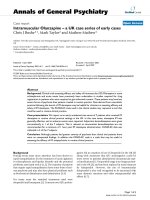
PMA induces a dose-dependent selective downregulation of CCR2Figure 2
PMA induces a dose-dependent selective downregulation of CCR2. (a). THP-1 cells were either untreated (lanes 1, 5
and 9) or treated with PMA at 1 nM (lanes 2, 6 and 10), 10 nM (lanes 3, 7 and 11) or 50 nM (lanes 4, 8 and 12) for 48 hours.
Messenger RNA was then prepared and RT-PCR performed using primers for CCR1 (lanes 1–4), CCR2 (lanes 5–8) and
GAPDH (lanes 9–12). M is a 100 bp DNA ladder. Similar results were obtained in seven other experiments. (b). THP-1 cells
were either left untreated or stimulated with PMA (50 nM) for the times indicated. Subsequently the cells were introduced
into a FACScan flow cytometer to measure cell surface expression of CCR1 (left panel) or CCR2 (right panel).
Journal of Inflammation 2005, 2:14 />Page 6 of 14
(page number not for citation purposes)
tration of 10% and incubated for 2 minutes at room tem-
perature. Subsequently, the cells were washed twice in
TBS, once in RPMI 1640 medium lacking FCS and antibi-
otics and once in RPMI 1640 complete medium. The cells
were then resuspended in RPMI 1640 complete medium,
stimulated with PMA and ionomycin (at the concentra-
tions indicated) and finally incubated at 37°C and 5%
CO
2
for 48 hours.
After the 48-hour incubation period, cell extracts were
made using the luciferase reporter lysis buffer (Promega).
Each lysate was subsequently assayed in the dual luci-
ferase reporter assay (Promega) following the manufac-
turer's instructions. Luciferase activity was determined
using a Monolight series 2010 luminometer (Analytical
Luminescence Laboratory) and then normalized to the
renilla control.
Results
Freshly isolated monocytes selectively downregulate
CCR2, but not CCR1, in culture
Human monocytes were isolated from blood leukopacks
and placed in culture for up to 5 days (Figure 1). During
this time these cells underwent changes in both morphol-
ogy and gene expression. Freshly isolated monocytes ini-
tially appeared small and round, but after 5 days in
culture they became adherent, and increased in both size
and granularity (Figure 1A). Next, we analyzed changes in
the expression of the macrophage differentiation markers
CD11b, CD36 and CD68 (Figure 1B). We found that
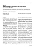
Sub-optimal concentrations of PMA, together with a modest calcium signal, also modulate CCR2Figure 3
Sub-optimal concentrations of PMA, together with a modest calcium signal, also modulate CCR2. (a). THP-1
cells were either unstimulated (lane1) or treated with PMA 1 nM (lane 2) or 50 nM (lane 3) for 48 hours. Alternatively, the
cells were treated with increasing concentrations of the calcium ionophore ionomycin alone (lanes 4–6) or in combination with
PMA 1 nM (lanes 7–9) also for 48 hours. Messenger RNA was then prepared and RT-PCR performed using primers for CCR1
(upper panel), CCR2 (middle panel) and GAPDH (lower panel). M represents markers, which are a 100 bp ladder. Similar
results were obtained in four other experiments. (b). THP-1 cells were either left untreated or stimulated with PMA (1 nM)
and ionomycin (1 µM) for the times indicated. Subsequently the cells were stained for expression of CCR1 (left panel) or
CCR2 (right panel) and analyzed by flow cytometry.
Journal of Inflammation 2005, 2:14 />Page 7 of 14
(page number not for citation purposes)
monocytes cultured for 5 days upregulated expression of
the integrin CD11b and the scavenger receptors CD36 and
CD68, consistent with a change in phenotype from
monocyte to macrophage (Figure 1B). Next, we wanted to
examine changes in the expression of chemokine recep-
tors as monocytes differentiated into macrophages. Using
primers specific for CXCR1-5 and CCR1-CCR9, we per-
formed semi-quantitative analysis of receptor mRNA
expression (Figure 1C). Initially, however, we determined
the efficacy and specificity of the primers by analyzing
genomic DNA samples prepared from freshly isolated
monocytes (Figure 1C, panel I). In all cases a single band
The PKC-inhibitor staurosporine blocks PMA, but not PMA plus ionomycin, induced downregulation of CCR2Figure 4
The PKC-inhibitor staurosporine blocks PMA, but not PMA plus ionomycin, induced downregulation of CCR2.
(a). THP-1 cells were either untreated (lanes 1, 2, 7, 8, 13 and 14) or preincubated with 50 nM staurosporine (lanes 3, 5, 9, 11,
15 and 17) or 10 nM staurosporine (lanes 4, 6, 10, 12, 16 and 18) for 2 hours. Subsequently, the cells were stimulated with 50
nM PMA (lanes 2, 5, 6, 8, 11, 12, 14, 17 and 18) for a further 46 hours. Messenger RNA was then prepared and RT-PCR per-
formed using primers for CCR1 (lanes 1–6), CCR2 (lanes 7–12) and GAPDH (lanes 13–18). M is a 100 bp DNA ladder. Similar
results were obtained in three other experiments. (b). THP-1 cells were either untreated (lanes 1, 3, 5, 7, 9 and 11) or prein-
cubated with 200 nM staurosporine (lanes 2 and 4, 6 and 8 and 10 and 12) for 2 hours. Subsequently the cells were stimulated
with a combination of 1 nM PMA and 1 µM ionomycin (lanes 3 and 4, 7 and 8 and 11 and 12) for a further 46 hours. Messenger
RNA was then prepared and RT-PCR performed using primers for CCR1 (lanes 1–4), CCR2 (lanes 5–8) and GAPDH (lanes 9–
12). M is a 100 bp DNA ladder. Similar results were obtained in three other experiments.
Journal of Inflammation 2005, 2:14 />Page 8 of 14
(page number not for citation purposes)
of the anticipated size was observed indicating that the
primers were specific for the desired chemokine receptor.
This data further suggested that a lack of chemokine recep-
tor expression observed in freshly isolated monocytes and
monocytes cultured for up to five days was a true result,
rather than as a reflection of inappropriate primer design.
Subsequently, we performed semi-quantitative analysis of
receptor mRNA expression on freshly isolated monocytes
and monocytes cultured for up to five days (Figure 1C,
panel II). Under these conditions, freshly isolated mono-
cytes showed strong expression of CCR1, CCR2, CCR5,
CXCR2 and CXCR4 mRNAs, and trace levels of CCR4 and
CCR7 mRNA. Expression of CCR1, CCR2, CCR5, and
CXCR4 mRNAs remained elevated after two days in cul-
ture, while that of CXCR2 decreased and that of CCR7
temporarily increased. However, after five days in culture
CCR2 mRNA expression but not that of CCR1, CCR5 or
CXCR4 was dramatically downregulated (Figure 1C,
panel II). Indeed, levels of CCR5 and CCR1 mRNA actu-
ally increased over those observed in freshly isolated
monocytes. To confirm the specificity of this effect we
subsequently compared cell surface expression of these
chemokine receptors in cultured monocytes and freshly
isolated monocytes by flow cytometry (Figure 1D). In
agreement with our mRNA data, expression of CCR2 pro-
tein, but not CCR1, CCR5 and CXCR4 was rapidly down-
regulated during monocyte maturation. Negligible cell
surface expression of CCR7 protein was observed at any of
the time points examined, while CXCR2 cell surface
expression remained curiously elevated despite downreg-
ulation of CXCR2 mRNA, suggesting that the half-life of
this protein is actually quite long (Figure 1D).
These results indicate that one consequence of monocyte
maturation is the selective downregulation of CCR2 gene
expression followed by a loss of CCR2 protein from the
surface of the cell. While the actual physiological role of
this process is unknown, it is likely that CCR2 down-reg-
ulation may be involved in restricting 'reverse-migration'
of differentiated monocytes back into the blood stream,
and thus facilitating capture within the tissues.
PMA-treatment of monocytes induces selective
downregulation of CCR2
Based on the above results we decided to further examine
the regulation of CCR2 expression in monocyte matura-
tion using the human monocyte cell line, THP-1 and
CCR1 as a control. Treatment of these cells with the PKC-
activating phorbol ester PMA for 48 hours is a widely
accepted procedure for maturing monocytes [27,28]. Cells
treated in this way undergo phenotypic changes consist-
ent with their maturation into macrophages [27-30] (also
compare Figures 1 and 6).
Next, we wanted to determine how treatment of the
monocyte cell line, THP-1, with PMA affected the expres-
sion of CCR2 in these cells. Thus, monocytes were stimu-
lated with PMA (at the concentrations indicated) for 48
hours and RNA prepared as described above. Our results
(Figure 2A) show that CCR2 was selectively down-regu-
lated in a dose dependent manner, whereas expression of
CCR1 (the other main CC receptor on monocytes) and
the house-keeping gene GAPDH remained unaffected.
PMA (50 nM) was sufficient to completely abrogate CCR2
expression (Figure 2A, lane 8), whilst 10 nM PMA reduced
expression of this chemokine receptor by approximately
75% (Figure 2A, lane 7). Treatment of THP-1 cells with 1
nM PMA did not affect expression of CCR2 (Figure 2A,
lane 6).
Subsequently, we examined whether PMA modulated the
cell surface expression of CCR1 and CCR2 by FACS anal-
ysis. THP-1 cells were again stimulated with PMA (50 nM)
for the times indicated, before being stained with the
Staurosporine blocks PMA, but not PMA plus ionomycin, induced downregulation of CCR2 promoter activityFigure 5
Staurosporine blocks PMA, but not PMA plus iono-
mycin, induced downregulation of CCR2 promoter
activity. THP-1 cells were transfected with either 5 µg of
vector alone (pGL3-basic; lane 1) or with 5 µg of the pGL3-
1335 construct (lanes 2–7). In addition, each sample was also
co-transfected with 2 µg of pRL-SV40 (renilla) to act as an
internal control. Cells were then either left untreated (lanes
1–4) or pretreated with staurosporine (100 nM) for 2 hours
(lanes 5–7). Next, the THP-1 cells were stimulated with a
combination of PMA alone (lanes 3 and 6) or PMA plus iono-
mycin (lanes 4 and 7) for a further 46 hours. Subsequently,
cell extracts were prepared and assayed for both luciferase
and renilla activity. After normalization to the renilla control,
the CCR2 transcriptional activity was determined relative to
the pGL3-basic vector, which was arbitrarily assigned a value
of 1. Similar results were obtained in two other experiments
Journal of Inflammation 2005, 2:14 />Page 9 of 14
(page number not for citation purposes)
appropriate antibodies and then analyzed by flow cytom-
etry (Figure 2B). Whereas the levels of CCR1 remained
high throughout the duration of the experiment, CCR2
protein expression decreased dramatically. The majority
of the expression was lost by 24 hours and by 48 hours vir-
tually no CCR2 was found on the surface of the cultured
THP-1 cells (compare Figure 2B, left and right panels).
Thus, THP-1 cells treated with PMA (50 nM) mimics the
differentiation process observed in cultured monocytes.
Two distinct signal transduction pathways regulate CCR2
expression during monocyte maturation
Our initial observations suggested that while PMA (50
nM) completely abrogated CCR2 expression, sub-optimal
concentrations of this phorbol ester (1 nM) had no effect
(Figure 2A). We wondered, therefore, whether the addi-
tion of a calcium signal (such as ionomycin) together with
the sub-optimal concentration of PMA might provide a
sufficiently strong stimulus to affect the expression of
IFN-γ plus M-CSF promotes a similar differentiation phenotype to that observed using pharmacologic stimuliFigure 6
IFN-γ plus M-CSF promotes a similar differentiation phenotype to that observed using pharmacologic stimuli.
(a). THP-1 cells were either left untreated (upper panel) or treated with 500 U/ml IFN-γ plus 5 ng/ml M-CSF (middle panel) or
50 nM PMA (lower panel) for 48 hours. Subsequently, the cells were photographed using a Nikon Diaphot Camera set up and
Axon Imaging Workbench software. Magnification is at 40 ×. (b). THP-1 cells were either left untreated or treated for 48
hours with either 50 nM PMA (PMA) or 500 U/ml IFN-γ plus 5 ng/ml M-CSF (I+M) as indicated. Subsequently, these cells were
stained with antibodies to macrophage markers CD36 (upper panel), CD11b (middle panel) and CD68 (lower panel) and then
analyzed by flow cytometry.
Journal of Inflammation 2005, 2:14 />Page 10 of 14
(page number not for citation purposes)
CCR2. Thus, we incubated monocytes with PMA (1 nM)
and ionomycin at the concentrations indicated for 48
hours, and then analyzed CCR2 expression. Our data
indicated that ionomycin alone does not affect expression
of CCR2 (Figure 3A, middle panel, lanes 4–6). However,
in the presence of a sub-optimal PMA signal (1 nM), there
was a selective dose-dependent reduction in CCR2 expres-
sion (Figure 3A, middle panel, lanes 7–9). At the same
time, similar concentrations of PMA and ionomycin did
not affect the levels of CCR1 nor GAPDH (Figure 3A
upper and lower panels). Monocytes treated with PMA (1
nM) plus ionomycin (1 µM) were also observed to adopt
an adherent phenotype and to increase in size similar to
the changes in morphology observed in freshly isolated
monocytes (data not shown). Furthermore, cell surface
expression of CCR2, but not CCR1, was found to be
downregulated in the presence of PMA (1 nM) plus iono-
mycin (1 µM) after 48 hours (Figure 3B). Thus, sub-opti-
mal concentrations of PMA together with a modest
calcium signal combine to mediate a maturation pheno-
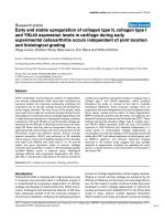
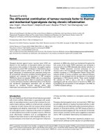
IFN-γ plus M-CSF promotes specific down-regulation of CCR2Figure 7
IFN-γ plus M-CSF promotes specific down-regulation of CCR2. (a). THP-1 cells were either untreated (lane 1, upper,
middle and lower panels) or treated with 500 U/ml IFN-γ plus 5 ng/ml M-CSF (lane 2 upper, middle and lower panels) or 50 nM
PMA (lane 3 upper, middle and lower panels) for 48 hours. Messenger RNA was then prepared and RT-PCR performed using
primers for CCR1 (upper panel), CCR2 (middle panel) and GAPDH (lower panel). M is a 100 bp DNA ladder. Similar results
were obtained in three other experiments. (b). THP-1 cells were transfected with either 5 µg of vector alone (pGL3-basic) or
with 5 µg of the pGL3-1335 construct. In addition, each sample was also transfected with 2 µg of pRL-SV40 (renilla) to act as
an internal control. Cells were then either left untreated or treated with either 500 U/ml IFN-γ plus 5 ng/ml M-CSF or 50 nM
PMA. Subsequently, cell extracts were prepared and assayed for both luciferase and renilla activity. After normalization to the
renilla control, CCR2 transcriptional activity was calculated relative to the pGL3-basic vector, which was arbitrarily assigned a
value of 1. Similar results were obtained in two other experiments.
0
2
4
6
8
10
12
14
16
18
20
1234567
Transcriptional activity (Fold Induction)
4
20
16
12
8
0
LANE
1324567
pGL3-BASIC
pGL3-1335
PMA
IFN-γ
γγ
γ plus M-CSF
STAURO
+
-
+
+
+
+
+
+++++
-
-
-
-
-
-
-
-
-
-
-
-
-
-
-+
+
+
B
321
GAPDH
CCR2
IFN-
γ
γ
γ
γ +MCSF
CCR1
CON
PMA
M
Lane
A
Journal of Inflammation 2005, 2:14 />Page 11 of 14
(page number not for citation purposes)
type in monocytes that also includes the selective down-
regulation of CCR2.
To determine whether the selective downregulation of
CCR2 observed in PMA versus PMA plus ionomycin
treated cells represented the same or two different signal-
ing pathways, we performed an experiment using the
broad-spectrum kinase inhibitor, staurosporine (Figure
4). We preincubated THP-1 cells with staurosporine at the
concentrations indicated for two hours, and then stimu-
lated with either PMA (50 nM; Figure 4A) or PMA (1 nM)
plus ionomycin (1 µM; Figure 4B) for 48 hours. Stau-
rosporine alone (at concentrations up to 200 nM) did not
significantly inhibit expression of CCR2 (Figure 4A, lanes
9 and 10 and Figure 4B, lane 6) nor CCR1 (Figure 4A,
lanes 3 and 4 and Figure 4B, lane 2). Furthermore, the
inhibitor did not abrogate the downregulation of CCR2
mediated by PMA plus ionomycin (Figure 4B, compare
lanes 7 and 8). In contrast, staurosporine at 50 nM, but
not at 10 nM, blocked the loss of CCR2 in PMA (50 nM)
treated cells (Figure 4A, compare lanes 7, 8, 11 and 12).
Thus, these results identify at least two possible signal
transduction pathways present in monocytes that could
regulate the expression of CCR2 during monocyte differ-
entiation.
CCR2 expression is regulated at the level of transcription
Having established that CCR2 is down-regulated during
monocyte differentiation, we next wanted to determine
whether the regulation occurs at the level of RNA stability
or at the level of transcription. We, therefore, cloned a
1335 bp fragment of the CCR2 promoter using the
sequence described by Yamamoto and colleagues [26].
This fragment was then subcloned into the mammalian
expression vector pGL3 upstream of the luciferase gene,
generating the pGL3-1335 construct. In addition to the
sequences upstream of the TATA box, pGL3-1335
included 115 bp of the 5'UTR, which contains the two
tandem C/EBP repeats that are thought to be necessary for
the basal expression of the CCR2 gene [26].
Subsequently, we transfected this construct into the THP-
1 cells using DEAE/dextran and either left the cells
untreated, or treated them with PMA (50 nM), or PMA (1
nM) plus ionomycin (1 µM) for 48 hours in the presence
or absence of staurosporine (100 nM). Cells were then
lyzed and assayed for transcriptional activity. Our results
showed that the pGL3-1335 construct, itself, gave a 13-
fold induction over the background construct lacking the
CCR2 promoter (Figure 5, compare lanes 1 and 2). Fur-
thermore, both PMA and PMA plus ionomycin strongly
abrogated this transcriptional activity (Figure 5 lanes 3
and 4) suggesting that the dual signal transduction path-
ways activated by PMA and PMA plus ionomycin medi-
ated regulation of CCR2 expression at the level of
transcription. In the presence of staurosporine, inhibition
of CCR2 promoter activity mediated by PMA, but not
PMA (1 nM) plus ionomycin (1 µM), was abrogated (Fig-
ure 5, compare lanes 6 and 7). Thus, these data indicate
that the PMA (but not the PMA plus ionomycin) mediated
inhibition of CCR2 promoter activity is ultimately regu-
lated by one or more staurosporine-sensitive transcription
factors.
Treatment with IFN-
γ
and M-CSF produces a similar
differentiation phenotype to that seen with PMA and
ionomycin
The above results reflect a phenotype induced by pharma-
cologic agents and we next wanted to ensure that this phe-
notype is applicable to physiologic agents also. To that
end, THP-1 cells treated with IFN-γ plus M-CSF have
already been shown to promote monocyte maturation,
although it has yet to be confirmed that these agents reg-
ulate CCR2 expression at the level of transcription [32].
Initially, though, we wanted to demonstrate that mono-
cytes treated with IFN-γ plus M-CSF showed changes in
morphology similar to that observed with freshly isolated
monocytes (compare Figures 1 and 6). After 48 hours
treatment with IFN-γ plus M-CSF, monocytes became
adherent and increased in size similar to that observed for
freshly isolated monocytes in culture (compare Figure 1A
and Figure 6A middle panel). PMA-treated monocytes
also underwent similar changes in morphology (Figure
6A, lower panel). Furthermore, flow cytometric studies
revealed that monocytes treated with either IFN-γ plus M-
CSF or PMA strongly upregulated the macrophage matu-
ration markers CD11b, CD36 and CD68 (Figure 6B). Sim-
ilar results were observed for cells treated with PMA plus
ionomycin (data not shown). Thus, monocytes treated
with a panel of physiologic and pharmacologic stimuli
promote maturation to the macrophage phenotype as
determined by changes in morphology and upregulation
of macrophage maturation markers.
Next, we wanted to determine whether IFN-γ plus M-CSF
induced the differentiation-associated downregulation of
CCR2 (Figure 7). Therefore, monocytes were treated with
IFN-γ (500 U/ml) plus M-CSF (5 ng/ml) for 48 hours and
CCR2 mRNA was examined (Figure 7A). Our results
showed that IFN-γ plus M-CSF did selectively downregu-
late CCR2, but not CCR1 in a manner analogous to that
observed for PMA and PMA plus ionomycin (Figure 7A
upper and middle panels). A similar pattern was also
observed when transcriptional activity was examined (Fig-
ure 7B). Here, PMA completely down-modulated CCR2
transcription, while the combined effects of IFN-γ plus M-
CSF reduced this activity by approximately 70%. In the
presence of staurosporine, the inhibition of CCR2 pro-
moter activity mediated by IFN-γ (500 U/ml) plus M-CSF
Journal of Inflammation 2005, 2:14 />Page 12 of 14
(page number not for citation purposes)
(5 ng/ml) was abrogated in a manner analogous to that
observed for PMA (Figure 7B lanes 6 and 7).
Taken together, these data suggest that PMA (50 nM),
PMA plus ionomycin and IFN-γ plus M-CSF mediate sim-
ilar changes in the monocyte phenotype during matura-
tion of these cells. Thus, the monocyte cell line, THP-1, is
a useful model system with which to investigate the
underlying regulatory mechanisms governing chemokine
receptor expression during monocyte differentiation.
Discussion
In this paper we demonstrate that a major consequence of
monocyte maturation into macrophages is the selective
downregulation of the chemokine receptor, CCR2, but
not the related CCR1. We have further shown that there
are multiple stimuli, which can selectively down-modu-
late CCR2 expression, including high concentrations of
PMA (50 nM), or low PMA (1 nM) plus ionomycin (1
µM), or IFN-γ (500 U/ml) plus M-CSF (5 ng/ml). Each of
these stimuli regulate the expression of CCR2 at the level
of transcription, although it appears that at least two dif-
ferent signal transduction pathways are involved based on
the ability of staurosporine to interfere with these proc-
esses. Treatment of THP-1 monocytes with staurosporine
abrogated the ability of PMA and IFN-γ plus M-CSF to
downregulate CCR2. By contrast, staurosporine was una-
ble to block PMA plus ionomycin mediated downregula-
tion of CCR2 expression. Thus, this study provides
evidence that there is dynamic and selective regulation of
the CCR2 gene during monocyte differentiation.
Our results indicate that treatment of THP-1 cells with
either PMA alone (50 nM) or PMA (1 nM) plus ionomy-
cin (1 µM) promotes a differentiation phenotype that is
characterized by morphological changes and altered
CCR2 gene expression. Indeed, these observations have
already been noted by other researchers studying mono-
cyte differentiation [27,28,32]. In particular, we show that
THP-1 cells rapidly become adherent and their morphol-
ogy changes from the typical round shape of monocytes to
spindle-shaped cells with pseudopodia, which are charac-
teristic of macrophages. At the same time there was also
an increase in the size and granularity of the cells. In addi-
tion, we demonstrated an up-regulation in expression of
genes associated with monocyte differentiation, notably
CD11b, CD36 and CD68. Concomitantly, the expression
of CCR2, but not CCR1, was selectively downregulated,
suggesting that the loss of this chemokine receptor is a
consequence of monocyte differentiation. This downregu-
lation was observed at the level of cell surface receptor
expression, mRNA expression, and transcription. Clearly,
these are specific regulatory events since the levels of
CCR1 mRNA are not affected by either combination of
pharmacologic agents.
However, when THP-1 cells were treated with PMA (50
nM) or PMA plus ionomycin in the presence of stau-
rosporine, differential results were obtained: PMA-medi-
ated modulation of CCR2 was sensitive to the inhibitory
effects of staurosporine (50 nM), whereas staurosporine
concentrations as high as 200 nM failed to block PMA
plus ionomycin-induced downregulation of CCR2. Stau-
rosporine alone did not promote the loss of either CCR2
or CCR1. These results indicate that staurosporine defines
a dichotomy in the regulation of CCR2 expression by
PMA (50 nM) versus PMA plus ionomycin that had not
previously been appreciated.
Staurosporine, itself, is a broad-spectrum inhibitor of pro-
tein kinases including PKA, PKC, and PKG. PMA has clas-
sically been shown to act almost exclusively through PKC
and this would explain why staurosporine was able to
block the PMA-induced downregulation of CCR2. By
inference, PMA plus ionomycin would appear to act
through a signal transduction pathway that is not inhib-
ited by staurosporine and presumably this means that sec-
ond messengers other than PKA, PKC and PKG are
involved. To that end, calcineurin, a calcium-sensitive
phosphatase may be a target for PMA plus ionomycin
[33]. An increase in the intracellular calcium concentra-
tion (such as that afforded by the presence of ionomycin)
promotes a conformational change in calcineurin, which
then dephosphorylates and activates the transcription fac-
tor NFAT facilitating its translocation to the nucleus. In
addition, it has been shown that PMA enhances the cal-
cium sensitivity of NFAT, thus creating a synergistic signal
[33,34]. This synergy may result from de novo synthesis
and post-translational modification of another transcrip-
tion factor termed activating protein-1, AP-1 [33,34].
Indeed, NFAT proteins show a characteristic ability to co-
operate with AP-1 in DNA-binding and transactivation
[33,34]. Interestingly, in the region of the CCR2 promoter
that we cloned there are two putative binding sites for AP-
1 (core binding motif TGA(C/G)TCA) and three putative
binding sites for NFAT (core binding motif GGAAA) as
determined by the MatInspecter transcription factor bind-
ing site analysis program. It has also been suggested that
additional transcription factors including OCT1 and C/
EBP can act synergistically with NFAT and again there are
multiple binding sites for each of these DNA-binding pro-
teins in the CCR2 promoter, although at this stage we
have no evidence to suggest that they are involved in the
physiological regulation of CCR2 gene expression.
A requirement for co-operation and cross-talk between
these two pharmacologic agents is further supported by
the fact that ionomycin alone (at concentrations as high
as 1 µM) was unable to down-modulate CCR2.
Journal of Inflammation 2005, 2:14 />Page 13 of 14
(page number not for citation purposes)
Some reports have suggested that CCL2 could be involved
in the early stages of CCR2 protein down-modulation,
while other studies indicate that the differentiation proc-
ess itself, is a major factor in the selective loss of CCR2
gene expression [8,32]. Numerous cytokines are known to
be involved in monocyte activation and differentiation,
among them M-CSF and IFN-γ [32,35,36]. M-CSF is a lin-
eage-specific hematopoetic growth factor that stimulates
monocyte differentiation [35,36]. The c-fms proto-onco-
gene encodes a high affinity receptor for M-CSF [37] and
it has been shown that THP-1 cells express this protein
and that it is up-regulated during differentiation. How-
ever, cells stimulated with M-CSF alone for 48 hours did
not lose expression of CCR2 (data not shown).
Conversely, IFN-γ alone, which is constitutively expressed
by monocyte lineage cells and which promotes matura-
tion of monocytes to macrophages [38], did significantly
reduce expression of CCR2, although the cells did not
become adherent and neither did they change their mor-
phology (data not shown). Interestingly, IFN-γ has been
demonstrated to up-regulate levels of M-CSF in mono-
cytes during maturation [38] and when both IFN-γ and M-
CSF were added, THP-1 cells did become adherent,
changed their morphology and selectively lost CCR2, but
not CCR1 – all of which are characteristics of the mono-
cyte differentiation phenotype. These results are in keep-
ing with the studies published by Tangirala and
colleagues, who reported similar phenomena in THP-1
cells [32]. In addition, our studies also demonstrated that
the regulatory effects mediated by IFN-γ plus M-CSF
occurred at the level of transcription, where a significant
down-regulation in CCR2 promoter activity was observed.
Moreover, in the presence of staurosporine, IFN-γ plus M-
CSF was unable to down-regulate levels of CCR2. This
result probably reflects the fact that IFN-γ signals exten-
sively through the JAK-STAT pathway, and studies have
suggested that staurosporine can block phosphorylation
of Janus kinases [39,40]. In addition, we have found two
putative binding sites in the CCR2 promoter for STAT
transcription factors which would further support the
contention that these transcription factors may be impor-
tant in the regulation of IFN-γ mediated downregulation
of CCR2.
Conclusion
This study demonstrates that expression of the chemokine
receptor CCR2 is exquisitely correlated with monocyte
maturation. Freshly isolated monocytes express high lev-
els of both CCR2 RNA and protein, whereas monocyte-
derived macrophages express neither CCR2 RNA nor pro-
tein. Conversely, levels of the closely-related chemokine
receptor CCR1 remained stable and elevated throughout
monocyte maturation. An analysis of the biochemical and
molecular mechanisms underlying the regulated expres-
sion of CCR2 revealed the existence of several signaling
pathways that selectively down-modulate CCR2 gene
expression during monocyte differentiation; this expres-
sion was largely regulated at the level of transcription. Sig-
naling through PMA and IFN-γ plus M-CSF, but not PMA
plus ionomycin was abrogated by prior treatment of the
THP-1 cells with staurosporine. Although the physiologi-
cal role of this process is not well understood, it is likely
that CCR2 down-regulation may be involved in restricting
the 'reverse-migration' of differentiated monocytes back
into the blood stream. This in turn facilitates the retention
of differentiated monocytes within inflamed tissues. Thus,
by improving our understanding of the regulatory mecha-
nisms that govern CCR2 expression on monocyte lineage
cells, we can better appreciate how monocyte recruitment
and activation is controlled during chronic inflammatory
pathologies such as atherosclerosis.
Competing interests
Brett Premack is currently employed as the Director of
Technology at Chemocentryx. Dr. Premack is the holder
of stocks within this company. This company investigates
the role of chemokines and their receptors as potential
therapeutics. One of these projects is to investigate the
role of CCR2 antagonists in cardiovascular disease and a
phase I clinical trial is ongoing. At the time this study was
undertaken Dr. Premack was an unpaid consultant for
Chemocentryx. Neither Roderick Phillips nor Marin Lutz
has a competing interest in this work.
Authors' contributions
RJP wrote the manuscript and performed all of the exper-
iments except Figure 1A and 1C. ML performed experi-
ments featured in Figure 1A and 1C. BP conceived of the
study, and participated in its design and coordination and
helped to draft the manuscript. All authors read and
approved the final manuscript.
RJP was supported by the American Heart Association
grant 9960044Y
References
1. Rossi D, Zlotnik A: The biology of chemokines and their receptors.
Annu Rev Immunol 2000, 18:217-242.
2. Murphy PM, Baggiolini M, Charo IF, Hebert CA, Horuk R, Matsushima
K, Miller LH, Oppenheim JJ, Power CA: International union of
pharmacology. XXII. Nomenclature for chemokine recep-
tors. Pharmacol Rev 2000, 52:145-176.
3. Premack BA, Schall TJ: Chemokine receptors: gateways to
inflammation and infection. Nat Med 1996, 2:1174-1178.
4. Baggiolini M: Chemokines and leukocyte traffic. Nature 1998,
392:565-568.
5. Murphy PM: International Union of Pharmacology. XXX.
Update on chemokine receptor nomenclature. Pharmacol Rev
2002, 54:227-229.
6. Power CA, Wells TN: Cloning and characterization of human
chemokine receptors. Trends Pharmacol Sci 1996, 17:209-213.
7. Charo IF, Myers SJ, Herman A, Franci C, Connolly AJ, Coughlin SR:
Molecular cloning and functional expression of two mono-
cyte chemoattractant protein 1 receptors reveals alterna-
Publish with Bio Med Central and every
scientist can read your work free of charge
"BioMed Central will be the most significant development for
disseminating the results of biomedical research in our lifetime."
Sir Paul Nurse, Cancer Research UK
Your research papers will be:
available free of charge to the entire biomedical community
peer reviewed and published immediately upon acceptance
cited in PubMed and archived on PubMed Central
yours — you keep the copyright
Submit your manuscript here:
/>BioMedcentral
Journal of Inflammation 2005, 2:14 />Page 14 of 14
(page number not for citation purposes)
tive splicing of the carboxyl-terminal tails. Proc Natl Acad Sci U
S A 1994, 91:2752-2756.
8. Fantuzzi L, Borghi P, Ciolli V, Pavlakis G, Belardelli F, Gessani S: Loss
of CCR2 expression and functional response to monocyte
chemotactic protein (MCP-1) during the differentiation of
human monocytes: role of secreted MCP-1 in the regulation
of the chemotactic response. Blood 1999, 94:875-883.
9. Neote K, DiGregorio D, Mak JY, Horuk R, Schall TJ: Molecular
cloning, functional expression, and signaling characteristics
of a C-C chemokine receptor. Cell 1993, 72:415-425.
10. Brown Z, Gerritsen ME, Carley WW, Strieter RM, Kunkel SL, West-
wick J: Chemokine gene expression and secretion by
cytokine-activated human microvascular endothelial cells.
Differential regulation of monocyte chemoattractant pro-
tein-1 and interleukin-8 in response to interferon-gamma.
Am J Pathol 1994, 145:913-921.
11. Goebeler M, Yoshimura T, Toksoy A, Ritter U, Brocker EB, Gillitzer
R: The chemokine repertoire of human dermal microvascu-
lar endothelial cells and its regulation by inflammatory
cytokines. J Invest Dermatol 1997, 108:445-451.
12. Marfaing-Koka A, Devergne O, Gorgone G, Portier A, Schall TJ,
Galanaud P, Emilie D: Regulation of the production of the
RANTES chemokine by endothelial cells. Synergistic induc-
tion by IFN-gamma plus TNF-alpha and inhibition by IL-4
and IL-13. J Immunol 1995, 154:1870-1878.
13. Martin T, Cardarelli PM, Parry GC, Felts KA, Cobb RR: Cytokine
induction of monocyte chemoattractant protein-1 gene
expression in human endothelial cells depends on the coop-
erative action of NF-kappa B and AP-1. Eur J Immunol 1997,
27:1091-1097.
14. Kumar AG, Ballantyne CM, Michael LH, Kukielka GL, Youker KA,
Lindsey ML, Hawkins HK, Birdsall HH, MacKay CR, LaRosa GJ, Ros-
sen RD, Smith CW, Entman ML: Induction of monocyte chem-
oattractant protein-1 in the small veins of the ischemic and
reperfused canine myocardium. Circulation 1997, 95:693-700.
15. Wysocki SJ, Zheng MH, Smith A, Lamawansa MD, Iacopetta BJ, Rob-
ertson TA, Papadimitriou JM, House AK, Norman PE: Monocyte
chemoattractant protein-1 gene expression in injured pig
artery coincides with early appearance of infiltrating mono-
cyte/macrophages. J Cell Biochem 1996, 62:303-313.
16. Ross R: The pathogenesis of atherosclerosis: a perspective for
the 1990s. Nature 1993, 362:801-809.
17. Berliner JA, Navab M, Fogelman AM, Frank JS, Demer LL, Edwards PA,
Watson AD, Lusis AJ: Atherosclerosis: basic mechanisms. Oxi-
dation, inflammation, and genetics. Circulation 1995,
91:2488-2496.
18. Cai JP, Hudson S, Ye MW, Chin YH: The intracellular signaling
pathways involved in MCP-1-stimulated T cell migration
across microvascular endothelium. Cell Immunol 1996,
167:269-275.
19. Randolph GJ, Furie MB: A soluble gradient of endogenous
monocyte chemoattractant protein-1 promotes the
transendothelial migration of monocytes in vitro. J Immunol
1995, 155:3610-3618.
20. Ross R: Cell biology of atherosclerosis. Annu Rev Physiol 1995,
57:791-804.
21. Lusis AJ: Atherosclerosis. Nature 2000, 407:233-241.
22. Boring L, Gosling J, Cleary M, Charo IF: Decreased lesion forma-
tion in CCR2-/- mice reveals a role for chemokines in the ini-
tiation of atherosclerosis. Nature 1998, 394:894-897.
23. Charo IF, Peters W: Chemokine receptor 2 (CCR2) in athero-
sclerosis, infectious diseases, and regulation of T-cell polari-
zation. Microcirculation 2003, 10:259-264.
24. Gu L, Okada Y, Clinton SK, Gerard C, Sukhova GK, Libby P, Rollins
BJ: Absence of monocyte chemoattractant protein-1 reduces
atherosclerosis in low density lipoprotein receptor-deficient
mice. Mol Cell 1998, 2:275-281.
25. Rollins BJ: Chemokines and atherosclerosis: what Adam
Smith has to say about vascular disease. J Clin Invest 2001,
108:1269-1271.
26. Yamamoto K, Takeshima H, Hamada K, Nakao M, Kino T, Nishi T,
Kochi M, Kuratsu J, Yoshimura T, Ushio Y: Cloning and functional
characterization of the 5'-flanking region of the human
monocyte chemoattractant protein-1 receptor (CCR2)
gene. Essential role of 5'-untranslated region in tissue-spe-
cific expression. J Biol Chem 1999, 274:4646-4654.
27. Tontonoz P, Nagy L, Alvarez JG, Thomazy VA, Evans RM: PPAR-
gamma promotes monocyte/macrophage differentiation
and uptake of oxidized LDL. Cell 1998, 93:241-252.
28. Rovera G, Santoli D, Damsky C: Human promyelocytic leukemia
cells in culture differentiate into macrophage-like cells when
treated with a phorbol diester. Proc Natl Acad Sci U S A 1979,
76:2779-2783.
29. Naito M, Umeda S, Yamamoto T, Moriyama H, Umezu H, Hasegawa
G, Usuda H, Shultz LD, Takahashi K: Development, differentia-
tion, and phenotypic heterogeneity of murine tissue macro-
phages. J Leukoc Biol 1996, 59:133-138.
30. Yesner LM, Huh HY, Pearce SF, Silverstein RL: Regulation of
monocyte CD36 and thrombospondin-1 expression by solu-
ble mediators. Arterioscler Thromb Vasc Biol 1996, 16:1019-1025.
31. Seager Danciger J, Lutz M, Hama S, Cruz D, Castrillo A, Lazaro J, Phil-
lips R, Premack B, Berliner J: Method for large scale isolation,
culture and cryopreservation of human monocytes suitable
for chemotaxis, cellular adhesion assays, macrophage and
dendritic cell differentiation. J Immunol Methods 2004,
288:123-134.
32. Tangirala RK, Murao K, Quehenberger O: Regulation of expres-
sion of the human monocyte chemotactic protein-1 recep-
tor (hCCR2) by cytokines. J Biol Chem 1997, 272:8050-8056.
33. Rao A, Luo C, Hogan PG: Transcription factors of the NFAT
family: regulation and function. Annu Rev Immunol 1997,
15:707-747.
34. Macian F, Lopez-Rodriguez C, Rao A: Partners in transcription:
NFAT and AP-1. Oncogene 2001, 20:2476-2489.
35. Lenny N, Westendorf JJ, Hiebert SW: Transcriptional regulation
during myelopoiesis. Mol Biol Rep 1997, 24:157-168.
36. Clarke S, Gordon S: Myeloid-specific gene expression. J Leukoc
Biol 1998, 63:153-168.
37. Nienhuis AW, Bunn HF, Turner PH, Gopal TV, Nash WG, O'Brien SJ,
Sherr CJ: Expression of the human c-fms proto-oncogene in
hematopoietic cells and its deletion in the 5q- syndrome. Cell
1985, 42:421-428.
38. Scheibenbogen C, Andreesen R: Developmental regulation of
the cytokine repertoire in human macrophages: IL-1, IL-6,
TNF-alpha, and M-CSF. J Leukoc Biol 1991, 50:35-42.
39. Fiorucci G, Percario ZA, Marcolin C, Coccia EM, Affabris E, Romeo
G: Inhibition of protein phosphorylation modulates expres-
sion of the Jak family protein tyrosine kinases. J Virol 1995,
69:5833-5837.
40. Callus BA, Mathey-Prevot B: Interleukin-3-induced activation of
the JAK/STAT pathway is prolonged by proteasome inhibi-
tors. Blood 1998, 91:3182-3192.