Báo cáo y học: "Early Migrating leukocytes are the source of Peroxiredoxin V during inflammation in the airway" potx
Bạn đang xem bản rút gọn của tài liệu. Xem và tải ngay bản đầy đủ của tài liệu tại đây (2.15 MB, 12 trang )
BioMed Central
Page 1 of 12
(page number not for citation purposes)
Journal of Inflammation
Open Access
Research
Migrating leukocytes are the source of Peroxiredoxin V during
inflammation in the airways
Raisa I Krutilina
3
, Andrei V Kropotov
1,3
, Christian Leutenegger
2,3
and
Vladimir B Serikov*
3
Address:
1
Institute of Cytology Russian Academy of Sciences, St. Petersburg, 194021, Russia,
2
University of California, Davis, Davis, CA 95616,
USA and
3
Children's Hospital Oakland Research Institute, Oakland, CA 94609, USA
Email: Raisa I Krutilina - ; Andrei V Kropotov - ;
Christian Leutenegger - ; Vladimir B Serikov* -
* Corresponding author
Abstract
Background: We characterized changes in expression of the antioxidant protein Peroxiredoxin
V (PRXV) during airway inflammation.
Methods: Studies in anesthetized rats and mice; PRXV expression determined by Western blot
analyses and immunohistochemistry; PRXV m-RNA expression determined by Taq-Man RT-PCR.
Results: Bacterial lung inflammation did not change expression of PRXV in murine epithelia but
produced massive influx of leukocytes highly expressing PRXV. Endotoxin and f-MLP induced
leukocyte migration in rat trachea but did not change mRNA levels and PRXV protein expression
in tracheal epithelial cells. In primary airway cell culture (cow), alveolar epithelial cells A549, or co-
culture of A549 with murine macrophages RAW264.7, exposure to live bacteria increased
expression of PRXV, which required serum. PRXV was secreted in vitro by epithelial and immune
cells.
Conclusion: Inflammation increased expression of PRXV in airways by at least 2 mechanisms: cell
population shift by massive influx of leukocytes expressing PRXV, and moderate post-
transcriptional up-regulation of PRXV in epithelial cells.
Background
To ensure adequate protection against oxidative stress
during states of pulmonary disease, several antioxidant
systems have evolved in the epithelial cells of mammalian
airways [1-4]. Peroxiredoxins I-VI (PRX I-VI) are a group
of potent antioxidant proteins that are the subject of
much research [5-10]. PRXs neutralize reactive oxygen by
transferring electrons from thioredoxins or cyclophilins.
The six PRXs differ in their intracellular distribution and
are thought to serve different functions and be regulated
by different mechanisms. PRXV is one of the key enzymes
of cellular antioxidant defense, as it is a potent protector
against DNA damage and also has other functions [11-
14].
Toxic insults to the respiratory tract down-regulate synthe-
sis of the PRXV protein. We have recently demonstrated in
vivo in rat tracheal epithelial cells that cigarette smoke
extract (CSE) directly down-regulated expression of PRXV,
which is one mechanism of cigarette smoke toxicity [15].
Published: 04 October 2006
Journal of Inflammation 2006, 3:13 doi:10.1186/1476-9255-3-13
Received: 29 March 2006
Accepted: 04 October 2006
This article is available from: />© 2006 Krutilina et al; licensee BioMed Central Ltd.
This is an Open Access article distributed under the terms of the Creative Commons Attribution License ( />),
which permits unrestricted use, distribution, and reproduction in any medium, provided the original work is properly cited.
Journal of Inflammation 2006, 3:13 />Page 2 of 12
(page number not for citation purposes)
Exposure of isolated tracheal segment to CSE significantly
reduced mRNA levels for PRXV and the amount of PRXV
protein in the epithelium. In cultures of the tracheal epi-
thelial cell lines, primary airway cell culture, and the alve-
olar epithelial cells A549, CSE significantly decreased
transepithelial electrical resistance, expression of PRXV
protein, and significantly induced glutathione and pro-
tein oxidation. Similarly, when respiratory tract toxicity
was induced in mice with naphthalene, the loss of the
Clara cell population was associated with a significant
decrease in PRXV expression [16]. In contrast, previous
reports had indicated that PRXV was over-expressed in the
lung during inflammation induced by endotoxin [17].
However, experiments in vitro in which pro-inflammatory
cytokines were added to human alveolar or bronchial epi-
thelial cells did not result in an up-regulated expression of
PRXV [18]. Neither the mechanism by which PRXV is up-
regulated during inflammation in tissues of the lung nor
the identity of the cells that are the source of PRXV pro-
duction are known.
We therefore investigated the effects of gram-negative bac-
terial inflammation on expression of PRXV in lung, lung
epithelial cells, and immune cells in vivo and in vitro. Our
first aim was to determine whether inflammation in vivo
influences expression of PRXV in the bronchial epithe-
lium and alveoli. Our second aim was to use an in vivo
model of inflammation to investigate whether changes of
transcription or translation of PRXV in the tracheal epithe-
lium, if they occurred, were a direct response to bacterial
pathogen lipopolysaccharide (LPS) by these cells or
whether the increased level of PRXV was induced by leu-
kocyte migration. Our third aim was to determine in vitro
whether exposure of the airway and alveolar epithelial
cells to live bacteria, either alone or in co-culture with
murine macrophages RAW264.7 changes the level of
PRXV mRNA as well as protein expression and secretion.
We found that both in vivo and in vitro inflammation
induced by bacteria resulted in an increased expression of
PRXV in the airway epithelium by at least 2 different
mechanisms: massive influx of activated leukocytes,
which highly express PRXV, and moderate translational
up-regulation of PRXV in the epithelial cells.
Methods
1. In vivo studies
Experiments in animals were performed according to pro-
tocols approved by the Institutional Animal Use Commit-
tee of the Children's Hospital Oakland Research Institute
and Institute of Cytology, RAS.
Experiments in mice
Bone-marrow transplantation
Recipient mice (n = 12) were given a sub-lethal dose of
whole-body irradiation (5.05 Gy) the day before trans-
plantation. While under general anesthesia (Pentobarbi-
tal, 25 mg/kg IP), the mice were infused with 10
6
whole
bone-marrow cells in 0.2 ml of PBS into the jugular vein.
Bacterial lung injury
In the experimental group, six chimeric mice received
intratracheal instillation of PBS (n = 3, control) or 7 × 10
6
cfu of E. coli K12 JM109 in 50 µl of PBS; the chimeric
model has been described previously [16]. As a secondary
control group for the bacterial inflammation study, 3
non-chimeric C57BL/6 mice received 7 × 10
6
cfu of E. coli,
while 3 non-chimeric mice without known lung pathol-
ogy were used as controls. These mice were euthanized
and studied 1–2 weeks after the E. coli instillation.
Experiments in rats
Perfusion of rat trachea
An anesthetized Sprague-Dawley rat model of an in situ
perfusion of isolated tracheal segment with an intact
blood supply was used, as described previously [19].
Experimental groups: Control group (n = 6): In the control
group, tracheal segments were filled with PBS and sam-
pled at 2 and 4 hours thereafter. Induced leukocyte migra-
tion (n = 4): In this group, 5 × 10
-8
M f-MLP (final
concentration) was added to tracheal lumen in PBS and
samples were taken at 4 hours. Endotoxin model (n = 4): In
this group, LPS E. coli O55:B5 at a concentration of 100
µg/ml was applied to the inner trachea for 4 hours.
At the end of the experiment in all groups, tracheal lumen
was thoroughly washed, and samples of the epithelial
layer from the tracheas were cut out, frozen in liquid
nitrogen, and further used for RT-PCR or immunohisto-
chemical analyses to determine expression of mRNA or
PRXV protein.
2. In vitro cell culture experiments
Cell culture techniques used have been described previ-
ously [20].
A549 (ATCC) cells were grown in Hank's F12 K medium
with 2 mM L-glutamine, 10% fetal bovine serum (FCS)
(Life Technologies, Gaithersburg, MD), and streptomy-
cin/penicillin. Co-culture experiments were performed in
DMEM with or without 10% heat-inactivated FCS. P. aer-
uginosa PAO1 was added for 12–24 hours to the apical
surface at a concentration of 5 × 10
7
cfu/ml. Following
exposure, cells were washed 3 times with PBS and then
either fixed with 4% paraformaldehyde for 24 hours for
IHC or collected for Western blot analyses in cell lysis
Journal of Inflammation 2006, 3:13 />Page 3 of 12
(page number not for citation purposes)
buffer on ice. Experiments were performed in triplicate in
3 different cultures.
Bronchial epithelial cells Calu-3 (ATCC) (gift of Dr. T.
Machen, University of California, Berkeley) were grown
on the internal surface of polycarbonate membranes (0.3
µm pore size, 6.5 mm diameter) in Transwells (Costar,
Cambridge, MA) with an air-liquid interface. These cells
were similarly exposed to PAO1 for 12 hours at a concen-
tration of 5 × 10
7
cfu/ml. TER, a measure of tight junc-
tional permeability, was measured with a voltmeter
(EVOMX-G, World Precision Instruments, Sarasota, FL).
CTE cells were grown and studied similarly. Primary cul-
tures of cow tracheal epithelial (CTE) cells was performed
as follows: Surface of the cow tracheas was scored into
thin strips and those were separated from the underlying
cartilage rings and placed in cold phosphate buffered
saline (PBS) + PSFG (Penicillin, Streptomycin, Fungizone,
Gentamycin). Strips were placed in 40 ml of Hank's BSS,
Ca
2+
/Mg
2+
free + PSFG with 1 mg/ml protease (Sigma Co),
and digested overnight at 4°C. Strips were then resus-
pended in DME H21/F-12 mix + 5% FCS + PSFG, shaken
vigorously to pull the cells off. The cell suspension was
centrifuged for 10 minutes at 1000 rpm. The cells were
plated 10
6
cells/cm
2
on 3 µ pore polycarbonate mem-
branes and grown in DME-H21/F-12 mix with PSFG and
a mixture of growth factors consisting of transferrin, insu-
lin, triiodothyronine, hydrocortisone, endothelial cell
growth supplement, and epidermal growth factor. As CTE
cells were more resistant to PAO1 than were the Calu-3,
exposure to 5 × 10
7
cfu/ml of bacteria was extended to 12
hours.
RAW 264.7 (ATCC) were grown in RPMI-1640 with 15%
FCS, THP-1 (ATCC) were grown in RPMI-1640 medium
with 2 mM L-glutamine adjusted to contain 1.5 g/L
sodium bicarbonate, 4.5 g/L glucose, 10 mM HEPES and
1.0 mM sodium pyruvate and supplemented with 0.05
mM 2-mercaptoethanol, 10% FCS.
3. Western Blot analyses and immunohistochemistry
These were performed as described previously [20]. Anti-
bodies used: Anti-Green Fluorescent Protein rabbit IgG,
1:50, (Molecular Probes, Eugene, OR), rat anti-mouse
CD45 antibody 1:10 (Calbiochem San Diego, CA), sec-
ondary anti-rat, anti-rabbit antibodies (Molecular
Probes,, Eugene, OR). Our own anti-PRXV rabbit anti-
body [12] at 1:200 dilution was used for PRXV staining.
4. Analyses of PRXV mRNA expression
Taq-Man analyses were performed at the University of
California, Davis, Lucy Whittier Molecular and Diagnostic
Core Facility, at the Department of Medicine and Epide-
miology by using standard techniques. For rat PRXV gene,
pre-developed TaqMan PCR assay (Rn00586040-m1) was
purchased from Applied BioSystems (Foster City, CA). In
order to determine the most stably transcribed house-
keeping gene, a housekeeping gene validation experiment
was conducted on a representative number of samples.
The housekeeping gene with the least standard deviation
in all treatment groups (HPRT1 or TFR2) was used to nor-
malize the target gene CT values. All gene transcriptions
were expressed and are presented here as an n-fold differ-
ence relative to the calibrator.
5. Statistical analyses
At least six different sections from each lung were used for
analyses. Cell counting was performed in 20 different ran-
domly selected visual fields. Numbers of GFP
+
cells were
determined as a percentage of the total number of cells
(counted by numbers of PI-stained nuclei). In antibody-
specific staining, numbers of ligand-positive cells were
expressed as a percentage of the total numbers of GFP
+
cells in 20 different visual fields. Fluorescence intensity in
cells was determined by built-in Zeiss LSM software
options. Western blot analyses was performed in samples
from 3 different cultures; results were quantified by pho-
tometry. Data are presented as the MEAN ± SE, statistical
significance by ANOVA or Student's t-test was established
at p < 0.05.
Results
1. Migrating leukocytes are the source of PRXV in the lung
We used a chimeric model to study the presence of leuko-
cytes in the lung during inflammation. Transplanted mice
demonstrated 30–50% bone marrow chimerism 3
months after transplantation. Engraftment of GFP
+
cells to
the lungs of control mice (non-injured lungs) was found
to be distinctive, but at a very low level (0.001–0.1%).
IHC and confocal microscopy allowed us to readily iden-
tify GFP
+
cells and determine the expression of PRXV (Fig-
ure 1).
Half of the mice subjected to LD
50
intratracheal instilla-
tion of live E. coli died from pneumonia within 1 week. In
the surviving mice, the peak of lung inflammation (7 days
after E. coli instillation) was predominantly associated
with the influx of GFP
+
leukocytes, which represented 16
± 3 % of total lung cells. 95% of GFP
+
cells in the lung
were CD45
+
cells.
Using this model, we first determined the level of expres-
sion of PRXV in the cells of the murine bronchial epithe-
lium (Figures 1 and 2). PRXV was abundantly expressed in
the bronchial epithelium of the lungs of control mice.
PRXV expression in the bronchial epithelial cells was sev-
eral-fold higher than in the cells of alveoli. We did not
observe significant changes in the level of PRXV expres-
sion in the bronchial epithelial cells during acute inflam-
mation (Figure 2). Similarly, we did not observe a
Journal of Inflammation 2006, 3:13 />Page 4 of 12
(page number not for citation purposes)
significant increase in PRXV expression in the cells of alve-
olar epithelial lining during inflammation. However, dur-
ing the development of inflammation, multiple
leukocytes appeared in the lung parenchyma, most of
which highly expressed PRXV (Figure 2). Therefore, infil-
tration of the lung parenchyma with leukocytes resulted
in an enhanced overall expression of PRXV at sites of
inflammation.
2. PRXV protein expression is up-regulated in rat tracheal
epithelium cells by f-MLP
We then used a perfused tracheal segment in vivo rat
model to determine whether short-term (4 hours) expo-
sure to f-MLP (induced leukocyte migration) or bacterial
(E. coli) LPS would enhance transcription and translation
of PRXV in the tracheal epithelium. Following exposure to
f-MLP or LPS, the tracheal segment was carefully washed
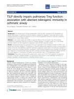
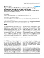
A-C: Bone marrow-derived GFP
+
cells infiltrated the lung following acute bacterial pneumoniaFigure 1
A-C: Bone marrow-derived GFP
+
cells infiltrated the lung following acute bacterial pneumonia. Confocal microscopy images of
the lung. Irradiated mice were transplanted with whole bone marrow from GFP
+
Tg mice. After induction of pneumonia by E.
coli instillation, lungs were fixed and stained for GFP with anti-GFP antibodies. A: Cryosection of the lung, which shows co-
localization of signal from Texas Red-labeled antibody against GFP (red, upper left panel) with GFP signal (green, upper right
panel). The lower panel is a combined image. B: Paraffin section of the lung from the same experiment. Lungs are co-stained for
DNA with Propidium Iodine (Upper left panel) and stained with anti-GFP antibody and secondary FITC labeled antibody
(Upper right panel). Lower left panel – tissue image in reflected light, lower right panel – combined image. C: Control staining
of paraffin-sectioned lungs with isotype primary antibody, no non-specific green fluorescence can be noted, same panel descrip-
tion as in B. D-E: PRXV was abundantly present in cells of the bronchial epithelium of mice, and acute bacterial inflammation
did not further significantly increase it. Confocal microscopy images of the cryosectioned lung, stained for PRXV with red-fluo-
rescent secondary antibody. D: – Non-inflamed control lung (cryosection), original magnification × 40, bar is 50 microns. Note
high expression of PRXV in the bronchial epithelium (blue arrow) but not in the alveoli (green arrow). E: Control staining with
isotype primary antibody; no non-specific red fluorescence is present. F: GFP
+
cells, which are present in high numbers in the
lung following pneumonia, highly express PRXV. Cryosection of the lung, stained for PRXV with Rhodamine-labeled antibodies
(red). Fluorescence intensity of the bronchial epithelium does not differ from control (Panel D). Note the presence of bright
green GFP
+
(or yellow due to superposition of green GFP and red PRXV signals) cells, which also highly express PRXV.
A
B
C
D
E
F
Journal of Inflammation 2006, 3:13 />Page 5 of 12
(page number not for citation purposes)
Following bacterial inflammation, GFP
+
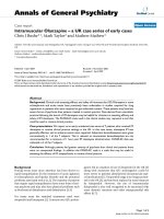
cells in the lung highly expressed PRXVFigure 2
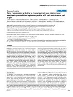
Following bacterial inflammation, GFP
+
cells in the lung highly expressed PRXV. Animals were transplanted with GFP
+
bone
marrow, and progeny of GFP
+
cells (green fluorescence) was located to the sites of inflammation in the bronchial epithelium.
Confocal microscopy images of the cryosectioned lung stained for PRXV with red fluorescent antibodies. A: GFP
+
cells in the
peribronchial interstitial spaces following inflammation of the lung, upper left panel – PRXV staining (red fluorescence), upper
right panel – GFP fluorescence (green), lower panel – combined image. B: GFP
+
cells in the wall of the large bronchus. Upper
left panel – PRXV staining (red fluorescence), upper right panel – GFP fluorescence (green), lower left panel – reflected light
image, lower right panel – combined image. C-D: Fluorescence intensity of PRXV label (red) co-localized with green GFP signal
in the lung tissues. At the bottom of each image the profile diagram of distribution of fluorescence intensity along selected seg-
ment (blue bar) is given. Red line is PRXV fluorescence intensity, green line is GFP fluorescence intensity. Original magnification
× 40. E: Summary results of relative PRXV fluorescence intensity in the bronchial epithelium (loose shade bar), alveolar walls
(dense shade bar), and GFP
+
cells (black bar) present in the lung from 3 different experiments. * – p < 0.05.
0
50
100
150
200
250
*
*
*
Experiment 3
Experiment 2
Experiment 1
Relative Fluorescence Intenstity
A B
C D
E
Journal of Inflammation 2006, 3:13 />Page 6 of 12
(page number not for citation purposes)
off the cells in the lumen. In our previous studies, 4 hours
of exposure of tracheal segment to f-MLP resulted in
enhanced leukocyte migration and increased permeability
[19,20]. We therefore used this time period to assess
expression of PRXV in the model of inflammation. In the
f-MLP model of inflammation, a 4-hour exposure of the
isolated tracheal segment to f-MLP provided a small
(32%) yet significant (p < 0.05) increase in the PRXV
expression in the cells of tracheal epithelium (from 182 ±
16 relative units in the control to 241 ± 3 relative units in
the experimental group), but not in mRNA levels (2.36 ±
0.23 in the control versus 1.51 ± 0.22 in the experimental
group). In the LPS model, we also did not observe statisti-
cally significant difference in PRXV mRNA levels in the
tracheal epithelium (4.71 ± 0.9 in the control versus 2.3 ±
0.7 in the LPS experiment model). There were no signifi-
cant differences in PRXV protein expression in the epithe-
lium (data not shown).
3. Live P. aeruginosa bacteria up-regulates expression, but
not transcription, of PRXV in cultured airway epithelium in
the presence of serum
Experiments were first performed in the alveolar epithelial
cell line A549, co-cultured with mouse macrophage cell
line RAW264.7, both with and without the presence of
serum. Western blot analyses demonstrated that co-cul-
ture of A549 with RAW264.7 and stimulation with PAO1
resulted in enhanced expression of PRXV only in the pres-
ence of serum, as shown in Figure 3. Results of quantita-
tive IHC are shown in Figure 4. In the presence of serum,
the addition of live P. aeruginosa modestly increased PRXV
expression in A549 cultures, as well as in co-cultures with
RAW264.7. P. aeruginosa bacteria itself were not positive
for PRXV staining. The levels of PRXV mRNA did not
change significantly in this system (data not shown). As
can be seen from Figures 3 and 4, RAW264.7 expressed
higher amounts of PRXV than the epithelial cells in cul-
ture, which is similar to our findings in vivo. However,
small amounts of RAWs in the co-culture (5:1 ratio of epi-
thelial cell/macrophages) did not significantly influence
the overall expression of PRXV in the co-culture system, as
epithelial cells were the predominant cell type.
Using the Calu-3 bronchial epithelial cell line, which per-
mits electrically resistant cell layers to be obtained, we
measured TER, mRNA levels, and the expression of PRXV.
We used TER as a measure of tight junctional electrical
permeability, a characteristic of the epithelial phenotype.
Following exposure to P. aeruginosa, the TER of these cells
significantly decreased (p < 0.05), indicating that – in this
model – addition of bacteria produced a considerable
damaging effect on the epithelial cell layers (Figure 5).
However, neither PRXV protein expression nor PRXV
mRNA levels changed after exposure to PAO1; the mean
relative PRXV protein expression following exposure to
PAO1 was 106 ± 25% in the presence of serum and 76 ±
22% without serum as compared to baseline (Figure 5A).
Unlike Calu-3, the primary cultures of cow tracheal epi-
thelium showed a pattern of increased PRXV expression
after exposure to bacteria which was similar to the pattern
shown by the A549 cells (Figure 5C).
Finally, using Western blot analyses of cell-conditioned
medium with and without serum, we studied the presence
of PRXV in the cell secretions of all cell lines that we used.
Actin was used as a marker of intracellular non-diffusible
proteins, and it was not found in the conditioned
medium. Calu-3 and THP-1 secreted the monomeric form
of PRXV into the medium (Figure 6A). THP-1, a human
acute monocytic leukemia cell line was used here as posi-
tive control for inflammatory reaction. In the medium
conditioned by the A549 cells, we observed only the PRXV
form with approximately 60 kDa weight, which probably
reflected polymer formation. We did not observe stimula-
tion of secretion by exposure to PAO1 in the medium with
serum (data not shown) or in the serum-free medium (68
± 21% of control) (Figure 6B).
Discussion
We investigated both in vivo and in vitro models of the
lung bacterial inflammation. In mice, rats, and cultures of
human airway epithelium cells, PRXV was abundantly
expressed under non-inflammatory control conditions. In
rats, neither the presence of endotoxins nor f-MLP-
induced migration of leukocytes in the tracheal epithe-
lium changed mRNA levels of PRXV; f-MLP slightly
increased expression of PRXV protein in the tracheal epi-
thelium. In mice, bacterial inflammation of the lung
resulted in a massive influx of leukocytes, which were the
source of the increased PRXV in the lung tissues. In pri-
mary airway cell culture (cow) and alveolar epithelial cells
A549, or co-culture of the epithelial cells with murine
macrophages RAW264.7, exposure to live bacteria mildly,
yet significantly, increased expression of PRXV protein.
Transcription of PRXV protein was not increased by expo-
sure to bacteria in the A549 or Calu-3 cells. PRXV was
secreted in vitro by both the epithelial and immune cells.
PRXV is a protein abundantly expressed under the base-
line conditions in the airway epithelium, and these obser-
vations suggest that the major pathophysiological
mechanism of its overall up-regulation in the lung during
gram-negative bacterial inflammation is a shift in tissue
cell populations due to migrating leukocytes. In the in
vitro cultured airway epithelia, expression of PRXV protein
was only moderately up-regulated in bacterial inflamma-
tion, while no transcriptional up-regulation was observed.
Journal of Inflammation 2006, 3:13 />Page 7 of 12
(page number not for citation purposes)
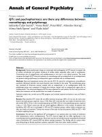
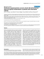
P. aeruginosa infection up-regulated expression of PRXV protein in cultures of the A549 epithelial cells, and co-cultures of A549 and RAW264.7, only in the presence of serumFigure 3
P. aeruginosa infection up-regulated expression of PRXV protein in cultures of the A549 epithelial cells, and co-cultures of A549
and RAW264.7, only in the presence of serum. A: Western blot analyses of PRXV expression in co-cultures of the A549 and
RAW 264.7 cells, stimulated with PAO1 without serum, actin used as control. No up-regulation of PRXV occurred in cells.
Note, that the amount of RAW264.7 used alone, was equal to the amount of cells, added to the A549 cells (5:1 – A549:RAW).
B: Expression of PRXV was up-regulated in the epithelial cells following contact with bacteria (PAO1) in the presence of serum
and in co-culture with immune cells (RAW 264.7). Western blot analyses of PRXV expression in co-cultures. Immunostaining
for actin used as control. C: Expression of PRXV is moderately up-regulated in the A549 cells by P. aeruginosa PAO1 and by co-
culture with RAW 264.7, with and without bacterial inflammation. Quantitative photometric data from Western blot analyses
performed in 3 separate cultures. * – p < 0.05.
A
B
C
A549
A549+PAO1
A549+ RAW
A549+ RAW+ PAO1
RAW
RAW+ PAO1
rPRXV
60 kDa
42 kDa
22 kDa
17 kDa
Actin
PRXV
0
10
20
30
40
50
60
A549
+RAW
+PAO1
*
*
*
A549
+RAW
A549
+PAO1
A549
Relative optical density
60 kDa
42 kDa
22 kDa
17 kDa
Actin
PRXV
A549
A549+PAO1
A549+ RAW
A549+ RAW+ PAO1
rPRXV
Journal of Inflammation 2006, 3:13 />Page 8 of 12
(page number not for citation purposes)
Our experimental finding that serum is required for the
effect of PAO1 on up-regulation of PRXV may have several
explanations. The most obvious is that recognition of bac-
teria by epithelial cells requires serum factors. Epithelial
cells, unlike immune cells, do not possess receptors of
innate immunity (Toll receptors and auxiliary proteins) in
sufficient quantity. It is known, that epithelial and
endothelial cells without the presence of immune cells are
activated with bacterial products like lipopolysaccharide
only in the presence serum. Generation of a response to
bacterial products in non-immune cells occur only at a
very high levels of bacterial product concentrations. We
did not observe up-regulation of PRXV in co-culture of the
epithelial and immune cells. Inflammatory reactions are
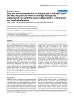
Co-culture of the alveolar epithelial cells with murine macrophages and stimulation by P. aeruginosa moderately upregulated PRXV expression, as determined by IHC and confocal microscopyFigure 4
Co-culture of the alveolar epithelial cells with murine macrophages and stimulation by P. aeruginosa moderately upregulated
PRXV expression, as determined by IHC and confocal microscopy. A-D: Typical confocal microscopy images of the A549 cul-
tures, stained for PRXV (green fluorescence, FITC labeled secondary antibody) and co-stained with Propidium Iodine for DNA.
A – control cultures (A549, no infection), B – cultures infected with PAO1; C – control co-cultures (A549 + RAW, no infec-
tion), D – cultures (A549+RAW264.7), infected with PAO1. At the bottom of each image, a diagram of the distribution of flu-
orescence intensity along the selected segment (red bar) is given. Green line is PRXV fluorescence intensity, red line is DNA
fluorescence intensity. Original magnification × 100. E: MEAN data of relative fluorescence intensity for PRXV staining in co-
cultures of the A549 and RAW264.7 cells. F: The RAW264.7 cells expressed higher amounts of PRXV than the A549 cells in
co-cultures. Confocal microscopy images of co-cultures – both types of cells are indicated by labeled arrows. Staining for
PRXV with FITC-labeled secondary antibody (green fluorescence). Co-staining – Propidium Iodine (red).
0
50
100
150
200
250
*
*
*
A549
+RAW
+PAO1
A549
+RAW
A549
+PAO1
A549
Relative fluorescence intensity
A549
R
A
W
A B C D
E
F
Journal of Inflammation 2006, 3:13 />Page 9 of 12
(page number not for citation purposes)
P. aeruginosa infection did not up-regulate expression of PRXV in the human bronchial epithelial cells Calu-3, as it did in the cow primary tracheal epithelial cell culturesFigure 5
P. aeruginosa infection did not up-regulate expression of PRXV in the human bronchial epithelial cells Calu-3, as it did in the
cow primary tracheal epithelial cell cultures. A: Western blot results of PRXV expression of the Calu-3 cell lysates (CELLS) and
the cell-conditioned medium (MEDIUM). The Calu-3 cells were stimulated with PAO1 bacteria either in the presence of FCS
or without it. PRXV was not upregulated in these cells, and its secretion in the medium was not changed. To confirm that
PAO1 induced alterations in Calu-3 layers, TER of epithelial layers was measured. B: Summary results of TER following expo-
sure of the Calu-3 epithelial cells to PAO1 with and without FCS. PAO1 induced a significant decrease in TER, which was more
pronounced in the presence of FCS. Open bar – initial TER, closed bar – TER after a 4-hour exposure to medium or bacteria.
In the "control" condition, cells were exposed only to the medium (or the medium with FCS) without bacteria. * – p < 0.05
compared to the initial value,
+
– p < 0.05 compared to the control without bacteria, n = 6. C: P. aeruginosa infection up-regu-
lated expression of PRXV protein in the primary cultures of the cow tracheal epithelial cells. C1-C2: Typical confocal micros-
copy images of the CTE cultures stained for PRXV (green fluorescence, FITC labeled secondary antibody) and co-stained with
Propidium Iodine for DNA. C1 – control cultures (no infection), C2 – cultures infected with PAO1 for 12 hours. At the bot-
tom of each image, a diagram of distribution of fluorescence intensity along the selected segment (blue bar) is given. Green line
is PRXV fluorescence intensity, red line is DNA fluorescence intensity. Original magnification × 63. C3: MEAN data are given
for fluorescence intensity in the control and PAO1-infected CTE cultures.
Calu-3
Recomb PRXV
Calu-3+PAO1 + Me
d
Calu-3+ Medium
Calu-3 +PAO1+ FCS
Calu-3+ FCS
Calu-3 + PAO1
Recomb PRXV
22 kDa
17kDa
22 kDa
17 kDa
CELLS
MEDIUM
0
50
100
150
200
250
300
350
400
+
+
*
*
*
Medium only
Medium + FCS
Control PAO1Control PAO1
TER, ohms x cm
2
0
50
100
150
200
*
CTE+PAO1
CTE
Relative fluorescence intensity
PAO1
CTE
C3
A B
C1
C2
Journal of Inflammation 2006, 3:13 />Page 10 of 12
(page number not for citation purposes)
complex even in this simplified in vitro model, with mul-
tiple loops of feedback regulation, both positive and neg-
ative. Likely, PAO1 caused activation of RAWs and
possibly apoptosis of these cells. Activated RAWs release
an array of pro-inflammatory cytokines, which might ini-
tiate apoptosis in epithelial cells and therefore decrease
PRXV expression. The fate of RAWs co-cultured with the
epithelial cells is difficult to estimate, but very likely RAWs
did not have much survival advantage in the medium
designed for epithelial cells.
Prior studies of PRX expression showed that PRXI, II, III,
V and VI are highly over-expressed in the human lung can-
cer cells [21]. Allergic inflammation in response to oval-
bumin induced overexpression of PRXI [22], which is also
well known to be induced by hyperoxia [23]. Stimulation
of the A549 cells and BEAS 2 B cells with hydrogen perox-
ide, menadione, tumor necrosis factor α, or transforming
growth factor β did not result in significant changes of
PRXV expression [18]. These in vitro results are in agree-
ment with our data.
In studies of secreted PRXV, we observed only a 60 kDa
band by Western blot analyses. Peroxiredoxins may form
polymers in an oxidized state. It is unlikely that the band
of interest was non-specific staining, simply because it was
observed only after stimulation, but not in control non-
stimulated cells and not in serum. Further investigations
are needed to define the mechanisms of PRXV polymeri-
zation in extra-cellular fluids.
Some insights into possible mechanisms of PRXV gene
regulation can be obtained by analysis of the PRXV gene
structure. The PRXV gene is located on human chromo-
some 11q13, which is a region of genetic linkage for
atopic hypersensitivity such as bronchial asthma. A 5' pro-
moter region (4 kb upstream of the first exon) contains 3
potential binding sites (hypoxia-response element HRE,
motifs ACGTG for hypoxia-inducible transcription factor
HIF-1 and one potential antioxidant/electrophile
response element (ARE/EpRE, motif TGACNNNGC).
Additional ARE/EpRE is also present within the first
intron, along with potential binding sites for transcription
factor NF-kappa-B (motif GGRNAKTCCC) and Alu-asso-
ciated retinoic acid-response element (RARE, motif
AGGTSMNNAGWTCR). Therefore, in theory, transcrip-
tion of this gene can be modulated in response to
hypoxia, inflammation, and oxidative stress by intrinsic
PRXV was secreted into the medium by the epithelial cellsFigure 6
PRXV was secreted into the medium by the epithelial cells. Cell-conditioned medium from different cell cultures (A549, Calu-
3, RAW264.7, THP-1) without FCS was analyzed by Western blot analyses for the presence of PRXV. Recombinant PRXV was
used as the control. In the medium conditioned by the A549 cells, only a high molecular-weight form of PRXV (either polymer
or possibly a glycosylated form) was present. The RAW 264.7 cells did not show appreciable amounts of PRXV secretion. B:
Upon stimulation with PAO1 without FCS, secretion of PRXV into the medium by the A549 or RAW 264. 7 cells showed no
change. Western blot analyses of cell-conditioned medium (without FCS) upon stimulation with PAO1. Western blot analyses
of the medium with FCS provided substantial non-specific staining, precluding illustration.
A549
Calu-3
RAW264.7
THP-1
Recombinant PRXV
60 kDa
42 kDa
22 kDa
17 kDa
A
A549
A549 PAO1
A549+ RAW
A549+ RAW+ PAO1
Recombinant PRXV
RAW
RAW+ PAO1
60 kDa
42 kDa
22kDa
17 kDa
B
Journal of Inflammation 2006, 3:13 />Page 11 of 12
(page number not for citation purposes)
regulatory elements. It should be noted, however, that the
functional activity of these potential transcription ele-
ments in the human PRXV promoter region has not been
confirmed experimentally.
According to our data from the in vivo studies, PRXV pro-
tein is already abundantly up-regulated, and it is not up-
regulated further by inflammation. One explanation is
that the mechanisms regulating PRXV transcription can-
not further increase expression in these cells, which are in
constant direct contact with pathogens, antigens, and oxi-
dants from the environment that are present in the bron-
chial tree in vivo.
Cells of the alveolar lining, however, are protected from
these stimuli. It is unclear why we did not observe up-reg-
ulation of PRXV in the alveolar epithelium in vivo during
bacterial inflammation of the airways, though we
observed mild up-regulation in the A549 cells in vitro.
Very likely, the model of lung bacterial inflammation that
we used (instillation of bacteria into the airways) affected
primarily the upper and conducting airways without caus-
ing massive inflammation in the alveolar spaces.
Adequate antioxidant protection in the lung is required
for normal function, especially under conditions of oxi-
dant stress caused by environmental factors [2]. Classic
antioxidant enzymes of the lung cells that reduce hydro-
gen peroxide are catalase and glutathione peroxidase,
while airway surface liquids and interstitial fluids are rich
in superoxide dismutase and glutathione. Other hydrogen
peroxide-reducing enzymes include thioredoxin-thiore-
ductase, peroxiredoxins, and glutaredoxins [3]. Under
physiologic conditions, superoxide dismutases and glu-
tathione peroxidase are much more efficient than perox-
iredixins in regulating the cell redox state. Under
conditions of high oxidative stress, however, enzymes like
thioredoxin may become physiologically important [1].
Hoshino and co-workers [24] showed that overexpression
of thioredoxin or administration of its recombinant form
protected mice against lung injury induced by pro-inflam-
matory cytokines and bleomycin. Adenovirus-mediated
transfer of 1-cys peroxiredoxin gene was shown to protect
mice from oxidative injury induced by exposure to oxygen
[8]. PRXV has multiple functions: in addition to its anti-
oxidant activity, PRXV is also a transcriptional co-repres-
sor [13,14] and an inhibitor of p53-dependent apoptosis
[10]. The anti-apoptotic activity of PRXV was demon-
strated in tendon cells [9].
Our results showed that acute inflammation in the lung
results in up-regulation of PRXV expression by different
mechanisms. In the bronchial epithelium, we observed
only a moderate rise in expression of this protein on a
post-transcriptional level. Activated leukocytes that move
to the lungs when inflammation occurs are a rich source
of PRXV. However, this is likely to be a mechanism of leu-
kocyte self-defense against self-induced oxidation rather
than the mechanism of tissue protection. Defining the
mechanism of PRXV regulation of expression in leuko-
cytes during activation by mitogens was not the aim of
present investigation and requires further study. Nonethe-
less, as PRXV is normally abundantly expressed in airways
and as mechanisms of further PRXV up-regulation in the
bronchial epithelium are limited, there is a basis for pro-
posing therapeutic administration of this protein in
recombinant form. Administration of PRXV in aerosol
form may have significant therapeutic potential, espe-
cially in conditions where PRXV expression is down-regu-
lated [15]. Inflammatory conditions are not the likely
candidates for such an intervention, however, as there is
no deficiency of PRXV in the airway epithelium during
inflammation.
Conclusion
In vivo and in vitro bacterial inflammation mildly up-regu-
lates expression of PRXV protein in the airway epithelial
cells. An increased influx of activated leukocytes to
inflamed tissues serves as a source of enhanced expression
of PRXV in the lung.
Abbreviations
BSA – bovine serum albumin;
CSE – cigarette smoke extract;
CTE – cow tracheal epithelium;
FCS – fetal calf serum;
FITC – fluorescein isothiocyanate;
f-MLP – formyl-methionyl-leucyl-phenylalanine;
PBS – phosphate-buffered saline;
PRX – peroxiredoxin;
TER – transepithelial electrical resistance.
Declaration of competing interests
The author(s) declare that they have no competing inter-
ests.
Authors' contributions
RIK carried out experiments in cell cultures, Western blot
analyses.
AVK carried out antibody development, experiments in
cell cultures.
Publish with BioMed Central and every
scientist can read your work free of charge
"BioMed Central will be the most significant development for
disseminating the results of biomedical research in our lifetime."
Sir Paul Nurse, Cancer Research UK
Your research papers will be:
available free of charge to the entire biomedical community
peer reviewed and published immediately upon acceptance
cited in PubMed and archived on PubMed Central
yours — you keep the copyright
Submit your manuscript here:
/>BioMedcentral
Journal of Inflammation 2006, 3:13 />Page 12 of 12
(page number not for citation purposes)
CL carried out Taq-man RT-PCR analyses.
VBS carried out animal experiments, IHC, study design,
and drafted the manuscript.
Acknowledgements
Research described in this manuscript was supported by Philip Morris USA
Inc. and by Philip Morris International. We would like to express our appre-
ciation of technical assistance of Hyon Choi with cell cultures.
References
1. Crapo JD: Redox active agents in inflammatory lung injury.
Am J Resp Crit Care Med 2003, 168:1027-1028.
2. Kinnula VL: Focus on antioxidant enzymes and antioxidant
strategies in smoking related airway diseases. Thorax 2005,
60:693-700.
3. Kinnula VL, Crapo JD: Superoxide dismutases in the lung and
human lung diseases. Am J Resp Crit Care Med 2003,
167:1600-1619.
4. Rahman I, MacNee W: Oxidant/antioxidant imbalance in smok-
ers and chronic obstructive pulmonary disease. Thorax 1996,
51:348-350.
5. Kim HS, Kang SW, Rhee SG, Clerch LB: Rat lung peroxiredoxins
I and II are differentially regulated during development and
by hyperoxia. Am J Physiol Lung Cell Mol Physiol 2001,
280:L1212-L1217.
6. Kinnula VL, Lehtonen S, Kaarteenaho-Wiik R, Lakari E, Paakko P,
Kang SW, Rhee SG, Soini Y: Cell specific expression of peroxire-
doxins in human lung and pulmonary sarcoidosis. Thorax
2002, 57:157-164.
7. Manevich Y, Feinstein SI, Fisher AB: Activation of the antioxidant
enzyme 1-CYS peroxiredoxin requires glutathionylation
mediated by heterodimerization with GST. PNAS 2004,
101:3780-3785.
8. Wang Y, Manevich Y, Feinstein SI, Fisher AB: Adenovirus-medi-
ated transfer of the 1-cys peroxiredoxin gene to mouse lung
protects against hyperoxic injury. Am J Physiol Lung Cell Mol Phys-
iol 2004, 286:L1188-L1193.
9. Yuan J, Murrell GAC, Trickett A, Landtmeters M, Knoops B, Wang
M-X: Overexpression of antioxidant enzyme peroxiredoxin 5
protects human tendon cells against apoptosis and loss of
cellular function during oxidative stress. Biochim et Biophys Acta
– Mol Cell Res 2004, 1693:37-45.
10. Zhou Y, Kok KH, Chun AC, Wong CM, Wu HW, Lin MC, Fung PC,
Kung H, Jin DY: Mouse peroxiredoxin V is a thioredoxin perox-
idase that inhibits p53-induced apoptosis. Biochem Biophys Res
Comm 2000, 24:921-927.
11. Kropotov AV, Grudinkin PS, Pleskach NM, Gavrilov BA, Tomilin NV,
Zhivotovsky BD: Down-regulation of peroxiredoxin V stimu-
lates formation of etoposide-induced double-strand DNA
breaks. FEBS Letters 2000, 572:75-79.
12. Kropotov AV, Sedova V, Ivanov V, Sazeeva N, Tomilin A, Krutilina RI,
Oei SL, Griesenbeck J, Buchlow G, Tomilin NV: A novel human
DNA-binding protein with sequence similarity to a subfamily
of redox proteins, which is able to repress RNA-polymerase-
III-driven transcription of the Alu-family retroposons in vitro.
Eur J Biochem 1999, 260:336-346.
13. Kropotov AV, Tomilin NV: A human B-box-binding protein
down-regulated in adenovirus 5-transformed human cells.
FEBS Letters 1996, 386:43-46.
14. Kropotov AV, Tomilin NV: Evidence for a regulatory protein
complex on RNA polymerase III promoter of human retro-
posons of Alu family. Genetica 1997, 98:223-233.
15. Serikov VB, Leutenegger C, Krutilina RI, Kropotov AV, Pleskach NM,
Jung Suh H, Tomilin NV: Cigarette Smoke Extract Inhibits
Expression of Peroxiredoxin V and Increases Airway Epithe-
lial Permeability. Inhalation Toxicology 2006, 18:79-92.
16. Serikov VB, Popov BV, Kropotov AV, Tomilin NV: Bone Marrow-
Derived Cells Restore Expression of Peroxiredoxin V in the
Airways Following Acute Naphthalene Injury in Mice. Cyto-
therapy 2005, 7:483-493.
17. Knoops B, Clippe A, Bogard C, Arsalane K, Wattiez R, Hermans C,
Duconseille E, Falmagne P, Bernard A: Cloning and characteriza-
tion of AOEB166, a novel mammalian antioxidant enzyme of
the peroxiredoxin family. J Biol Chem 1999, 274:30451-30458.
18. Lehtonen ST, Markkanen PMH, Peltoniemi M, Sang Won Kang, Kin-
nula VL: Variable over-oxidation of peroxiredoxins in human
lung cells in severe oxidative stress. Am J Physiol: Lung Cellular and
Molecular Physiology 2005, 288:L997-L1001.
19. Serikov VB, Jang JY, Widdicombe JH: An estimate of hydrostatic
pressure which drives fluid into airway lumen. J Appl Physiol
2002, 92:1702-1708.
20. Serikov VB, Choi H, Chmiel KJ, Wu R, Widdicombe JH: Role of ERK
in the increase in airway epithelial permeability during leu-
kocyte transmigration. Am J Resp Cell Mol Biol 2004, 30:261-270.
21. Lehtonen ST, Svensk AM, Soini Y, Paakko P, Hirvikoski P, Kang SW,
Saily M, Kinnula VL: Peroxiredoxins, a novel protein family in
lung cancer. Int J Cancer 2004, 111:514-21.
22. Fajardo I, Svensson L, Bucht A, Pejler G: Increased levels of
hypoxia-sensitive proteins in allergic airway inflammation.
Am J Respir Crit Care Med 2004, 170:477-484.
23. Das KC, Pahl PM, Guo XL, White CW: Induction of peroxire-
doxin gene expression by oxygen in lungs of newborn pri-
mates. Am J Respir Cell Mol Biol 2001, 25:226-232.
24. Tomoaki Hoshino, Hajime Nakamura, Masaki Okamoto, Seiya Kato,
Shinichi Araya, Keiko Nomiyama, Kotaro Oizumi, Young HA, Hisam-
ichi Aizawa, Junji Yodoi: Redox-reactive protein Thioredoxin
prevents proinflammatory cytokine- or Bleomycin-induced
lung injury. Am J Resp and Crit Care Medicine 2003, 168:1075-1083.