Báo cáo y học: "Anti-inflammatory potential of a malleable matrix composed of fermented whey proteins and lactic acid bacteria in an atopic dermatitis model" pot
Bạn đang xem bản rút gọn của tài liệu. Xem và tải ngay bản đầy đủ của tài liệu tại đây (419.83 KB, 10 trang )
BioMed Central
Page 1 of 10
(page number not for citation purposes)
Journal of Inflammation
Open Access
Research
Anti-inflammatory potential of a malleable matrix composed of
fermented whey proteins and lactic acid bacteria in an atopic
dermatitis model
Josée Beaulieu
1,2
, Claude Dupont
1
and Pierre Lemieux*
2
Address:
1
Institut national de la recherche scientifique, INRS-Institut Armand-Frappier, 531 boul. des Prairies, Laval, Québec, Canada, H7V 1B7
and
2
Technologie Biolactis, 500 boul. Cartier suite 218, Laval, Québec, Canada, H7V 5B7
Email: Josée Beaulieu - ; Claude Dupont - ; Pierre Lemieux* -
* Corresponding author
Abstract
Background: Over the last 10 years, whey proteins have received considerable attention in the
area of functional foods and nutraceuticals. In this paper, a novel fermented whey protein-based
product described as a gel-like Malleable Protein Matrix (MPM) has been tested for its anti-
inflammatory activity. Preliminary in vitro results have already indicated that MPM could exert such
an anti-inflammatory activity.
Methods: The systemic anti-inflammatory activity of the MPM was explored using the oxazolone-
induced atopic contact dermatitis mouse model (ACD). Parameters including ear thickness, side
effects as well as neutrophil extravasation were monitored.
Results: In the ACD model, the MPM exhibited an anti-inflammatory effect comparable to that of
hydrocortisone (positive control). Mice fed with MPM showed strong reduction of the ear
inflammation while no side effects, as compared to hydrocortisone, were observed. The MPM
seemed to reduce neutrophil extravasation in tissue as evidenced by blood polymorphonuclear
cells and ear myeloperoxidase content.
Conclusion: The anti-inflammatory activity demonstrated in the ACD model suggests that the
mechanism of action of the MPM is different than that of hydrocortisone and could become a
relevant product for people suffering from dermatological manifestations associated with immune
dysfunctions such as allergies, eczema, dermatitis, and autoimmune diseases.
Background
Modern life-styles which leads to obesity, stress and inac-
tivity, is a major cause of immunological diseases, partic-
ularly those associated with chronic inflammation which
are on the upswing during the last decade [1-3]. Many evi-
dences exist that functional foods have protective effects
on immune deficiency [4-6] including whey proteins,
which can modulate some immune functions [5]. Other
studies revealed that whey proteins possess a myriad of
activities including antioxidant activity attributed to
increasing glutathione content [7,8], anti-allergic, [9] anti-
inflammatory [9-11] and immunomodulatory activities
[12-19]. Whey proteins such as β-lactoglobulin (β-LG),
bovine serum albumin (BSA) and α-lactalbumin (α-LA)
have been shown to stimulate splenocyte proliferation,
increase interleukin-1 production by macrophages and
Published: 21 March 2007
Journal of Inflammation 2007, 4:6 doi:10.1186/1476-9255-4-6
Received: 9 June 2006
Accepted: 21 March 2007
This article is available from: />© 2007 Beaulieu et al; licensee BioMed Central Ltd.
This is an Open Access article distributed under the terms of the Creative Commons Attribution License ( />),
which permits unrestricted use, distribution, and reproduction in any medium, provided the original work is properly cited.
Journal of Inflammation 2007, 4:6 />Page 2 of 10
(page number not for citation purposes)
increase GSH production [18,19]. Whey peptides have
recently been shown to possess immunomodulatory
activities such as a stimulation of lymphocytes, an increas-
ing in phagocytosis process as well as in secretion of
immunoglobulin A (IgA) by Peyer's patches [5,13,17,20].
Lactoferrin (LF), a minor whey protein, has been exten-
sively studied. LF assists the phagocytosis process in neu-
trophils, increases production of interleukin-8 (IL-8) [13]
and stimulates immune cell production [15,19,21]. More-
over, LF has also demonstrated anti-inflammatory effects
in animal models by an inhibition of pro-Th1 cytokines
and an increasing in regulatory cytokine IL-10 production
[9,11]. More specifically, LF exerts its anti-inflammatory
effect during mouse atopic contact dermatitis (ACD) by
reducing ear thickness and infiltration of inflammatory
cells following a direct topical contact [11].
In addition, some Lactic Acid Bacteria (LAB) have shown
immunomodulatory and anti-inflammatory activities.
The genus Lactobacillus commonly used in many fer-
mented dairy products [22] is the most studied of these
probiotics [23]. The effects of LAB are very strain-depend-
ent but many lactobacilli act on Peyer's patches to stimu-
late IgA production, phagocytosis process and possess
anti-inflammatory and anti-allergic activities by reducing
the production of cytokines and immunoglobulin E (IgE)
[24-27]. Cytokine production is also strain-dependent as
some lactobacilli are able to increase Th1 profile while oth-
ers increase Th2 profile [28]. These results suggest that
lactobacilli could act both as immunostimulating and anti-
inflammatory agents. Some studies also indicate that the
effects of probiotics acting in synergy with food ingredi-
ents can be more intense than the probiotics alone [29].
Moreover, vitamins present in the MPM (niacin and ribo-
flavin) as well as calcium also possess immunomodula-
tory effects [30-32].
Considering the positive effects on the immune system of
both whey proteins and probiotic lactobacilli, a novel fer-
mented whey protein-based ingredient, called Malleable
Protein Matrix (MPM) [33], was tested for its immu-
nomodulatory activities [34]. It was previously demon-
strated that MPM stimulates production of blood
polymorphonuclear cells, cytokine IL-18 as well as glu-
tathione by white blood cells in healthy rat suggesting a
stimulation of innate immunity [33,34]. On the other
hand, MPM can also reduce the production of important
pro-inflammatory cytokines such as TNFα [33]. Moreo-
ver, it was shown in vitro that MPM reduces pro-inflamma-
tory cytokines and inhibits the cytokines production
following LPS stimulation on CaCo2 cells [33]. These
results suggested that MPM might also exhibit anti-
inflammatory properties when placed in the context of
inflammation.
The objective of this present study was to evaluate the sys-
temic anti-inflammatory potential of MPM and to deter-
mine how its complex composition may lead to
synergistic effects. For this purpose, the oxazolone-
induced atopic contact dermatitis mouse model (ACD)
was used. This ACD mouse model requires two distinct
phases [35]. First, the sensitization phase is initiated by
topical application of oxazolone, which permits the acti-
vation of T cells through Langerhans cells acting as an
antigen presenting cells. The elicitation phase is next
achieved by a subsequent topical application of oxa-
zolone, which initiate the inflammatory process by
recruiting activated T effector cells which in turn attract
inflammatory cells [36-38]. The inflammatory cells
recruited in this ACD model are principally macrophages,
which attract neutrophils in the early inflammatory phase
and monocytes as well as dendritic cells in the early and
late inflammatory phases. CD4+ T cells act as regulatory
cells and not as effector cells in the ACD model, in which
they control the intensity of inflammatory reaction
[39,40]. A similar dermatitis model has recently been used
to evaluate the anti-inflammatory activity of LF [11] and a
milk-product fermented by Lactobacillus casei [27].
Methods
Reagents
The Malleable Protein Matrix (MPM) was obtained from
Technologie Biolactis inc. (LaBaie, Qc, Canada). Briefly,
the MPM is obtained by a protein specific recuperation
procedure following the fermentation of sweet whey by a
proprietary Lactobacillus kefiranofaciens strain (R2C2) iso-
lated from kefir grains and adapted to grow in whey [33].
The composition of MPM is shown in Table 1. On a
humid basis (w/w), the MPM contains 80% water, 8%
protein, 6% minerals (2% calcium), 5% carbohydrate
(2.7% lactose) and less than 1% of fat. Lyophilized MPM
required reconstitution in water: 20 g of lyophilized MPM
was blended with 80 mL of water for 2 minutes at maxi-
mum speed (20% w/v). The final reconstituted product is
stable at 4°C for at least 1 month. Water-soluble hydro-
cortisone (HC) was obtained form Sigma-Aldrich Canada
(Oakville, On, Canada) and was diluted in deionized dou-
ble-distilled water to a final concentration of 10 mg/mL.
For the mouse ACD model, the 4-ethoxy-methylene-2-
phenyloxazol-5-one (oxazolone) (Sigma-Aldrich Canada)
was required at a concentration of 5% (w/v) in acetone to
cause inflammation.
Animals
CD-1 female mice were obtained from Charles River Lab-
oratories (St-Constant, Qc, Canada) and were used at 20
days of age for studies in the mouse ACD model. The ani-
mals were housed in filter top isolator cages in a room
kept at 20–23°C with humidity maintained between 35–
45% with a 12-hour light-dark cycle and free access to a
Journal of Inflammation 2007, 4:6 />Page 3 of 10
(page number not for citation purposes)
standard laboratory pelleted Rodent Lab Diet 5001 (Ren's
Feed & Supplies Limited, Oakville, On). The experimental
protocols used were approved by the Animal Care Com-
mittee of the INRS- Institut Armand-Frappier (Comité
Institutionnel des Soins aux Animaux et de leur Utilisa-
tion (CISAU)) and were performed in accordance with the
recommendations of the Canadian Council on Animal
Care as specified in the Guide to the Care and Use of
Experimental Animals (CISAU # 0306-01 and # 0410-01).
Mouse atopic contact dermatitis (ACD)
After a week adaptation in the animal facility, the mice
were separated in groups of 10 animals. The grouping was
randomized according to the weight of the rodents. The
murine model of ACD was based on those firstly
described by Garrigue et al. [41] and modified as follows:
abdomen hair of CD-1 mice was removed and the sensiti-
zation phase was done by the application of 100 μL of
oxazolone 5 % (w/v) in acetone on the hairless abdomen.
After four days, the elicitation phase (first challenge) was
initiated by the application of 50 μL of oxazolone 5% (w/
v) in acetone on the right ear (25 μL each side of the ear).
The second challenge was done 7 days after the first chal-
lenge with the same procedure. The ear thickness of the
mice was measured every day with a digital caliper (VWR,
Mont-Royal, Canada).
Dose-response curve
The dose-response curve has been done in the prophylac-
tic anti-inflammatory mouse ACD model. Groups of 10
CD-1 mice received each day by gavages (per os (p.o)),
100 μL of reconstituted lyophilized MPM at three doses
20% (w/v), 10% (w/v) and 5% (w/v), 100 μL of water or
100 μL of water-soluble hydrocortisone (10 mg/mL). The
mouse ACD was performed as described previously and
ear thickness was measured every day.
Table 1: Composition of MPM
Composition (g/100 g)
Protein 8.1
Lipids 0.9
Ash (minerals) 5.1
Carbohydrates 4.6
Lactose 2.7
Galactose 0.2
Minerals (mg/100 g)
Potassium 142.9
Sodium 175.2
Calcium 1600
Phosphorus 730.3
Selenium < 0.1
Magnesium 5.4
Oligo-elements (mg/100 g)
Copper 0.07
iron 0.24
Manganese 0.05
Zinc 0.13
Vitamins (mg or
μ
g/100 g)
Riboflavine (B2) 0.32 mg
Niacin (B3) 1.00 mg
Pyridoxine (B6) 0.04 mg
Cobalamine (B12) Not detected
Ascorbic acid (C) Not detected
Folic acid 5 μg
Bacterial count (CFU/100 g)
LAB 6 × 10
11
Journal of Inflammation 2007, 4:6 />Page 4 of 10
(page number not for citation purposes)
Prophylactic protocol – Mouse ACD
The prophylactic anti-inflammatory potential of MPM
was evaluated by the administration of MPM seven days
prior to sensitization. Groups of 10 CD-1 mice received
each day by gavages (per os (p.o)), 100 μL of reconstituted
lyophilized MPM, 100 μL of water or 100 μL of water-sol-
uble hydrocortisone (10 mg/mL). The mouse ACD was
performed as described previously and ear thickness was
measured every day. The mice's weight was measured
twice a week. The spleen's weight was measured at the end
of the protocol and was normalized in accordance to each
mouse's weight.
Therapeutic protocol – Mouse ACD
The therapeutic anti-inflammatory potential of MPM was
evaluated by the administration of MPM, soluble hydro-
cortisone or water as in the prophylactic protocol, but
only after the first challenge. The other parameters were
followed as described.
Evaluation of peripheral white blood cell counts
At the end of the prophylactic protocol of mouse ACD, the
blood of each mouse was taken and white blood cell
counts evaluated by flow cytometry. Briefly, the red blood
cells were lysed with Optilyse C (Beckman-Coulter, Fuller-
ton) in accordance with manufacturer's instructions. The
cell counts were obtained by passage of 20 μL of prepara-
tion in a Flow Cytometry Epics XL cytometer (Beckman
Coulter, Fullerton). The lymphocytes, monocytes and poly-
morphonuclears (PMN) were separated in accordance
with cell size and cell granulometry.
Evaluation of ears-myeloperoxidase (MPO) content
The method for the evaluation of MPO content was
adapted from those developed by Bradley et al. [42] and
Xia and Zweier [43]. The mice were sacrificed at the end of
prophylactic protocol by CO2 and the ears were immedi-
ately remove and frozen quickly in liquid nitrogen. The
ears were chopped up and added in 50 mM phosphate
potassium buffer, pH 6.0 supplemented with 0.5% hexa-
decyltrimethylammonium bromide (HTAB). The ears
were disrupted with three cycles of sonication (10 sec.) in
water-ice bath followed by three freeze-thaw cycles in
methanol-dry ice bath and another three cycles of sonica-
tion in water-ice bath. The homogenates were centrifuged
at 10 000 g for 15 min at 4°C and the supernatants were
conserved at -80°C until analyses. For the quantification
of MPO content, 100 μL of homogenates (or MPO stand-
ard from Sigma-Aldrich, Oakville, On) were mixed with 2.9
mL of 50 mM phosphate potassium buffer containing
0.117 mg/mL of o-dianisidine (Sigma-Aldrich, Oakville,
On) and 0.0005% hydrogen peroxide. The oxydation of o-
dianisidine kinetic was followed at 460 nm with a spectro-
photometer Varian Cary 300 (Varian, St-Laurent, QC) dur-
ing 5 min at 25°C.
Statistical analysis
The inflammatory mouse ACD experiments were per-
formed with groups counting 10 mice/group and two
independent experiments. The statistical analysis of data
was performed by the biostatistical service of INRS-Insti-
tut Armand-Frappier. Statistical analysis used was a
repeated measure one-way ANOVA test that permits the
comparison between groups during the entire experiment
independently of each day. When the ANOVA test was not
possible because of interactions between groups, a Stu-
dent test was run for comparison of groups at each day.
Results
MPM is a whey-fermented product, which by its composi-
tion, has a high potential as an anti-inflammatory agent.
The oxazolone-induced atopic contact dermatitis (ACD)
model was used for the demonstration of MPM's effect on
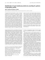
inflammatory diseases. Figure 1 shows an important
reduction of ear thickness in mice consuming MPM as
compared to that of the water control group. In the dose-
response curve experiment, it is demonstrated that MPM
possesses a higher anti-inflammatory effect when the con-
centration of product was 20% (Figure 1A). Conse-
quently, MPM has been used for all experimentations at
20%. In the prophylactic protocol (Figure 1B), the maxi-
mal reduction of ear thickness was in the order of 26% in
the MPM group and 35% in the hydrocortisone group as
compared to the water control group. This thickness
reduction was observed immediately after the first chal-
lenge and increases markedly after the second challenge.
The ANOVA test indicates that reduction of ear thickness
in the MPM group was statistically different of those from
water group (p < 0.07) for the entire experiment. How-
ever, the ANOVA between MPM and HC groups was not
possible because of interaction between the two groups.
However, Student test has confirmed that the difference
between both groups is not statistically different for the
entire experiment (with exception for day 4). This statisti-
cal analysis permits to conclude that the anti-inflamma-
tory effect of MPM is comparable to that of
hydrocortisone treatment. In the therapeutic protocol
(Figure 1C), the reduction of ear thickness was statistically
different only after the second challenge in the MPM
group compared to the water control group and reached a
maximal reduction of 37%. For the group treated with
hydrocortisone the maximal reduction of ear thickness
reached 40%. Using the prophylactic protocol, these anti-
inflammatory observations were confirmed in another
independent experiment with the same batch of MPM and
also with two other different batches of MPM. The results
obtained were similar and statistically significant as con-
firmed by the ANOVA analysis, indicating the reproduci-
bility of anti-inflammatory effect using different batches
of MPM (data not shown).
Journal of Inflammation 2007, 4:6 />Page 5 of 10
(page number not for citation purposes)
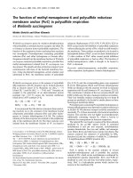
The consumption of hydrocortisone is associated with a
negative effect on mice growth which is clearly demon-
strated by the cessation of growth in the mice who
received hydrocortisone (Figure 2). The MPM demon-
strated an absence of detrimental effect on growth in com-
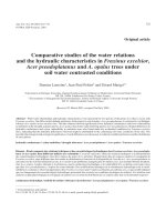
parison to water control group. Moreover, the
hydrocortisone treatment induced a spleen atrophy repre-
sented by a 50% reduction in spleen weight as compared
to water or MPM consumption (Figure 3). This spleen
atrophy indicates an immunosuppression of immune
cells after hydrocortisone treatment. No statistical differ-
ence was observed between the water and MPM group on
spleen weight suggesting no immunosuppression follow-
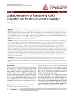
ing MPM consumption. The cell counts confirmed this
immunosuppression following hydrocortisone treatment
as demonstrated by the important reduction (approxi-
mately 50%) in circulating lymphocytes in comparison to
water control group (Figure 4). On the contrary, the MPM
consumption showed a tendency to increase lymphocyte
numbers. These results indicated that MPM consumption
Ear thickness of mice administered p.o. with the MPM, hydrocortisone or waterFigure 1
Ear thickness of mice administered p.o. with the MPM, hydrocortisone or water. A. Dose-response curve during
the prophylactic model : Administrations started 7 days prior sensitization and challenges with oxazolone (p < 0.07 for MPM
20% and hydrocortisone groups compared with water reference group in the ANOVA statistical analysis). Legend: Light-grey
bars: Water, Dark-grey bars: MPM 20%, White bars: MPM 10%, Hashed bars: MPM 5%, Black bars: Hydrocortisone. B. Prophy-
lactic model: Administrations started 7 days prior sensitization and challenges with oxazolone (p < 0.07 for MPM and hydro-
cortisone groups compared with water reference group in the ANOVA statistical analysis) Legend: Light-grey bars: Water,
Dark-grey bars: MPM, Black bars: Hydrocortisone. C. Therapeutic model: Administrations started after sensitization but during
oxazolone challenges (p < 0.05 for MPM and hydrocortisone groups compared to water reference group in the ANOVA statis-
tical analysis from day 8 until the end of experiment). Legend: Light-grey bars: Water, Dark-grey bars: MPM, Black bars: Hydro-
cortisone. (n = 10)
Journal of Inflammation 2007, 4:6 />Page 6 of 10
(page number not for citation purposes)
do not induce side effects generally associated with hydro-
cortisone treatment.
The polymorphonuclear (PMN) cell counts were higher in
MPM and hydrocortisone fed groups compared to the
water control group (Figure 4). The blood PMN counts
were 1.86 and 2.35 fold higher in MPM and hydrocorti-
sone respectively indicating a possible diminution of
PMN extravasation in the ear of mice. The diminution of
neutrophils extravasation as suggested by blood PMN
counts was confirmed by the reduction of neutrophil con-
tent in ear of 62.4% and 82.6% following MPM and
hydrocortisone treatment respectively, as measured by
myeloperoxidase (MPO) ear analysis (Figure 5).
Discussion
MPM contains a variety of ingredients including whey
proteins and peptides, LAB and their related exopolysac-
charides, group B vitamins and calcium (Table 1). All
these ingredients possess effects on the immune system
such as an interesting anti-inflammatory potential
[5,14,15,32,44,45]. In light of these components, the
MPM is believed to possess an anti-inflammatory poten-
tial, which may be amplified by the synergy of its individ-
ual components. Previous observations suggested that the
MPM could be an interesting treatment in inflammatory
diseases. Indeed, it was demonstrated that MPM reduced
production of cytokine TNFα in healthy rat [33]. This
cytokine is very important in the development of the ACD
disease and contributes in the amplification of inflamma-
tory reaction [46]. The reduction of this pro-inflammatory
cytokine following MPM consumption indicated its
potential in the inhibition of development of inflamma-
tory disease and reduction of its intensity. Moreover,
Circulating cell counts 17 days after the first oxazolone chal-lenge in the prophylactic ACD modelFigure 4
Circulating cell counts 17 days after the first oxa-
zolone challenge in the prophylactic ACD model. Leg-
end: Light-grey bars: Lymphocyte counts, Dark-grey bars:
PMN counts (* p < 0.05; ** p < 0.01) (n = 10)
Mice weight during the prophylactic ACD modelFigure 2
Mice weight during the prophylactic ACD model. Leg-
end: Light-grey bars: Water, Dark-grey bars: MPM, Black
bars: Hydrocortisone. (* p < 0.05) (n = 10)
Mice spleen weight at the sacrifice in the prophylactic ACD modelFigure 3
Mice spleen weight at the sacrifice in the prophylac-
tic ACD model. Legend: Light-grey bars: Water, Dark-grey
bars: MPM, Black bars: Hydrocortisone. (** p < 0.01) (n = 10)
Journal of Inflammation 2007, 4:6 />Page 7 of 10
(page number not for citation purposes)
MPM inhibited the production of cytokines in vitro on
CaCo2 cells stimulated with LPS [33] suggesting the inhi-
bition of development of inflammation following an
inflammatory stimulus.
The anti-inflammatory potential of MPM has been con-
firmed in these studies with the murine ACD model. This
model of inflammation has proven to be a sensitive and
useful tool to determine efficacy and potency of several
anti-inflammatory and immunosuppressive drugs used in
dermatological disorders such as dermatitis and psoriasis.
Glucocorticoids, such as hydrocortisone, are commonly
used to relieve skin and joint inflammation and have been
used as a positive control group in these experiments [35].
This model comprises two important phases in order to
examine inflammation: 1) Sensitization phase that is
developed by application of oxazolone on the abdomen,
allowing the recruitment of antigen presenting cells,
which capture and present the antigen (oxazolone) to
naive T lymphocytes that afterwards become active. 2)
Elicitation phase developed by the application of oxa-
zolone on the ear, which allows activation of T lym-
phocytes to move to the ear and recruit inflammatory cells
[35,47].
MPM and hydrocortisone administered p.o. either in a
prophylactic (Figure 1B) or a therapeutic fashion (Figure
1C) reduced the inflammation with similar efficiency as
demonstrated by the reduction of ear redness and thick-
ness. In the prophylactic protocol (Figure 1B), the reduc-
tion of ear inflammation was observed as soon as one day
after the first challenge and this protective effect was con-
served throughout the entire course of the experiment. On
the other hand, for therapeutic protocol, the anti-inflam-
matory effect following MPM consumption was apparent
only after the second challenge (Figure 1C). The effect of
MPM in this model (therapeutic protocol) showed that a
certain period of time is required to overcome existing
inflammation. This indicates that the MPM possesses an
anti-inflammatory effect in an existing disease and is not
only a preventive treatment. This therapeutic effect is
interesting because those who suffer from such disease
can consume MPM during crisis and will benefit of its
effect. This study has shown that the reduction of inflam-
mation by MPM consumption is not negligible as demon-
strated by the comparison with hydrocortisone treatment.
From that observation, we could speculate that the MPM
might also exerts a beneficial effect on the reduction of
skin itching and pain.
The MPM has a strong anti-inflammatory effect as demon-
strated by its ability to reduce dermatological inflamma-
tion to the same extent than that of hydrocortisone.
However in contrast to hydrocortisone, the MPM showed
no side effects generally associated to medication includ-
ing spleen atrophy, reduction in lymphocyte circulating
cells or deleterious effect on body weight gain (Figures 2,
3 and 4). Hydrocortisone exerts its anti-inflammatory
potential by suppression of immune cells. The reduction
of inflammation observed by hydrocortisone treatment
corresponded to a suppression of total immune cells (not
only those implicated in inflammation), which was seen
by the reduction in blood lymphocytes (Figure 4) and in
spleen weight (Figure 3) for the mice consuming hydro-
cortisone. Consequently, people treated by hydrocorti-
sone will be in a general immunosuppressed state and are
therefore, more susceptible to contract other diseases and
infection. No reduction in immune cells or spleen atrophy
was observed in the mice who consumed MPM in com-
parison with the control water group. In fact, a trend
showing immune stimulation by the MPM consumption
was observed as indicated by the tendency to increase
lymphocytes counts as well as spleen weight.
Atopic dermatitis is a disease that affects young children
consequently, the use of hydrocortisone would not be
advisable because of its inhibitory properties on growth
[48]. This inhibition in growth following hydrocortisone
consumption has been demonstrated in this study where
the growth of these young mice treated with hydrocorti-
sone was stopped during all the experiment (Figure 2) in
comparison with mice treated with MPM and water which
gained weight. Consequently, consumption of MPM by
children and young adult in replacement of hydrocorti-
Myeloperoxidase (MPO) contents in ears 18 days after the first oxazolone challenge during the prophylactic ACD modelFigure 5
Myeloperoxidase (MPO) contents in ears 18 days
after the first oxazolone challenge during the prophy-
lactic ACD model. (* p < 0.05) (n = 10)
Journal of Inflammation 2007, 4:6 />Page 8 of 10
(page number not for citation purposes)
sone as an anti-inflammatory product would be a good
alternative.
The absence of all these detrimental effects by MPM con-
sumption suggests that the mechanism of its anti-inflam-
matory action is different than that of hydrocortisone.
However, both hydrocortisone and MPM seem to inhibit
neutrophil extravasation and accumulation in inflamed
tissues as shown with a higher polymorphonuclear cells
(PMN) in circulation as well as a reduced MPO content in
ear (Figures 4 and 5). Results in figure 4 demonstrate an
inverse correlation between inflammation and PMN
counts where in the hydrocortisone and MPM groups, the
blood PMN counts is higher while the ear thickness is
lower than reference water group. These results are con-
sistent with those observed for ear MPO content (Figure
5). The MPO is an enzyme exclusively present in neu-
trophil granules and its enzymatic activity measured in a
tissue is in direct correlation of the levels of neutrophils in
a tissue [42]. The MPO results showed that the neutrophil
infiltration in ear of mice that received hydrocortisone
and MPM is reduced compared to the mice receiving
water. The blood PMN count parameter and ear MPO
content could be explained by the fact that in ACD, the
neutrophils (the most important group in PMN) move
from blood to ear because these cells are principally
responsible for inflammation [36-38]. The hydrocorti-
sone as well as the MPM seems to prevent the neutrophil
extravasation from blood to ear, reducing the ear inflam-
mation. However, the mechanism causing this inhibition
of neutrophil extravasation is different between these two
groups because of the absence of immunosuppression in
MPM group as seen by the absence of spleen atrophy as
well as blood lymphocyte counts (Figures 3 and 4). This
inhibition of neutrophil infiltration indicate that MPM
will be a good candidate for the treatment or prevention
of neutrophilic diseases such as, Sweet syndrome (a neu-
trophilic dermatose resulting of Crohn's disease compli-
cations) as well as chronic obstructive pulmonary disease
[49,50].
It is previously demonstrated that MPM enhances some
cytokines, blood PMN cells and glutathione production
by leukocytes [33] indicating that MPM exerts a definitive
immunomodulation. Its consumption could either be
beneficial in a context of stimulation of innate immunity
but detrimental in the context of inflammatory disease.
This present study reveals the interesting properties of
MPM in the reduction of inflammation confirming that
despites its innate immunity stimulation potential, MPM
act also as an anti-inflammatory agent. The complexity of
MPM components as well as the potential synergy
between its components could explain the properties of
MPM to be an immunomodulatory agent as well as to be
an anti-inflammatory agent in the context of inflamma-
tion. These two different immune situations suggest that
MPM act trough a regulatory mechanism explaining their
both immunomodulatory and anti-inflammatory proper-
ties. These results demonstrate that, as a new product, the
Malleable Protein Matrix reduces inflammation and
immune dysfunctions when consumed orally while main-
taining an appropriate immune system threshold. Experi-
ments to demonstrate the mechanism of action
responsible for the anti-inflammatory effect of the MPM
consumption and other parameters to determine how
specific cells are implicated and influenced by MPM con-
sumption in this ACD model are underway.
Conclusion
MPM possesses a strong anti-inflammatory effect compa-
rable to hydrocortisone when examined in the ACD
model. The anti-inflammatory effects of consumption of
MPM occur without the undesirable side effects normally
associated with hydrocortisone. Therefore, MPM would
be an alternative of choice for children and young adult
suffering from chronic inflammatory of various diseases
such as ACD. The consumption of the MPM could act as a
preventive or a therapeutic nutraceutical in the case of
inflammatory diseases like atopic dermatitis or related
diseases such as, psoriasis. Psoriasis is a chronic inflam-
matory disease with similar effects on the immune system
to that observed for ACD.
Competing interests
Technologie Biolactis (TB) was the industrial sponsor of a
Natural Science and Engineering Research Council of
Canada (NSERC) grant obtained by INRS (CD). Collabo-
rative research conventions and agreements intervened
between TB, INRS and NSERC. INRS is a minor share-
holder of TB (less than 1%) and does not have any vote.
The findings of the present study are covered by a patent
application (PCT CA2002/001899). JB was an on-site
scholar of Fond de Recherche en Santé du Québec (FRSQ)
and part of the scholarship was covered by TB.
Authors' contributions
JB design the animal studies, carried out the animal and
other experiments, perform the statistical analysis and
drafted the manuscript. CD participated in the design of
animal studies, data interpretation and the statistical anal-
ysis. CD revised the manuscript for the intellectual con-
tent and language. PL participated in the design of animal
studies, data interpretation and revised the manuscript for
the intellectual content and language. All authors read
and approved the final manuscript.
Acknowledgements
The authors wish to thank M. Roger Dubuc for the help in adaptation of
the MPO enzymatic assay and Lilianne Gueerts for the help in animal stud-
ies. We also thank Drs Alain Lamarre and Denis Girard for the intellectual
help in animal design as well as results interpretation. Jean-François
Journal of Inflammation 2007, 4:6 />Page 9 of 10
(page number not for citation purposes)
Lapointe revised the manuscript for the intellectual content and language
and participated in data interpretation. Marie Désy has done the statistical
analysis in INRS-IAF biostatistical service. This study was funded by the Nat-
ural Science and Engineering Research Council of Canada (NSERC) Strate-
gic Grant STP 246405-1. JB was a Ph.D. scholar of Fond de recherche en
santé du Québec (FRSQ)
References
1. Oeser A, Chung CP, Asanuma Y, Avalos I, Stein CM: Obesity is an
independent contributor to functional capacity and inflam-
mation in systemic lupus erythematosus. Arthritis Rheum 2005,
52:3651-3659.
2. Toker S, Shirom A, Shapira I, Berliner S, Melamed S: The associa-
tion between burnout, depression, anxiety, and inflamma-
tion biomarkers: C-reactive protein and fibrinogen in men
and women. J Occup Health Psychol 2005, 10:344-362.
3. Bruunsgaard H: Physical activity and modulation of systemic
low-level inflammation. J Leukoc Biol 2005, 78:819-835.
4. Calder PC, Kew S: The immune system: a target for functional
foods? British Journal of Nutrition 2002, 88:S165-S176.
5. Beaulieu J, Dupont C, Lemieux P: Whey proteins and peptides:
beneficial effects on immune health. Therapy 2006, 3:1-10.
6. Laiho K, Ouwehand A, Salminen S, Isolauri E: Inventing probiotic
functional foods for patients with allergic disease. Ann Allergy
Asthma Immunol 2002, 89:75-82.
7. Bounous G, Gold P: The biological activity of undenatured die-
tary whey proteins: role of glutathione. Clin Invest Med 1991,
14:296-309.
8. Kent KD, Harper WJ, Bomser JA: Effect of whey protein isolate
on intracellular glutathione and oxidant-induced cell death
in human prostate epithelial cells. Toxicol In Vitro 2003,
17:27-33.
9. Ward PP, Uribe-Luna S, Conneely OM: Lactoferrin and host
defense. Biochem Cell Biol 2002, 80:95-102.
10. Kano H, Mogami O, Uchida M: Oral administration of milk fer-
mented with Lactobacillus delbrueckii ssp. bulgaricus
OLL1037R-1 to DBA/1 mice inhibits secretion of proinflam-
matory cytokines. Cytotechnology 2002, 40:67-73.
11. Kimber I, Cumberbatch M, Dearman RJ, Headon DR, Bhushan M,
Griffiths CE: Lactoferrin: influences on Langerhans cells, epi-
dermal cytokines, and cutaneous inflammation.
Biochem Cell
Biol 2002, 80:103-107.
12. Cross ML, Gill HS: Modulation of immune function by a modi-
fied bovine whey protein concentrate. Immunol Cell Biol 1999,
77:345-350.
13. Miyauchi H, Hashimoto S, Nakajima M, Shinoda I, Fukuwatari Y, Hay-
asawa H: Bovine lactoferrin stimulates the phagocytic activity
of human neutrophils: identification of its active domain. Cell
Immunol 1998, 187:34-37.
14. Migliore-Samour D, Roch-Arveiller M, Tissot M, Jazziri M, Keddad K,
Giroud JP, Jolles P: Effects of tripeptides derived from milk pro-
teins on polymorphonuclear oxidative and phosphoinositide
metabolisms. Biochem Pharmacol 1992, 44:673-680.
15. Brix S, Bovetto L, Fritsche R, Barkholt V, Frokiaer H: Immunostim-
ulatory potential of beta-lactoglobulin preparations: Effects
caused by endotoxin contamination. Journal of Allergy and Clinical
Immunology 2003, 112:1216-1222.
16. Wong CW, Watson DL: Immunomodulatory effects of dietary
whey proteins in mice. J Dairy Res 1995, 62:359-368.
17. Gill HS, Doull F, Rutherfurd KJ, Cross ML: Immunoregulatory
peptides in bovine milk. Br J Nutr 2000, 84(Suppl 1):S111-117.
18. Bounous G, Kongshavn PA: Differential effect of dietary protein
type on the B-cell and T-cell immune responses in mice. J
Nutr 1985, 115:1403-1408.
19. Hakansson A, Andreasson J, Zhivotovsky B, Karpman D, Orrenius S,
Svanborg C: Multimeric alpha-lactalbumin from human milk
induces apoptosis through a direct effect on cell nuclei. Exp
Cell Res 1999, 246:451-460.
20. Matar C, Valdez JC, Medina M, Rachid M, Perdigon G: Immunomod-
ulating effects of milks fermented by Lactobacillus helveticus
and its non-proteolytic variant. J Dairy Res 2001, 68:601-609.
21. Clare DA, Catignani GL, Swaisgood HE: Biodefense properties of
milk: The role of antimicrobial proteins and peptides.
Current
Pharmaceutical Design 2003, 9:1239-1255.
22. Stiles ME, Holzapfel WH: Lactic acid bacteria of foods and their
current taxonomy. International Journal of Food Microbiology 1997,
36:1-29.
23. Ouwehand AC, Salminen S, Isolauri E: Probiotics: an overview of
beneficial effects. Antonie Van Leeuwenhoek 2002, 82:279-289.
24. Clancy R: Immunobiotics and the probiotic evolution. FEMS
Immunology and Medical Microbiology 2003, 38:9-12.
25. Erickson KL, Hubbard NE: Probiotic immunomodulation in
health and disease. J Nutr 2000, 130:403S-409S.
26. Kitazawa H, Watanabe H, Shimosato T, Kawai Y, Itoh T, Saito T:
Immunostimulatory oligonucleotide, CpG-like motif exists
in Lactobacillus delbrueckii ssp. bulgaricus NIAI B6. Int J Food
Microbiol 2003, 85:11-21.
27. Chapat L, Chemin K, Dubois B, Bourdet-Sicard R, Kaiserlian D:
Lactobacillus casei reduces CD8+ T cell-mediated skin
inflammation. Eur J Immunol 2004, 34:2520-2528.
28. Cross ML, Mortensen RR, Kudsk J, Gill HS: Dietary intake of
Lactobacillus rhamnosus HNOO1 enhances production of
both Th1 and Th2 cytokines in antigen-primed mice. Med
Microbiol Immunol (Berl) 2002, 191:49-53.
29. Kopp-Hoolohan L: Prophylactic and therapeutics uses of probi-
otics : a review. Journal of american diet association 2001,
101:229-238.
30. Reddy S, Young M, Ginn S: Immunoexpression of interleukin-
1beta in pancreatic islets of NOD mice during cyclophospha-
mide-accelerated diabetes: co-localization in macrophages
and endocrine cells and its attenuation with oral nicotina-
mide. Histochem J 2001, 33:317-327.
31. Grimble RF: Effect of antioxidative vitamins on immune func-
tion with clinical applications. Int J Vitam Nutr Res 1997,
67:312-320.
32. Meydani SN, Ha WK:
Immunologic effects of yogurt. Am J Clin
Nutr 2000, 71:861-872.
33. Simard E, Pilote D, Dupont C, Lajoie N, Quet M, Lemieux P, Goyette
P: Malleable protein matrix and uses thereof. United States Pat-
ent #60/341 2001.
34. Beaulieu J, Dubuc R, Beaudet N, Lapointe J-F, Dupont C, Lemieux P:
Immunomodulatory potential of a malleable matrix com-
posed of fermented whey proteins and lactic acid bacteria. J
Med Food 2006 in press.
35. Grabbe S, Schwarz T: Immunoregulatory mechanisms involved
in elicitation of allergic contact hypersensitivity. Immunol
Today 1998, 19:37-44.
36. Romani N, Schuler G: The immunologic properties of epider-
mal Langerhans cells as a part of the dendritic cell system.
Springer Semin Immunopathol 1992, 13:265-279.
37. Steinman RM, Witmer-Pack M, Inaba K: Dendritic cells: antigen
presentation, accessory function and clinical relevance. Adv
Exp Med Biol 1993, 329:1-9.
38. Bos JD, Kapsenberg ML: The skin immune system: progress in
cutaneous biology. Immunol Today 1993, 14:75-78.
39. Desvignes C, Etchart N, Kehren J, Akiba I, Nicolas JF, Kaiserlian D:
Oral administration of hapten inhibits in vivo induction of
specific cytotoxic CD8+ T cells mediating tissue inflamma-
tion: a role for regulatory CD4+ T cells. J Immunol 2000,
164:2515-2522.
40. Dubois B, Chapat L, Goubier A, Papiernik M, Nicolas JF, Kaiserlian D:
Innate CD4+CD25+ regulatory T cells are required for oral
tolerance and inhibition of CD8+ T cells mediating skin
inflammation. Blood 2003, 102:3295-3301.
41. Garrigue JL, Nicolas JF, Fraginals R, Benezra C, Bour H, Schmitt D:
Optimization of the mouse ear swelling test for in vivo and
in vitro studies of weak contact sensitizers. Contact Dermatitis
1994, 30:231-237.
42. Bradley PP, Priebat DA, Christensen RD, Rothstein G: Measure-
ment of cutaneous inflammation: estimation of neutrophil
content with an enzyme marker. J Invest Dermatol
1982,
78:206-209.
43. Xia Y, Zweier JL: Measurement of myeloperoxidase in leuko-
cyte-containing tissues. Anal Biochem 1997, 245:93-96.
44. Merlino LA, Curtis J, Mikuls TR, Cerhan JR, Criswell LA, Saag KG:
Vitamin D intake is inversely associated with rheumatoid
arthritis: results from the Iowa Women's Health Study.
Arthritis Rheum 2004, 50:72-77.
Publish with Bio Med Central and every
scientist can read your work free of charge
"BioMed Central will be the most significant development for
disseminating the results of biomedical research in our lifetime."
Sir Paul Nurse, Cancer Research UK
Your research papers will be:
available free of charge to the entire biomedical community
peer reviewed and published immediately upon acceptance
cited in PubMed and archived on PubMed Central
yours — you keep the copyright
Submit your manuscript here:
/>BioMedcentral
Journal of Inflammation 2007, 4:6 />Page 10 of 10
(page number not for citation purposes)
45. Ruas-Madiedo P, Hugenholtz J, Zoon P: An overview of the func-
tionality of exopolysaccharides produced by lactic acid bac-
teria. International Dairy journal 2002, 12:163-171.
46. Xu H, Bjarnason B, Elmets CA: Sensitization versus elicitation in
allergic contact dermatitis: potential differences at cellular
and molecular levels. Am J Contact Dermat 2000, 11:228-234.
47. Cavey D, Bouclier M, Burg G, Delamadeleine F, Hensby CN: The
pharmacological modulation of delayed type hypersensitiv-
ity (DTH) reactions to topical oxazolone in mouse skin.
Agents Actions 1990, 29:65-67.
48. Kalant H, Roschlau WHE: Adrenocorticotropic hormone and
adrenal steroids. In Principles of medical pharmacology Edited by:
Decker BC. Toronto; 1989:474-482.
49. Callen JP: Neutrophilic dermatoses. Dermatol Clin 2002,
20:409-419.
50. O'Donnell R, Breen D, Wilson S, Djukanovic R: Inflammatory cells
in the airways in COPD. Thorax 2006, 61:448-454.