Báo cáo y học: "CXC-chemokine regulation and neutrophil trafficking in hepatic ischemia-reperfusion injury in P-selectin/ICAM-1 deficient mice" docx
Bạn đang xem bản rút gọn của tài liệu. Xem và tải ngay bản đầy đủ của tài liệu tại đây (967.38 KB, 10 trang )
BioMed Central
Page 1 of 10
(page number not for citation purposes)
Journal of Inflammation
Open Access
Research
CXC-chemokine regulation and neutrophil trafficking in hepatic
ischemia-reperfusion injury in P-selectin/ICAM-1 deficient mice
Keith M Monson, Shadi Dowlatshahi and Elahé T Crockett*
Address: Department of Physiology, College of Human Medicine, Michigan State University, East Lansing, Michigan, USA
Email: Keith M Monson - ; Shadi Dowlatshahi - ; Elahé T Crockett* -
* Corresponding author
Abstract
Background: Neutrophil adhesion and migration are critical in hepatic ischemia and reperfusion
injury (I/R). P-selectin and the intercellular adhesion molecule (ICAM)-1 can mediate neutrophil-
endothelial cell interactions, neutrophil migration, and the interactions of neutrophils with
hepatocytes in the liver. Despite very strong preclinical data, recent clinical trials failed to show a
protective effect of anti-adhesion therapy in reperfusion injury, indicating that the length of injury
might be a critical factor in neutrophil infiltration. Therefore, the aim of this study was to assess
the role of P-selectin and ICAM-1 in neutrophil infiltration and liver injury during early and late
phases of liver I/R.
Methods: Adult male wild-type and P-selectin/ICAM-1-deficient (P/I null) mice underwent 90
minutes of partial liver ischemia followed by various periods of reperfusion (6, 15 h, and a survival
study). Liver injury was assessed by plasma level of alanine aminotransferase (ALT) and
histopathology. The plasma cytokines, TNF-α, IL-6, MIP-2 and KC, were measured by ELISA.
Results: Reperfusion caused significant hepatocellular injury in both wild-type and P/I null mice as
was determined by plasma ALT levels and liver histopathology. The injury was associated with a
marked neutrophil infiltration into the ischemic livers of both wild-type and P/I null mice. Although
the levels of ALT and neutrophil infiltration were slightly lower in the P/I null mice compared with
the wild-type mice the differences were not statistically significant. The plasma cytokine data of
TNF-α and IL-6 followed a similar pattern to ALT data, and no significant difference was found
between the wild-type and P/I null groups. In contrast, a significant difference in KC and MIP-2
chemokine levels was observed between the wild-type and P/I null mice. Additionally, the survival
study showed a trend towards increased survival in the P/I null group.
Conclusion: While ICAM-1 and P-selectin does not appear to be critical for neutrophil infiltration
and I/R injury in the liver, they may regulate CXC-chemokine production. Blockage of these
adhesion molecules may improve survival and remote organ injury that often accompanies liver I/
R injury, through chemokine regulation.
Background
Hepatic I/R injury can result from surgical resection or
transplantation of the liver, from portal triad cross-clamp-
ing for control of hemorrhage in hepatic trauma, or after
Published: 24 May 2007
Journal of Inflammation 2007, 4:11 doi:10.1186/1476-9255-4-11
Received: 30 January 2007
Accepted: 24 May 2007
This article is available from: />© 2007 Monson et al; licensee BioMed Central Ltd.
This is an Open Access article distributed under the terms of the Creative Commons Attribution License ( />),
which permits unrestricted use, distribution, and reproduction in any medium, provided the original work is properly cited.
Journal of Inflammation 2007, 4:11 />Page 2 of 10
(page number not for citation purposes)
hemodynamic shock. In these situations, after a period of
ischemia, the liver can be significantly injured upon its
reperfusion [1]. If the injury is severe enough, this can lead
to liver failure, systemic inflammatory response syn-
drome, acute respiratory distress syndrome, and multiple
organ dysfunction syndrome, which are all associated
with high rates of morbidity and mortality.
Hepatic I/R injury occurs in a biphasic pattern: The acute
injury phase is characterized by hepatic injury occurring
within 1–6 h after reperfusion, associated with Kupffer
cell activation, and generation of reactive oxygen species
(ROS) and the pro-inflammatory cytokines [2,3]. This is
followed by the subsequent subacute-phase response that
is characterized by a massive neutrophil infiltration, peak-
ing 9–24 h following reperfusion. Neutrophil adhesion
and migration is dependent on selectins, β 2 integrins
(i.e., CD18: Mac-1, LFA-1) and members of the immu-
noglobulin gene superfamily adhesion molecules such as
ICAM-1 [4-6]. The adherence of neutrophils to hepato-
cytes can be mediated by Mac-1/ICAM-1, Mac-1/
unknown ligand(s) and lymphocyte function-associated
antigen (LFA-1)/ICAM-1 [4-6].
Studies of endotoxin-induced liver injury have suggested
an adherence-dependent neutrophil induced hepatocyte
injury [7], while others indicated an adherence-independ-
ent cytotoxicity of the hepatocytes [8]. P-selectin and
ICAM-1 involvement in neutrophil infiltration and I/R
injury has been documented in several studies [9-13].
Likewise, other studies have reported a lack of significant
role of these adhesion molecules in the liver I/R injury
[7,13-15]. Additionally, recent clinical trials of anti-adhe-
sion therapy in an attempt to reduce injury associated
with traumatic shock and reperfusion injury failed to
show a significant benefit, despite very strong preclinical
data [16]. In an effort to understand the disparity between
the preclinical and clinical trial studies, it was noted that
the lengths of injury in the clinical setting were longer
than those of the preclinical studies. It appears that the
underlying mechanism of neutrophil infiltration with a
short period of insult is different from those of injury
associated with a longer period of insult. Therefore, we
examined the role P-Selectin and ICAM-1 in liver reper-
fusion injury, at various lengths of reperfusion time. In the
current study, we sought to test whether or not hepatic I/
R injury would be attenuated in P/I null mice after longer
periods of reperfusion, at time points most consistent
with the neutrophil-mediated phase of liver injury.
Methods
All chemicals were purchased from Sigma Chemical (St.
Louis, MO), unless otherwise noted.
Animals
Adult male mice (i.e., 8–10 wk) were used in this study.
All animals received humane care in compliance with the
Guide for the Care and Use of Laboratory Animals (National
Institutes of Health Publication No. 85-23, revised 1985).
Experimental protocols were approved by the Michigan
State University Animal Use and Care Committee.
Gene-targeted double mutant mice deficient in P-selectin
and ICAM-1 (P/I double mutant), C57BL/6-
Icam1
tm1Bay
Selp
tm1Bay
, were used in this study. The breed-
ing pairs of double-knockout mice were purchased from
Jackson Laboratory (Bar Harbor, ME) and bred under the
guidance of University Laboratory Animal Resources at
Michigan State University. The wild-type mice were male
C57BL/6. Before and after surgery, all the animals had
unlimited access to food and water.
A murine model of lobar hepatic ischemia, as previously
described by our laboratory, was used [13]. The experi-
mental procedures were performed under aseptic condi-
tions. Adult male mice (8–10 wk) weighing between 23–
30 g were anesthetized with inhaled methoxyflurane (Bax-
ter Caribe, Inc., Guayama, PR) followed by an intraperito-
neal injection (35 mg/kg body wild-type) of sodium
pentobarbital (Abbott Laboratories, North Chicago, IL). A
midline laparotomy was performed. The ligamentous
attachments of the left lateral and median lobes were care-
fully divided. The left lateral and median lobes were freed.
The portal circulation to both of these lobes was carefully
dissected and the portal vein and hepatic artery supplying
the median and left lateral lobes were then interrupted
with an atraumatic vascular clamp (Accurate Surgical and
Scientific Instruments Corporation, Westbury, NY). The
left lateral lobe was also rotated 180 degrees counter-
clockwise on its vascular pedicle to eliminate any poten-
tial perfusion that might occur with an imperfect clamp
occlusion. The caudate and right lateral lobes, as well as
the papillary and quadrate processes, retained an intact
portal and arterial inflow and venous outflow to prevent
intestinal venous congestion. This procedure resulted in
the induction of ischemia to approximately 65–70 per-
cent of the liver. The mortality due to the surgical proce-
dure was minimal (< 1–2%). After 90 minutes of partial
hepatic ischemia the clamp was removed, the left lobe was
rotated back 180 degrees clockwise, and reperfusion was
initiated. The midline laparotomy was closed in a single
layer fashion using 5-0 nylon suture. Sterile lactated
Ringer's solution (0.8 ml) was administered subcutane-
ously to compensate for operative blood and fluid losses.
Animals were divided into two groups; the test group
underwent I/R and the sham group underwent the same
anesthesia and midline laparotomy dissection of the por-
tal vessels and liver, but without vascular occlusion. Mice
were euthanized after 6 and 15 h of reperfusion and the
Journal of Inflammation 2007, 4:11 />Page 3 of 10
(page number not for citation purposes)
blood and liver tissue were collected and processed, as
described below. Additionally, a survival study was car-
ried on in which the length of survival from the start of
reperfusion was recorded up to three weeks at the time the
mice were euthanized.
Peripheral blood and tissue procurement
Blood samples were collected from the right ventricle via
a left anterior thoracotomy in a sterile heparinized syringe
containing 50 μl of heparin (100 USP Units/ml). The
blood samples were centrifuged and plasma were col-
lected and stored at -30°C until further use. Portions of
the ischemic and non-ischemic liver lobes were fixed in
buffered 10% formalin, embedded in paraffin, and used
for hematoxylin and eosin (H&E) staining. Other por-
tions of ischemic and non-ischemic liver lobes were snap
frozen in liquid nitrogen and stored at -70°C, until use for
immunohistochemistry staining, and MPO analysis.
Demonstration of hepatocellular injury by determination
of plasma alanine aminotransferase levels
The plasma ALT levels were determined spectraphotomet-
rically, as previously described [13]. The ALT values are
expressed in international units per liter (IU/L).
Histopathological studies
H&E staining was performed on tissue sections prepared
at 5-μm intervals. A pathologist, blinded to the experi-
mental procedure of the mice, examined the histopathol-
ogy of the hepatic tissue sections.
Immunohistochemistry for ICAM-1 expression and
neutrophil sequestration
ICAM-1 expression of the hepatic tissue was detected by
an immunohistochemistry technique as previously pub-
lished by our laboratory [13]. Briefly, cryosections (5-um
thick) from ischemic and nonischemic hepatic lobes fixed
in acetone were stained using an anti-mouse ICAM-1 anti-
body (3E2, IgG, Pharmingen, San Diego, CA) and a
biotin-conjugated goat anti-hamster IgG secondary anti-
body (Pharmingen). ICAM-1 molecules were visualized
using a Vectastain avidin-biotin complex reagent and 3,3'-
diaminobenzidine chromogen kits (Vector Lab, Inc., Bur-
lingame, CA). The tissue sections were examined using a
Nikon light microscope interfaced with a Spot 24-Bit Dig-
ital Color Camera. Similarly, immunohistochemical
staining for neutrophils was performed using a primary
antibody (IgG
2a
) specific to the mouse neutrophil (Cedar-
lane International Distributor, Ontario, Canada).
Plasma cytokine concentrations
Plasma TNF-α, IL-6, KC, and MIP-2 levels were deter-
mined in a 96-well Nunc-Immuno microplate (VWR Sci-
entific, Chicago, IL), using a sandwich e
nzyme-linked
i
mmunosorbent assay (ELISA) technique, as previously
described [17]. The capture antibody was a polyclonal
anti-mouse TNF-α, IL-6, KC, or MIP-2 specific goat IgG
(R&D Systems, Minneapolis, MN) and the detection anti-
body was a biotinylated polyclonal anti-mouse TNF-α, IL-
6, KC or MIP-2 specific goat IgG, (R&D Systems). All
plasma samples were tested in duplicate. The minimal
detectable protein concentration was 20 pg/ml.
Demonstration of neutrophil recruitment by
myeloperoxidase (MPO) assay
Liver MPO content was measured according to the previ-
ously published method by our laboratory [17]. Briefly,
the frozen liver tissues were homogenized using a Tissue
Tearor, centrifuged and the pellets were resuspended in
the buffer. The MPO activity was determined using a
tetramethylbenzidine substrate kit (ImmunoPure, Pierce,
Rockford, IL) and read at 450 nm using a human MPO as
a standard. One unit of MPO activity was defined as the
quantity of enzyme degrading 1 μmol peroxide/min at
25°C.
Statistical analysis
All data are expressed as means ± SEM. Comparison
between two groups was performed using an unpaired
Student t-test. Comparisons between multiple groups and
various time points were performed using a Kruskal-Wal-
lis One-Way Analysis of Variance (ANOVA) followed by a
Bonferroni test. Survival data was assessed using the Kap-
lan-Meier log rank test. Analysis was performed using the
Number Cruncher Statistical System (Number Cruncher
Statistical Systems, Kaysville, UT). P ≤ 0.05 was considered
significant.
Results
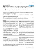
Verification of ICAM-1 deficiency in P/I null mice
The absence of ICAM-1, and P-selectin expression in the
P/I null mice was confirmed in randomly selected litter
mice by specific immunohistochemical staining and
reverse transcriptase polymerase chain reaction (RT-PCR),
as previously published by our laboratory [13]. The
ICAM-1 expression was determined in all the animals
used in this study. ICAM-1 was constitutively expressed in
the wild-type control mice as indicated by brown staining
along endothelial lining of the sinusoids, and hepatic vas-
culatures (Figure 1A). The ICAM-1 expression was mark-
edly increased in wild-type mice following hepatic I/R
(Figure 1C). In contrast, in P/I null liver tissue, ICAM-
expression was absent (Figures 1B and 1D). The RT-PCR
data are not shown.
Demonstration of hepatocellular injury by changes in
plasma ALT levels
Hepatic I/R caused significant hepatocellular damage as
demonstrated by plasma ALT levels. The plasma ALT lev-
els of both wild-type and P/I null mice after 90 minutes of
Journal of Inflammation 2007, 4:11 />Page 4 of 10
(page number not for citation purposes)
ischemia followed by 6 and 15 h of reperfusion were sig-
nificantly elevated when compared to their respective
sham-operated mice (Figure 2). ALT levels were negligible
in both groups by the survival time-point. Although there
was no statistically significant difference in ALT levels
between the wild-type and P/I null mice at either time
point of reperfusion (i.e. 6 and 15 h), P/I null mice
showed decreased ALT levels compared to the wild-type
mice.
Demonstration of hepatocellular injury by histopathology
The histopathologic injury of the liver tissue was evalu-
ated based on sinusoidal congestion, cytoplasmic vacuoli-
zation, hepatocellular necrosis, and neutrophil
infiltration. The liver sections from the sham-operated
mice displayed minimal/no necrosis, similar to that of the
non-operated control mice (Figure 3A and 3B). Addition-
ally, there was no apparent evidence of hepatic injury due
to ischemia alone (i.e., at zero hour of reperfusion; image
not shown). However, reperfusion of the ischemic liver
induced an extensive hepatocellular necrosis, sinusoidal
congestion, and neutrophil infiltration after 6 and 15 h of
reperfusion in both wild-type and P/I null mice (Figures
3C, 3D, 3E, and 3F). There was sparing of the periportal
areas with progressively increased injury approaching the
central vein. In general, it appeared that the wild-type I/R
mice exhibited larger areas of coagulative necrosis when
compared to the P/I null mice. The injury was associated
with a marked number of neutrophils infiltrated into the
midzonal region of ischemic liver after 6 and 15 h of
reperfusion in both wild-type and P/I null mice, which
was confirmed with in situ immunohistochemical stain-
ing of the neutrophils (Figure 3, last row). There were a
minimal number of neutrophils present in the livers of
sham-operated mice and in the non-ischemic lobes of
mice subjected to I/R, indicating that ischemia was a pre-
requisite for reperfusion injury to occur. Further, neu-
trophil infiltration was quantitated by measuring the liver
MPO content (Figure 4). The liver MPO levels of both
wild-type and P/I null mice after 90 minutes of ischemia
followed by 6 and 15 h of reperfusion were significantly
elevated compared to the sham-operated mice at the cor-
responding time points. Further, there was no statistically
significant difference in MPO levels between the wild-type
and P/I null mice, at either time point.
Plasma TNF-
α
, IL-6, MIP-2, and KC levels
In order to determine whether plasma cytokine/chemok-
ine levels correlated with tissue injury, the plasma
cytokine/chemokine levels were measured using an
ELISA. As figure 5 displays, the plasma levels of all
cytokines (i.e., TNF-α, IL-6, MIP-2, and KC) were signifi-
cantly increased in response to I/R, in both the wild-type
Time-course of plasma ALT levels following hepatic I/RFigure 2
Time-course of plasma ALT levels following hepatic
I/R. Mice were subjected to 90 minutes of ischemia followed
by reperfusion with various lengths of time. "Control" indi-
cates mice that underwent no surgical procedure. "Sham"
indicates mice that underwent surgical procedure with no
vascular occlusion followed by reperfusion, while "I/R"
denote mice that underwent I/R surgical procedure. Values
are expressed as mean ± SEM. (*) Sham-operated mice were
statistically different from the I/R groups (i.e., p ≤ 0.05). Sham
and P/I group (n = 3–16 mice per each data time point).
0
1000
2000
3000
4000
5000
6000
Plasma ALT Levels (UI/L)
Control
Sham 6h
Sham 15h
IR 6h
IR 15h
IR survl
P/ I
WT
Null
*
*
*
*
Immunohistochemical Staining of Liver for ICAM-1 Expres-sionFigure 1
Immunohistochemical Staining of Liver for ICAM-1
Expression. Positive ICAM-1 staining (brown color) is dem-
onstrated in wild-type mice along the vasculature and sinu-
soids, but is notably absent in the P/I null mice. (A) Control,
wild-type mouse, (B) Control, P/I null mouse, (C) Wild-type
mouse after 90 minutes of ischemia and 6 h of reperfusion,
(D) P/I null mouse after 90 minutes of ischemia and 6 h of
reperfusion.
Wild-type P/I Null
A
B
C
D
Journal of Inflammation 2007, 4:11 />Page 5 of 10
(page number not for citation purposes)
and P/I null groups, and reached their maximum at 6 h of
reperfusion, which then declined to baseline by 15 h of
reperfusion. The data related to the wild-type is consistent
with our previous studies, where a similar pattern was
observed in wild-type mice subjected to hepatic I/R [20].
As Figure 5A. shows, hepatic IR caused significant produc-
tion of TNF-α, by 6 h of reperfusion in both wild-type and
P/I mice, which declined by 15 h of reperfusion. However,
the data indicates no significant difference in I/R-induced
TNF-α production between the wild-type and P/I mice.
This data is consistent with plasma ALT data, which
showed a maximal increase in ALT levels at I/R 6 h, fol-
lowed by a decrease in levels by I/R 15 h of reperfusion. A
similar pattern was observed in plasma IL-6 levels (Figure
5B). In contrast, chemokine production showed a differ-
ent pattern, in that the P/I null mice had significantly
lower levels of plasma KC and MIP-2 at I/R 6 h than the
wild-type mice (Figure 5C. and 5D).
Survival study
Ten wild-type and ten P/I null mice were subjected to 90
minutes of partial hepatic ischemia and were observed
post-operatively and their approximate times of death
were recorded. At the end of three weeks, all surviving
mice were euthanized. All the P/I null mice survived the
full three weeks while only 7 out of 10 of the wild-type
mice survived that length of time. The Kaplan-Meier log
rank test showed no statistically significant difference
between the two groups (p = 0.067), although a trend
towards improved survival in the P/I null group was
apparent (Figure 6).
Discussion
Neutrophil infiltration plays an important role in reper-
fusion tissue injury, which is mediated by adhesion mol-
ecules such as selectins, β2-integrins, and ICAM-1. It has
been suggested that inhibition of the adhesion molecules
would prevent neutrophil infiltration, thus providing pro-
tection against organ injury caused by I/R. Currently
though, there is a disparity between preclinical and clini-
cal trial data, and it has been suggested that this disparity
may be the result of the length of insult used in previous
studies. Thus, the current study examined the role of P-
selectin and ICAM-1, adhesion molecules involved in
cytokine production, neutrophil infiltration, and hepato-
cellular injury, following hepatic I/R injury after short and
longer periods of insult. Transgenic P/I null and wild-type
mice were subjected to 90 minutes of warm liver ischemia
followed by various periods of reperfusion. Hepatic I/R
caused significant hepatocellular injury at 6 and 15 h of
reperfusion in both wild-type and P/I null mice, which
was associated with a marked increase in neutrophil infil-
tration to the ischemic liver. The difference between the
two mouse groups was moderate and statistically insignif-
Hepatic histopathology following I/RFigure 3
Hepatic histopathology following I/R. Wild-type and P/I
null mice subjected to the sham operation or 90 minutes of
liver ischemia followed by various reperfusion times. The
ischemic liver sections were prepared and stained with H&E.
Figures A and B represent the sham mice; there is essentially
normal hepatic histology; and C, D, E, and F represent mice
subjected to I/R. A pattern of reperfusion damage is evident
by loss of hepatocytes in the pericentral and midzonal
regions, with relative sparing of the periportal areas. Note
the presence of neutrophils in the midzonal region around
the central vein. Figures G and H show immunohistochemi-
cal staining of neutrophils using a specific anti-neutrophil anti-
body, i.e., subjected to 90 minutes of ischemia followed 6 h
of reperfusion. Neutrophils are indicated by dark brown
color stain. (A) Wild-type mouse, 6 hour sham; (B) P/I null
mouse, 6 hour sham; (C and E) Wild-type mice subjected to
90 minutes of ischemia followed by 6 and 15 h of reperfusion
respectively; (D and F) P/I null mouse subjected to 90 min-
utes of ischemia followed by 6 and 15 h of reperfusion,
respectively.
Wild-type P/I Null
B
D
F
H
C
A
E
G
Journal of Inflammation 2007, 4:11 />Page 6 of 10
(page number not for citation purposes)
icant. In contrast, there was a significant difference in
CXC-chemokine production in that the P/I null mice had
significantly lower levels CXC-chemokines than their
wild-type mice counterparts. Additionally, P/I null mice
showed a favorable trend to survival. These findings sug-
gested that while P-selectin and ICAM-1 do not play a crit-
ical role for neutrophil infiltration and liver injury, it may
regulate chemokine production and confer a survival
advantage.
The data of the present study is consistent with previously
reported studies that demonstrated no attenuation of neu-
trophil infiltration in hepatic sinusoids despite blocking a
number of different adhesion molecules [14,18-20]. Stud-
ies have also shown that neutrophil infiltration was
largely independent of the adhesion molecules, despite
the presence of adhesion molecules on endothelial cells
lining the hepatic sinusoids and vasculature [21,22]. In
contrast, other studies have shown that neutrophil infil-
tration was dependent on the adhesion molecules and
that hepatocellular injury was reduced by anti-adhesion
antibody treatment [10,21]. These studies collectively
indicate that the role of adhesion molecules is tissue and
stimulus specific. As discussed below, there are a number
of possible explanations as to why P-selectin and ICAM-1
deficiency did not appear to be critical for neutrophil infil-
tration and hepatocellular injury following liver I/R.
Although P-selectin is considered a critical adhesion mol-
ecule in initial tethering and rolling of neutrophils on
endothelial cells, several studies suggest that P-selectin is
unlikely to play an important role in hepatic injury
through neutrophil sequestration or transendothelial
migration. First, P-selectin is not expressed on the sinusoi-
dal endothelium [22,23], where the predominant neu-
trophil extravasation takes place in the liver [7]. Second,
within the liver venules, leukocytes can use other adhe-
sion molecules such as α-4 integrin, independent of the
selectins, and finally, within the liver sinusoids, no known
selectin molecules or α-4 integrin molecules appear to
play a dominant role in leukocyte recruitment [24]. Nev-
ertheless, it should be noted that P-selectin might partici-
pate in I/R injury through its role in platelet aggregation
and binding to the neutrophils [25]. Other factors such as
swelling of the endothelial lining cells, vasoconstriction
of the sinusoids, and, stiffening and decreased deforma-
bility of the neutrophils, may also contribute to the
mechanical trapping of neutrophils in hepatic sinusoids.
[26,27].
The study presented in this article suggests an ICAM-1
independent mediated neutrophil infiltration into the
ischemic liver, though it has to be noted that P/I null mice
are not true ICAM-1 knockouts. The P/I null mice may
have had low levels of alternatively spliced forms of
ICAM-1 that could have been up-regulated on the vascular
endothelium, and thereby promoted neutrophil migra-
tion [28,29]. However, this possibility is remote, since the
3E2 mAb that was used in the present study corresponds
to the common form of ICAM-1. Further, the lack of
ICAM-1, per se, is not a critical factor that results in dys-
functional β
2
-integrin-mediated migration. Finally, other
adhesion molecule(s), ligand(s), and/or yet unknown
counter-receptor(s) might also mediate neutrophil infil-
tration. For example, ICAM-2, a ligand for β2-integrins,
and α4-integrins (α4β1/VLA-4 and α4β1/VCAM-1), could
be potential candidates [30-34]. In addition, neutrophils
also express CD11d/CD18 and α9-integrin, which both
bind to VCAM-1, and could possibly play an important
role in neutrophil extravasation, at sites of inflammation
[35]. The importance of α4- and α 9-integrin/VCAM-1
pathways in neutrophil infiltration in I/R-induced hepatic
injury remains unclear. Further, other proteins are recog-
nized to act as ligands for β
2
-integrins such as those pro-
duced during coagulation and complement pathway
activation, which could mediate neutrophil adhesion and
infiltration into the ischemic liver [36-39]. Therefore, evi-
dence supports this study's finding that ICAM-1 deficiency
does not play a key role in neutrophil infiltration and
hepatic injury, and that other compensatory mechanisms
exist to fulfill the role of ICAM-1.
Hepatic Myeloperoxidase Levels following I/R in Wild-type and P/I null miceFigure 4
Hepatic Myeloperoxidase Levels following I/R in
Wild-type and P/I null mice. Mice were subjected to the
sham operation or to 90 minutes of liver ischemia with vari-
ous reperfusion times (i.e., 6, and 15 hrs). The ischemic liver
was collected and its MPO content was determined for the
reperfusion time points indicated. Values are expressed as
the mean ± SEM. (*) Sham-operated mice were statistically
different from the I/R groups (i.e., p ≤ 0.05). Comparison
between Wild-type and P/I null at each time points indicated
no significant differences.
Null
0
.2
.4
.6
.8
1
Sham 6 h Sham 15 h IR 6 h IR 15 h
P/I
WT
*
*
*
*
Liver MPO Content (U/ml)
Journal of Inflammation 2007, 4:11 />Page 7 of 10
(page number not for citation purposes)
Inflammatory cytokines such as TNF-α and IL-6 have been
shown to play key roles in the pathophysiology of hepatic
I/R injury [2,17,40]. TNF-α is the proximal cytokine that
is expressed following hepatic I/R, and correlates with
hepatic reperfusion injury. IL-6 is a multifunctional
cytokine that is both pro-mitogenic and anti-apoptotic for
hepatocytes, and is considered a marker for tissue injury
severity [41,42]. The data from this study corroborates
this as it was found that TNF-α and IL-6 levels paralleled
ALT plasma levels (Figure 5A. and 5B). There was no sig-
nificant difference in plasma TNF-α, and IL-6 levels
between the wild-type and P/I null mice.
The CXC-chemokine production was also examined in
this study. Plasma MIP-2 and KC levels in the sham
groups were constant and minimal, and a significant
increase was induced by hepatic I/R in both wild-type and
P/I null mice (Figure 5C. and 5D). However, in contrast to
the plasma TNF-α and IL-6, a significant difference was
observed between the wild-type and P/I null mice CXC-
chemokine levels after 6 h of reperfusion. This is a novel
observation and the exact mechanism to explain the
reduced chemokine production in P/I null mice in
response to hepatic I/R is not known, though it may be
postulated that the adhesion molecule deficiency may
Time-course of plasma TNF-α, IL-6, KC, and MIP-2 levels following various reperfusion times, after the onset of 90 minutes of ischemiaFigure 5
Time-course of plasma TNF-α, IL-6, KC, and MIP-2 levels following various reperfusion times, after the onset
of 90 minutes of ischemia. "Control" indicates mice that underwent no surgical procedure. "Sham" indicates mice that
underwent surgical procedure with no vascular occlusion followed by reperfusion, while "I/R" indicates mice that underwent
surgical procedure with vascular occlusion for 90 minutes followed by reperfusion for various lengths of time. Values are
expressed as mean ± SEM. *P < 0.05 wild-type group vs. P/I group (n = 3–16 mice per each time point/group). A: TNF-α data;
B: IL-6 data; C: KC data; D: MIP-2 data.
0
1000
2000
3000
4000
5000
6000
7000
8000
9000
Plasma KC (pg/ml)
Control
Sham 6h
Sham 15h
IR 6h
IR 15h
IR srvl
P/ I
WT
0
50
100
150
200
250
300
350
Plasma TNF (pg/ml)
Control
Sham 6h
Sham 15h
IR 6h
IR 15h
IR srvl
P/ I
WT
0
500
1000
1500
2000
2500
3000
3500
Plasma IL-6 (pg/ml)
Control
Sham 6h
Sham 15h
IR 6h
IR 15h
IR srvl
P/ I
WT
0
500
1000
1500
2000
2500
3000
3500
Plasma MIP-2 (pg/ml)
Control
Sham 6h
Sham 15h
IR 6h
IR 15h
IR srvl
P/ I
WT
C
D
*
*
A
B
Null
Null
Null
Null
Journal of Inflammation 2007, 4:11 />Page 8 of 10
(page number not for citation purposes)
play a role. The genetic knockout mice have altered
expression of other molecules which may have reflected
the expression of the chemokines. In support of this
study, a recent report showed significantly lower chemok-
ine production (i.e. KC) in P/E-selectin deficient mice
than their wild-type counterparts [43]. In addition, a
recent study highlights the role of selectins and non-
integrin collagen receptors in chemokine production and
function through p38 mitogen-activated protein kinase
and NF-κB activation [44]. Further studies are necessary to
examine the role of these adhesion molecules in chemok-
ine regulation and their pathophysiologic role in different
organ systems.
Previous studies have suggested a direct association
between CXC-chemokines, neutrophil recruitment and
liver injury. Specifically, blockage of CXC-chemokines
with antibodies was associated with neutrophil infiltra-
tion and liver injury in the rat and mouse models of warm
hepatic I/R [2,40]. This is in part consistent with the wild-
type data presented in this study, in that the CXC-chem-
okine levels correlated with liver injury and neutrophil
infiltration during the early-phase of hepatic I/R (i.e. 6 h
of reperfusion). However, during the late-phase of hepatic
I/R (i.e. 15 h of reperfusion), the CXC-chemokines were at
baseline levels, while neutrophil infiltration was maxi-
mal. The neutrophil infiltration may have been mediated
by other more potent chemoattractants (e.g. C5a, LTB
4
)
and mediators (e.g. apoptotic cells). This hypothesis is
supported by Dorman et al's study, in which a CXC-inde-
pendent neutrophil infiltration into the liver was found in
response to apoptotic cells in a mouse model of endotox-
imia [45]. They showed that wild type as well as the
CXCR2 -/- mice had similar neutrophil infiltration and
liver injury. There are other potential factors to explain
why neutrophil trafficking was not associated with chem-
okine production. One possible explanation is that the
generated CXC-chemokine in P/I null mice was at its opti-
mal concentration to mediate neutrophil infiltration and
liver injury. Further, other inflammatory mediators may
have been involved in neutrophil infiltration (e.g. C5a,
LTB
4
). Finally, the nature of hepatic sinusoidal endothe-
lium, which is fenestrated, may have allowed direct adhe-
sion of neutrophils to the hepatocytes, resulting in liver
damage. Future studies are necessary to examine the
potential role of these various factors in neutrophil infil-
tration in hepatic I/R injury.
The survival data presented in this study showed that
although not statistically significant the P/I null mice
exhibited a favorable trend toward increased survival than
their wild-type counterparts. The data also suggested that
the potential survival advantage of P/I null mice was not
a result of decreased hepatic injury. Since local organ
injury appeared to be similar between both groups, it is
likely that the P/I null mice were less susceptible to the
systemic manifestations of hepatic I/R injury, such as
acute respiratory distress syndrome, and multiple organ
dysfunction syndrome [46]. It has yet to be elucidated
though, whether the decreased CXC-chemokines had a
potential role in favoring the survival. A previously pub-
lished study demonstrated that P-selectin inhibition
improved the survival of mice subjected to warm intesti-
nal I/R, in which T lymphocytes (with Th2 profile) played
a central role [47]. This is further supported by a study that
has implicated CD4+T-lymphocytes as key regulators in I/
R-induced inflammatory response in the liver [48]. The
profile of Th1 and Th2 cytokines in P/I null mice has not
been studied and as such, future studies are warranted to
examine the role of T lymphocytes, in their contribution
to increased survival.
In summary, the results of this study suggest that P-selec-
tin and ICAM-1 adhesion molecules do not play a critical
role in mediating neutrophil infiltration and liver injury
caused by hepatic I/R. However, these adhesion molecules
may play a role in CXC-chemokine regulation, which may
exhibit other functions than chemotactic activities. Inhibi-
tion of these adhesion molecules may enhance overall
survival by playing a role in the systemic organ injury that
often ensues following liver I/R.
Abbreviations
ICAM-1, Intercellular adhesion molecule-1; I/R,
ischemia/reperfusion; IL-6, interleukin-6;LFA-1, lym-
phocyte function-associated antigen; mAbs, monoclonal
Kaplan-Meier Survival CurveFigure 6
Kaplan-Meier Survival Curve. Mice were subjected to 90
minutes of liver ischemia followed by a 3-week period of
reperfusion. The plot shows two curves. The solid line rep-
resents the survival curve for the P/I null mice (10/10 mice
survived) and the broken line represents the survival curve
for the wild-type mice (7/10 mice survived). Wild-type sur-
vival was not significantly different from P/I null survival (p =
0.067) although a trend is apparent.
Journal of Inflammation 2007, 4:11 />Page 9 of 10
(page number not for citation purposes)
antibodies; MPO, Myeloperoxidase; P/I null mice, P-
selectin/ICAM-1-deficient mice; TNF, tumor necrosis fac-
tor; WT, wild-type.
Competing interests
The author(s) declare that they have no competing inter-
ests.
Authors' contributions
This study represents parts of the Research Thesis project
performed by KM under the direction of EC. KM carried
out the surgical operation, collection of samples, analysis
and interpretation of the MPO and ALT data, as well as
drafting the manuscript. SD participated in the analysis of
the cytokine data and the preparation of the manuscript.
EC was responsible for conceiving, supervising the design
and performance of the project, as well as preparation of
the manuscript. All authors read and approved the manu-
script.
Acknowledgements
The authors would like to thank the Department of Surgery – College of
Human Medicine, and the McLaren Foundation at Michigan State University
for providing funding. The authors would also like to thank Crystal
Remelius, and Karen Hess for their technical assistance in cytokine analysis,
and immuno-staining of the tissue samples in this research project.
References
1. Carden DL, Granger DN: Pathophysiology of ischaemia-reper-
fusion injury. JPathol 2000, 190:255.
2. Colletti LM, Kunkel SL, Walz A, Burdick MD, Kunkel RG, Wilke CA,
Strieter RM: The role of cytokine networks in the local liver
injury following hepatic ischemia/reperfusion in the rat.
Hepatology 1996, 23:506.
3. Jaeschke H, Farhood A: Neutrophil and Kupffer cell-induced
oxidant stress and ischemia-reperfusion injury in rat liver.
Am J Physiol 1991, 260:G355.
4. Jaeschke H, Hasegawa T: Role of neutrophils in acute inflamma-
tory liver injury. Liver Int 2006, 26:912-9.
5. Lobb RR: Integrin-Immunoglobulin superfamily interactions
in endothelial-leukocyte adhesion. In Adhesion – Its role in inflam-
matory disease Edited by: Harlan JM, Liu DY. Freeman Press;
1992:1-18.
6. Crockett-Torabi E, Sullenbarger B, Smith CW, Fantone JC: Neu-
trophil activation through L-selectin and Mac-1. J Immunol
1995, 154:2291.
7. Chosay JG, Essani NA, Dunn CJ, Jaeschke H: Neutrophil margina-
tion and extravasation in sinusoids and venules of liver dur-
ing endotoxin-induced injury. Am J Physiol 1997, 272:G1195.
8. Ganey PE, Bailie MB, VanCise S, Colligan ME, Madhukar BV, Robinson
JP, Roth RA: Activated neutrophils from rat injured isolated
hepatocytes. Lab Invest 1994, 70:53.
9. Dulkanchainun TS, Goss JA, Imagawa DK, Shaw GD, Anselmo DM,
Kaldas F, Wang T, Zhao D, Busuttil AA, Kato H, Murray NG, Kupiec-
Weglinski JW, Busuttil RW: Reduction of hepatic ischemia/
reperfusion injury by a soluble P-selectin glycoprotein lig-
and-1. Ann SurgI 1998, 227:832.
10. Farhood A, McGuire GM, Manning AM, Miyasaka M, Smith CW, Jae-
schke H: Intercellular adhesion molecule 1 (ICAM-1) expres-
sion and its role in neutrophil-induced ischemia-reperfusion
injury in rat liver. J Leukoc Biol 1995, 57:368.
11. Singh I, Zibari GB, Zizzi H, Granger DN, Cruz L, Gonsales E, McDon-
ald JC, Brown MF: Anti-P-selectin antibody protects against
hepatic ichemia-reperfusion injury.
Transplant Proc 1998,
30:2324.
12. Bullard DC, Qin L, Lorenzo I, Quinlin WM, Doyle NA, Bosse R, Vest-
weber D, Doerschuk CM, Beaudet AL: P-selectin/ICAM-1 double
mutant mice: acute emigration of neutrophils into the peri-
toneum is completely absent but is normal into pulmonary
alveoli. J Clin Invest 1995, 95:1782.
13. Young CS, Palma JM, Mosher BD, Harkema J, Naylor DF, Dean RE,
Crockett E: Hepatic ischemia/reperfusion injury in P-selectin
and intercellular adhesion molecule-1 double-mutant mice.
Am Surg 2001, 67:737.
14. Essani NA, Fisher MA, Simmons CA, Hoover JL, Farhood A, Jaeschke
H: Increased P-selectin gene expression in the liver vascula-
ture and its role in the pathophysiology of neutrophil-
induced hepatic injury in murine endotoxin shock. J Leukoc
Biol 1998, 63:288.
15. Wong J, Johnston B, Lee SS, Bullard DC, Smith CW, Beaudet AL,
Kubes P: A minimal role for selectins in the recruitment of
leukocytes into the inflamed liver microvasculature. J Clin
Invest 1997, 99:2782.
16. Harlan JM, Winn RK: Leukocyte-endothelial interactions: Clin-
ical trials of anti-adhesion therapy. Crit Care Med 2002, 30:S214.
17. Mosher BD, RE Dean, J Harkema, D Remick, J Palma, Crockett E:
Inhibition of chemokines production by kupffer cells
decreased hepatic ischemia/reperfusion injury in mice. J Sur-
gical Research 2001, 99:201.
18. Essani NA, Fisher MA, Farhood A, Manning AM, Smith CW, Jaeschke
H: Cytokine-induced upregulation of hepatic intercellular
adhesion molecule-1 messenger RNA expression and its role
in the pathophysiology of murine endotoxin shock and acute
liver failure. Hepatology 1995, 21:1632.
19. Hill J, Lindsay T, Rusche J, Valeri CR, Shepro D, Hechtman HB: A
Mac-1 antibody reduces hepatic and lung injury but not neu-
trophil sequestration after intestinal ischemia-reperfusion.
Surgery 1992, 112:
166.
20. Jaeschke H, Farhood A, Fisher MA, Smith CW: Sequestration of
neutrophils in the hepatic vasculature during endotoxemia is
independent of beta 2 integrins and intercellular adhesion
molecule-1. Shock 1996, 6:351.
21. Essani NA, McGuire GM, Manning AM, Jaeschke H: Differential
induction of mRNA for ICAM-1 and selectins in hepatocytes,
Kupffer cells and endothelial cells during endotoxemia. Bio-
chem Biophys Res Commun 1995, 211:74.
22. Wisse E: An ultrastructural characterization of the endothe-
lial cell in the rat liver sinusoid under normal and various
experimental conditions, as a contribution to the distinction
between endothelial and Kupffer cells. J Ultrastruct Res 1972,
38:528.
23. Steinhoff G, Behrend M, Schrader B, Duijvestijn AM, Wonigeit K:
Expression patterns of leukocyte adhesion ligand molecules
on human liver endothelia. Lack of ELAM-1 and CD62 induc-
ibility on sinusoidal endothelia and distinct distribution of
VCAM-1, ICAM-1, ICAM-2, and LFA-3. Am J Pathol 1993,
142:481.
24. Fox-Robichaud A, Kubes P: Molecular mechanisms of tumor
necrosis factor alpha-stimulated leukocyte recruitment into
the murine hepatic circulation. Hepatology 2000, 31:1123-7.
25. Yadav SS, Howell DN, Steeber DA, Harland RC, Tedder TF, Clavien
PA: P-Selectin mediates reperfusion injury through neu-
trophil and platelet sequestration in the warm ischemic
mouse liver. Hepatology 1999, 29:1494-502.
26. McCuskey RS, Urbaschek R, Urbaschek B: The microcirculation
during endotoxemia. Cardiovasc Res 1996, 32:752.
27. Worthen GS, Schwab B III, Elson EL, Downey GP: Mechanics of
stimulated neutrophils: cell stiffening induces retention in
capillaries. Science 1989, 245:183.
28. King PD, Sandberg ET, Selvakumar A, Fang P, Beauder AL, Dupont B:
Novel isoforms of murine intercellular adhesion molecule-1
generated by alternative RNA splicing.
J Immunology 1995,
154:6080-6093.
29. van Den Engel NK, Heidenthal E, Vinke A, Kolb H, Martin S: Circu-
lating forms of intercellular adhesion molecule (ICAM)-1 in
mice lacking membranous ICAM-1. Blood 2000, 95:1350-5.
30. Huang MT, Larbi KY, Scheiermann C, Woodfin A, Gerwin N, Haskard
DO, Nourshargh S: ICAM-2 mediates neutrophil transmigra-
tion in vivo: evidence for stimulus specificity and a role in
PECAM-1-independent transmigration. Blood 2006,
107:4721-7.
31. Molla M, Gironella M, Miquel R, Tovar V, Engel P, Biete A, Pique JM,
Panes J: Relative roles of ICAM-1 and VCAM-1 in the patho-
Publish with BioMed Central and every
scientist can read your work free of charge
"BioMed Central will be the most significant development for
disseminating the results of biomedical research in our lifetime."
Sir Paul Nurse, Cancer Research UK
Your research papers will be:
available free of charge to the entire biomedical community
peer reviewed and published immediately upon acceptance
cited in PubMed and archived on PubMed Central
yours — you keep the copyright
Submit your manuscript here:
/>BioMedcentral
Journal of Inflammation 2007, 4:11 />Page 10 of 10
(page number not for citation purposes)
genesis of experimental radiation-induced intestinal inflam-
mation. Int J Radiat Oncol Biol Phys 2003, 57:264-73.
32. Johnston B, Kubes P: The alpha4-integrin: an alternative path-
way for neutrophil recruitment. Immunol Today 1999,
20:545-50.
33. Bowden RA, Ding ZM, Donnachie EM, Petersen TK, Michael LH, Bal-
lantyne CM, Burns AR: Role of alpha4 integrin and VCAM-1 in
CD18-independent neutrophil migration across mouse car-
diac endothelium. Circ Res 2002, 90:562-9.
34. Ibbotson GC, Doig C, Kaur J, Gill V, Ostrovsky L, Fairhead T, Kubes
P: Functional alpha4-integrin: a newly identified pathway of
neutrophil recruitment in critically ill septic patients. Nat
Med 2001, 7:465-70.
35. Taooka Y, Chen J, Yednock T, Sheppard D: The integrin
alpha9beta1 mediates adhesion to activated endothelial cells
and transendothelial neutrophil migration through interac-
tion with vascular cell adhesion molecule-1. J Cell Biol 1999,
145:413-20.
36. Suzuki K, Watanabe T, Sakurai S, Ohtake K, Kinoshita T, Araki A,
Fujita T, Takei H, Takeda Y, Sato Y, Yamashita T, Araki Yoshihiko,
Sendo F: A novel glycosylphosphatidyl inositol-anchored pro-
tein on human leukocytes: A possible role for regulation of
neutrophil adherence and migration. J Immunol 1999,
162:4277-4284.
37. Gross TJ, Leavell KJ, Peterson MW: CD11b/CD18 mediates the
neutrophil chemotactic activity of fibrin degradation prod-
uct D domain. Thromb Haemost 1997, 77:894-900.
38. Siegbahn A: Cellular consequences upon factor VIIa binding to
tissue factor. Haemostasis 2000, 2:41-7.
39. Bombeli T, Schwartz BR, Harlan JM: Adhesion of activated plate-
lets to endothelial cells: evidence for a GPIIbIIIa-dependent
bridging mechanism and novel roles for endothelial intercel-
lular adhesion molecule 1 (ICAM-1), alphavbeta3 integrin,
and GPIbalpha. J Exp Med 1998, 187:329.
40. Lentsch AB, Yoshidome H, Cheadle WG, Miller FN, Edwards MJ:
Chemokine involvement in hepatic ischemia/reperfusion
injury in mice: Roles for macrophage inflammatory protein-
2 and KC. Hepatology 1998,
27:563-568.
41. Teoh N, Field J, Farrell G: IL-6 is one likely mediator of the
hepatoprotective and pro-proliferative effects of ischaemic
preconditioning. J Hepatol 2006, 45:20-7.
42. Jin X, Zimmers TA, Perez EA, Pierce RH, Zhang Z, Koniaris LG: Par-
adoxical effects of short- and long-term interleukin-6 expo-
sure on liver injury and repair. Hepatology 2006, 43:474-84.
43. Li Z, Burns AR, Smith CW: Two waves of neutrophil emigration
in response to corneal epithelial abrasion: distinct adhesion
molecule requirements. Invest Ophthalmol Vis Sci 2006,
47:1947-55.
44. Matsuyama W, Wang L, Farrar WL, Faure M, Yoshimura T: Activa-
tion of discoidin domain receptor 1 isoform b with collagen
up-regulates chemokine production in human macrophages:
role of p38 mitogen-activated protein kinase and NF-kappa
B. J Immunol 2004, 172:2332-40.
45. Dorman RB, Gujral JS, Bajt ML, Farhood A, Jaeschke H: Generation
and functional significance of CXC chemokines for neu-
trophil-induced liver injury during endotoxemia. Am J Physiol
Gastrointest Liver Physiol 2005, 288:G880-6.
46. Liu DL, Jeppsson B, Hakansson CH, Odselius R: Multiple-system
organ damage resulting from prolonged hepatic inflow inter-
ruption. Arch Surg 1996, 131:442.
47. Farmer DG, Shen XD, Amersi F, Anselmo D, Ma JP, Ke B, Gao F, Dry
S, Fernandez S, Shaw GD, McDiarmid SV, Busuttil RW, Kupiec-Weg-
linski J: CD62 blockade with P-Selectin glycoprotein ligand-
immunoglobulin fusion protein reduces ischemia-reper-
fusion injury after rat intestinal transplantation. Transplanta-
tion 2005, 79:44-51.
48. Zwacka RM, Zhang Y, Halldorson J, Schlossberg H, Dudus L, Engel-
hardt JF: CD4(+) T-lymphocytes mediate ischemia/reper-
fusion-induced inflammatory responses in mouse liver. J Clin
Invest 1997, 100:279-89.