Báo cáo y học: "Treatment of experimental colitis in mice with LMP-420, an inhibitor of TNF transcription" pptx
Bạn đang xem bản rút gọn của tài liệu. Xem và tải ngay bản đầy đủ của tài liệu tại đây (1.08 MB, 13 trang )
BioMed Central
Page 1 of 13
(page number not for citation purposes)
Journal of Inflammation
Open Access
Research
Treatment of experimental colitis in mice with LMP-420, an
inhibitor of TNF transcription
Laura P Hale* and George Cianciolo
Address: Department of Pathology, Duke University Medical Center, Durham, NC, USA
Email: Laura P Hale* - ; George Cianciolo -
* Corresponding author
Abstract
Background: LMP-420 is a boronic acid-containing purine nucleoside analogue that
transcriptionally inhibits TNF production but is non-cytotoxic to TNF-producing cells.
Methods: This study investigated the efficacy of LMP-420 as an anti-inflammatory agent in acute
and chronic colitis induced by oral administration of dextran sulfate sodium (DSS) to mice and in
chronic colitis following piroxicam administration to IL-10-deficient mice. The severity of colon
inflammation was assessed histologically. TNF levels were measured by enzyme immunoassay.
Results: Administration of DSS for 7 days resulted in severe acute colitis that was associated with
a marked increase in stool and colon tissue TNF levels. Initiation of therapy with intraperitoneal
(i.p.) LMP-420 on day 4 of DSS exposure decreased colonic TNF to near normal levels on day 7.
However, neither i.p. nor oral treatment with LMP-420 affected the development or severity of
acute DSS colitis. Initiation of LMP-420 therapy after 3 cycles of DSS administration to establish
chronic colitis also had no effect on the severity of chronic colitis. Analysis of colonic TNF
combined with longitudinal analysis of TNF and TNF receptor (TNF-RII) levels in stool during the
development of chronic DSS colitis demonstrated that the initially elevated colonic TNF levels
returned to normal despite intense on-going inflammation in mice with chronic colitis. RAG-2
-/-
mice deficient in T and B cells also developed severe ongoing colitis in response to 3 cycles of DSS,
but showed marked differences vs. wild type mice in stool TNF and TNF-RII in response to DSS
exposure. Systemic and oral LMP-420 treatment for 16 days decreased colonic TNF levels in IL-
10-deficient mice with chronic colitis, with a trend to decreased histologic inflammation for oral
LMP-420.
Conclusion: These studies demonstrate that short-term treatment with a transcriptional inhibitor
of TNF production can decrease systemic and local colonic levels of TNF but may not decrease the
histologic severity of colitis. Longer term studies using colitis models that are more dependent on
TNF elevation should be performed to more accurately assess the potential of LMP-420 for therapy
of inflammatory bowel disease.
Introduction
Inflammatory bowel diseases such as Crohn's disease
(CD) and ulcerative colitis (UC) are hypothesized to
result from abnormal immune responses to antigens
Published: 10 March 2008
Journal of Inflammation 2008, 5:4 doi:10.1186/1476-9255-5-4
Received: 11 July 2007
Accepted: 10 March 2008
This article is available from: />© 2008 Hale and Cianciolo; licensee BioMed Central Ltd.
This is an Open Access article distributed under the terms of the Creative Commons Attribution License ( />),
which permits unrestricted use, distribution, and reproduction in any medium, provided the original work is properly cited.
Journal of Inflammation 2008, 5:4 />Page 2 of 13
(page number not for citation purposes)
derived from intestinal microbiota (reviewed in [1,2])
that are perpetuated by ongoing exposure to these anti-
gens in the intestine. A number of pro-inflammatory
cytokines and chemokines have been demonstrated to be
elevated in colonic mucosa and/or leukocytes derived
from human inflammatory bowel disease (IBD) patients
(reviewed in [3,4]). These include IL-1, IL-6, IL-12, IFN-γ,
monocyte chemoattractant protein-1 (MCP-1; also called
JE or C-C chemokine ligand 2 (CCL2), and tumor necrosis
factor (TNF).
TNF is a major regulator of inflammation. The murine/
human chimeric monoclonal antibody infliximab (Remi-
cade™; Centocor; Malvern, PA, USA) neutralizes TNF
activity by binding with high affinity to both soluble and
membrane-bound TNF [5,6]. Infliximab binding to mem-
brane-bound TNF renders those cells susceptible to lysis
by complement or effector cells [6]. Infliximab binding
also induces apoptosis of activated T cells from CD
patients [7]. Etanercept (Enbrel™; Immunex Corp; Seattle,
WA, USA) is a dimeric fusion protein consisting of the
extracellular ligand binding domain of the human p75
TNF receptor linked to the Fc portion of human IgG1.
Etanercept binds specifically to TNF and blocks its interac-
tion with naturally occurring cell surface TNF receptors.
Cells expressing transmembrane TNF bind etancercept
but are not lysed in vitro in the presence or absence of
complement [8]. Both the anti-TNF drugs infliximab and
etanercept have been shown to be beneficial in rheuma-
toid arthritis and psoriasis [9-11]. Infliximab has been
shown to significantly decrease inflammatory activity and
to be an effective maintenance therapy in patients with
CD or UC and to enhance closing of fistulas in CD [12-
16]. However, an authoritative randomized controlled
trial of etanercept failed to demonstrate efficacy in CD
when used at the same doses effective for treatment of
rheumatoid arthritis [17]. The mechanisms responsible
for the differential efficacy of infliximab vs. etanercept in
IBD remain unclear. However, destruction of TNF-pro-
ducing cells by infliximab (but not etanercept) could pro-
duce a generalized immunosuppressive effect that might
contribute to its efficacy in IBD [7,18].
Disadvantages of infliximab treatment include the high
cost of therapy, the need for administration by intrave-
nous infusion, development of anti-chimeric antibodies
that limit drug effectiveness, increased susceptibility to
severe opportunistic infections [19,20], and the reactiva-
tion of tuberculosis [21-23]. Its relatively long plasma
half-life (10.5 days) makes it difficult to stop drug action
if adverse effects occur. An orally active small molecule
that inhibits production of TNF and other pro-inflamma-
tory cytokines without generalized immunosuppression
would be predicted to overcome many of the disadvan-
tages associated with currently available TNF antagonists.
Because such a drug would also allow determination of
how local vs. systemic TNF inhibition and cytotoxicity
toward TNF-producing cells contribute to efficacy in IBD
therapy, the data generated would also have broad appli-
cability toward improving IBD therapies.
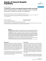
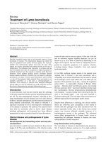
LMP-420 (MW = 284.5; Figure 1) is a purine-based small
molecule that bears a boronic acid side chain. It is a more
potent analogue of a parent compound that was originally
identified by a cell-based screen for agents that altered
cytokine production by human monocytes [24]. LMP-420
strongly inhibits production of both TNF mRNA and pro-
tein [25] and is non-toxic in vitro toward cells present in
the colon, including lymphocytes, monocytes, and intes-
tinal epithelial cells. It does not bind to or interfere with
the activity of pre-formed TNF (unpublished data). LMP-
420 can be readily administered by injection. However, its
physicochemical stability, combined with a chemical
structure favoring retention in the gastrointestinal tract,
suggested that LMP-420 might also have direct local activ-
ity within the intestine when given orally. Based on its in
vitro activity profile, we hypothesized that in vivo therapy
with LMP-420 would decrease intestinal production of
pro-inflammatory cytokines including TNF and thus
decrease intestinal inflammation in IBD. Here, we present
results obtained with the dextran sulfate sodium (DSS)
murine models of acute and chronic colitis and chronic
colitis in IL-10-deficient (IL-10
-/-
) mice.
Materials and methods
Animal studies
Specific pathogen-free wild type female C57BL/6 mice
were obtained from Jackson Laboratories (Bar Harbor,
ME, USA) and typically used at an age of 6–8 weeks. Start-
ing weights (mean ± SD) for acute and chronic DSS stud-
ies were 18.8 ± 1.0 g and 19.2 ± 0.9 g. RAG-2
-/-
female mice
on the C57BL/6 background were obtained from Taconic
Farms (Germantown, NY, USA) (mean weight 22.5 ± 1.2
g). Mice were housed in polycarbonate micro-isolator or
Chemical structure of LMP-420Figure 1
Chemical structure of LMP-420. (2-amino-6-chloro-9
[5(dihydroxyborylpentyl]-purine).
Journal of Inflammation 2008, 5:4 />Page 3 of 13
(page number not for citation purposes)
individually ventilated cages and were allowed access to
food and water ad libitum. All animal studies were
approved by the Duke University Institutional Animal
Care and Use Committee.
A bacterial lipopolysaccharide (LPS) challenge model was
used to test the ability of LMP-420 to block TNF produc-
tion in vivo. Mice were treated for 16 days with 2 mg (100
mg/kg) LMP-420 given intraperitoneally (i.p.) once daily
or with LMP-420 doses of up to 145/mg/kg/day given
orally mixed in food. The i.p. dose of LMP-420 used was
chosen to represent the highest parenteral dose reasona-
ble given the solubility of the drug (~10 mg/ml in 5%
sorbitol). The 16 day treatment period was chosen to
assess the toxicity of repeated daily dosing of LMP-420
and because a treatment period of 16 days or longer is typ-
ically needed to result in histologically detectable differ-
ences in inflammatory activity. Vehicle- or LMP-420
treated mice were challenged with a lethal dose of 0.5 mg
LPS (from E. coli strain O111:B4; catalog#L-2630, Sigma-
Aldrich, St. Louis, MO, USA) given 4 hrs after the last
LMP-420 dose. Mice were euthanized 2 hrs after LPS chal-
lenge to measure TNF levels in serum and colon tissues.
Acute colitis was induced by addition of 3% DSS (40 kDa
molecular weight; obtained from ICN, Costa Mesa, CA,
USA) into the drinking water for 7 days. Chronic colitis
was induced by 3 cycles of DSS administration, each con-
sisting of 5 days DSS followed by 16 days of recovery. The
clinical severity of colitis was assessed by daily observa-
tions for weight loss, stool consistency, and the presence
of gross bleeding. Freshly passed stool pellets were tested
periodically for occult blood using Hemoccult Sensa II
cards (Beckman Coulter, Palo Alto, CA, USA).
Specific pathogen and Helicobacter-free female IL-10-defi-
cient (IL-10
-/-
) mice on the C57BL/6 background (strain
name = B6.129P2-Il10
tm1Cgn
/J; stock # 002251) were
obtained from Jackson Laboratories (Bar Harbor, ME,
USA). Powdered food containing 200 ppm piroxicam was
administered for 7 days at 6 – 7 wks of age (weights = 18.6
± 1.6 g, mean ± SD) to accelerate the development of
chronic colitis [26]. Piroxicam was discontinued and mice
were treated for 16 days with a dose range of LMP-420 (0,
5, 15, or 45 mg/kg/day) given i.p. once daily or food con-
taining LMP-420 that delivered mean LMP-420 doses of 0,
41, 62, or 138 mg/kg/day.
In vitro studies
Thioglycollate-stimulated macrophages were obtained for
in vitro studies by peritoneal lavage (ice-cold PBS; 0.1%
BSA; 10 u/ml sodium heparin) of euthanized mice
injected intraperitoneally 3–4 days earlier with 1 ml of
sterile thioglycollate broth (DIFCO; Voight Global Distri-
bution, Kansas City, MO, USA). Peritoneal exudates were
centrifuged, washed once with PBS and resuspended in
complete RPMI medium containing 10% heat-inactivated
(56°C, 45 min) fetal bovine serum (FBS). Macrophages
were isolated by subsequent overnight adherence to plas-
tic by incubation at 37°C in humidified 5% CO
2
, then
plated into 96 well plates at 1 × 10
5
cells/well in
RPMI1640 with 5% heat-inactivated FBS, 100 u/ml peni-
cillin, 100 µg/ml streptomycin and the indicated concen-
tration of LMP-420. Cells were incubated for 2 hrs, LPS (1
µg/ml; from E. coli strain O111:B4, Sigma-Aldrich, St.
Louis, MO, USA) was added, and after an additional 24
hrs TNF content of the media was measured.
Murine splenocytes were prepared from spleens harvested
from normal mice after euthanasia. Briefly, the spleens
were removed aseptically and placed in plastic Petri dishes
containing ~10 ml of tissue culture medium. The medium
was drawn up in a 27 gauge needle on a 10-ml syringe and
then repeatedly injected under the splenic capsule, forcing
leukocytes from the splenic tissue into the medium. The
leukocytes were pelleted by centrifugation (15 min, 200 ×
g, 4°C), washed once with PBS and contaminating red
blood cells removed by a brief (< 20 s) hypotonic lysis in
9 volumes of sterile H
2
O followed by the addition of 1
volume of 10× PBS. The leukocytes were washed once
with complete RPMI medium and resuspended to a con-
centration of 1 × 10
6
/ml. For studies of LPS stimulation,
splenocytes were cultured for 2 hrs in 96 well plates at 2 ×
10
5
cells/well in RPMI1640 with 5% heat-inactivated FBS,
100 u/ml penicillin, 100 µg/ml streptomycin with the
indicated concentrations of LMP-420. One µg/ml LPS was
added and TNF content of the media was measured after
an additional 24 hrs of culture. For studies using CD3
stimulation, splenocytes (1 × 10
6
/ml) were incubated
with media (RPMI1640 with 5% heat-inactivated FBS,
100 u/ml penicillin, 100 µg/ml streptomycin) or the indi-
cated concentration of LMP-420 in media for 2 hrs at
37°C in polypropylene tubes and then 200 ml of cell sus-
pension was added to each well of a 96-well BioCoat™
antimurine CD3 plate (BD Biosciences, Franklin Lakes,
NJ, USA). The TNF content of culture supernatants was
measured at both 24 and 48 hr time points for CD3-stim-
ulated splenocytes.
Treatment with LMP-420
LMP-420 was custom synthesized by Scynexis Inc.
(Research Triangle Park, NC, USA) under provisions of a
Material Transfer Agreement between LeukoMed (Raleigh,
NC, USA) and Duke University. For i.p. injections, a 10
mg/ml stock solution was prepared in 5% sorbitol, pH 9.0
in sterile water and further diluted as necessary to deliver
the desired dose in a volume of 0.2 ml. For oral delivery,
LMP-420 was mixed with powdered rodent diet contain-
ing 20 µg omeprazole per 3.5 g food to minimize gastric
degradation of LMP-420. All control mice in oral dosing
Journal of Inflammation 2008, 5:4 />Page 4 of 13
(page number not for citation purposes)
studies also received food containing omeprazole. The
method of serial dilutions was used to ensure uniform
mixing of drugs into powdered food. The weight of food
consumed daily was recorded and averaged to determine
the mean dose received per kg body weight during ther-
apy. For studies involving both i.p. and oral administra-
tion of LMP-420, the doses listed are as administered.
Serum and tissue levels of LMP-420 were not measured in
this study.
Tissue analysis
Mice were euthanized by CO
2
asphyxiation in accordance
with the American Veterinary Medical Association Panel
on Euthanasia. The entire colon (cecum to anus) was
removed and colon length was measured from the ileoce-
cal junction to the anus. The colon was then divided into
segments representing the cecum, proximal, mid-, distal,
and terminal colon/rectum. Colon tissue obtained from
the proximal ends of the mid-, distal, and terminal colon
segments was harvested for TNF measurement. Five colors
of permanent tissue marking dye (Bradley Products,
Bloomington, MN, USA) were used to specifically identify
each colonic segment. These tissues were fixed in Carnoy's
solution for 2 – 4 hrs, then processed and embedded into
paraffin.
The severity of inflammation seen in hematoxylin and
eosin-stained sections was scored independently by a
pathologist blinded to treatment group. Histologic scores
were calculated as described, using a scale that takes into
account mucosal changes in 5 different bowel segments,
including hyperplasia and ulceration, degree of inflam-
mation, and % of each bowel segment affected by these
changes [[26]; modified from [27]]. Using this scale, the
maximum score is 75 and a score > 12 indicates colitis.
Cytokine/cytokine receptor measurements
Stool for cytokine analysis was collected before DSS expo-
sure began (day 0), at the end of each 5 day cycle of DSS
administration (day 5, 26, and 47, and after 16 days of
recovery prior to beginning the next DSS cycle (days 21,
42, and 63). Freshly obtained stool was kept on ice until
it was homogenized at 100 mg stool/ml buffer in PBS con-
taining 1% bovine serum albumin, 0.1% Kathon (a
microbiocide; Supelco, Bellefonte, PA, USA), and Protease
Inhibitor Cocktail (Sigma-Aldrich, St. Louis, MO, USA).
Insoluble material was removed by centrifugation at 4°C
at 15,000 × g in a microfuge for 10 min. Stool extracts
were filtered at 0.2 µm then stored at -20°C until assayed.
Fresh colon samples were homogenized at 100 mg tissue/
ml buffer using a PowerGen 125 tissue grinder (Fisher Sci-
entific, Suwanee, GA, USA) and the BioPlex Cell Lysis Kit
(BioRad, Hercules, CA, USA) according to the manufac-
turer's instructions. Colon tissue extracts were then frozen
at -20°C until analysis. TNF and TNF-RII were quantitated
in tissue culture supernatants, stool, and colon tissue
extracts by enzyme immunoassay using Duo-Set reagents
(R&D Systems, Minneapolis, MN, USA). Results were
expressed as pg/ml for culture supernatants or as pg/100
mg tissue or stool.
Statistical analysis
Statistical comparison of groups was performed using Stu-
dent's t test or analysis of variance (ANOVA). A value of p
≤ 0.05 was considered to be significant.
Results
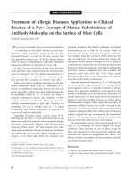
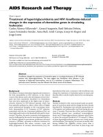
LMP-420 inhibits TNF response to LPS in vitro
LMP-420 very effectively inhibits production of TNF
mRNA and protein when applied topically in vitro to mac-
rophages and lymphocytes, cell types that are present in
the colonic lamina propria (Figure 2). This suggests that if
it is not degraded, LMP-420 could have local (topical)
activity in the gastrointestinal tract when administered
orally. The concentration of LMP-420 required to inhibit
50% of the TNF synthesized (IC
50
) by LPS-stimulated
murine monocytes in vitro is ~500 nM (Figure 2). The cor-
responding IC
50
for TNF production by human peripheral
blood mononuclear cells is ~50 nM (25). The molecular
basis for the ~10-fold increased sensitivity of human vs.
murine cells to LMP-420 is unclear, but suggests that LMP-
420 is likely to have greater anti-inflammatory activity in
humans than in mice.
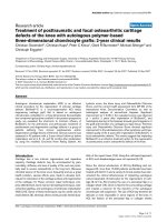
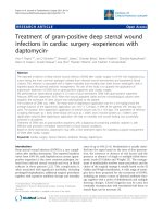
LMP-420 inhibits TNF response to LPS in vivo
Based upon its in vitro profile, LMP-420 was hypothesized
to be an efficient inhibitor of TNF in vivo. The degree of
systemic and colonic TNF blockade that resulted from dif-
ferent LMP-420 in vivo dosing protocols was first assessed
in wild type mice using a bacterial lipopolysaccharide
(LPS) challenge model. Mice treated with 2 mg (100 mg/
kg) LMP-420 given intraperitoneally (i.p.) experienced
transient behavioral depression consistent with hypoten-
sion, but recovered to normal behavior within 20 minutes
after injection. TNF was below the limit of detection (< 10
pg/ml) in the serum of mice not challenged with LPS. As
expected, serum TNF levels were markedly increased to
1109 ± 87 pg/ml (mean ± SEM; n = 9) upon LPS chal-
lenge. However, pre-treatment with i.p. LMP-420
decreased LPS-induced serum TNF levels by 42% (Figure
3A; p = 0.0.001) to 641 ± 57 pg/ml serum (n = 3). In con-
trast to the lack of detectable TNF in the serum of control
mice, detectable levels of TNF (66 ± 30 pg/100 mg tissue)
were present in colonic tissue of control mice at baseline
prior to LPS stimulation. Systemic challenge with LPS
increased levels of TNF present in colonic tissue to 388 ±
37 pg/100 mg tissue (n = 9; increase of 488%; p =
0.0.001). Pre-treatment with LMP-420 significantly
decreased total colonic TNF content by 28% to 281 ± 27
pg/100 mg tissue (n = 3) (Figure 3A; p = 0.0.045).
Journal of Inflammation 2008, 5:4 />Page 5 of 13
(page number not for citation purposes)
Although these mice were treated with 2 mg (100 mg/kg)
LMP-420 for 16 days prior to challenge, similar levels of
TNF inhibition were observed when LPS challenge
occurred 4 hrs after a single dose of LMP-420 (data not
shown).
Contrary to the toxicity seen when 100 mg/kg LMP-420
was administered i.p., no adverse effects were seen when
doses of LMP-420 up to 145 mg/kg/day were adminis-
tered orally in food for 16 days. LPS challenge was per-
formed for mice that received 75 mg/kg LMP-420 orally in
food for 5 days prior to LPS challenge. This oral dose pro-
vided similar decreases in LPS-induced serum (-43%) and
total colonic TNF levels (-29%; Figure 3B) as were seen
with mice given 100 mg/kg via the i.p. route (Figure 3A).
LMP-420 markedly decreases colonic TNF when given i.p.
after initiation of acute DSS colitis
The LPS challenge experiments demonstrated that both
i.p. and orally-administered LMP-420 significantly inhib-
ited in vivo TNF production in mice both systemically, as
indicated by serum TNF and locally in the colon. Therapy
with the anti-TNF antibody drug infliximab has been
shown to provide clinical benefit for at least a subset in
humans with IBD. To determine the efficacy of LMP-420
therapy in a murine model of IBD, C57BL/6 mice were
given 3% DSS in drinking water for 7 days. Therapy with
1 mg LMP-420 or vehicle given i.p. once daily was begun
on day 4 of DSS exposure, when symptoms of weight loss,
decreased stool consistency, and stool bleeding indicated
the onset of severe acute colitis. The 1 mg i.p. dose was
chosen to minimize the hypotension and behavioral
depression that was seen when 2 mg was administered in
the LPS challenge study. On day 7 of DSS exposure, DSS-
exposed mice treated with vehicle had markedly increased
levels of colonic TNF (409 ± 107 pg/100 mg colon tissue;
n = 5) compared to mice who were not exposed to DSS
(65 ± 5 pg/100 mg colon tissue; n = 5) (p = 0.03). DSS-
exposed mice that were treated with i.p. LMP-420 demon-
strated a significant (85%) decrease in colonic TNF levels
(117 ± 8 pg/100 mg tissue; n = 5; p = 0.05 vs. vehicle-
treated mice), that represented near normalization of
their colonic levels of TNF compared with control mice
that were not exposed to DSS. TNF was not detected in the
serum of either vehicle- or LMP-420-treated DSS-exposed
or control mice at this time point.
LMP-420 has no effect on histologic severity of acute or
established chronic DSS colitis
Despite the efficacy of i.p. LMP-420 in decreasing colonic
levels of TNF induced by DSS exposure, there was no dif-
ference in clinical or histologic severity of colitis observed
on day 7 in mice treated i.p. with vehicle- vs. LMP-420
(data not shown). However, the 3 day treatment period
used was likely too short to allow healing. Longer treat-
LMP-420 inhibits production of TNF protein by murine lym-phocytes and macrophagesFigure 2
LMP-420 inhibits production of TNF protein by
murine lymphocytes and macrophages. A. A 2 hr pre-
treatment with LMP-420 markedly decreases TNF produc-
tion by thioglycollate-elicited macrophages (MΦ) (n = 3) 24
hrs after exposure to LPS. The IC50 threshold (the concen-
tration that inhibits 50% of the TNF produced) is indicated
by the dotted line and is slightly less than 1 µM for this assay.
* indicates p ≤ 0.03. B. Pre-treatment with LMP-420 also
markedly decreases TNF production by splenocytes (n = 3)
24 hrs after LPS exposure. The IC50 threshold for this assay
(dotted line) was slightly less than 1 µM. C. Pre-treatment
with LMP-420 also significantly decreased TNF production by
splenocytes (n = 8) at both 24 and 48 hrs after stimulation
with CD3 antibody. The IC50 for CD3-stimulated T cells at
48 hrs (dotted line) was slightly greater than 1 µM. * indi-
cates p < 0.03 and ** indicates p ≤ 0.003 vs. CD3-stimulated
control not exposed to LMP-420.
Journal of Inflammation 2008, 5:4 />Page 6 of 13
(page number not for citation purposes)
ments were not possible in the acute DSS model due to
severe weight loss that required euthanasia for humane
reasons. To determine if LMP-420 treatment could pre-
vent the development of acute DSS colitis, LMP-420 was
administered i.p. for 5 days prior to as well as throughout
the 7 day exposure to DSS. Oral administration of LMP-
420 was not used for these studies, since mice rapidly
decrease their food consumption when acute colitis devel-
ops and it was not possible to maintain a consistent oral
dose using drug mixed with food. Systemic (i.p.) treat-
ment with LMP-420 given prior to and during DSS expo-
sure did not alter the clinical or histologic severity of
colitis (Figure 4A, B). The severe acute inflammation
induced by DSS tends to produce fibrosis that can be
objectively monitored by measuring colon lengths. LMP-
420 treatment also did not affect colonic shortening
induced by DSS (Table 1).
Next, we determined whether LMP-420 treatment would
influence the severity of chronic colitis initiated by multi-
ple cycles of DSS exposure. Treatment with a dose range of
LMP-420 was begun immediately upon discontinuation
of the 3d DSS cycle when all mice had severe chronic col-
itis. The LMP-420 dose range was 0, 5, 15, 45 mg/kg/day
for i.p. administration and 0, 26, 63, and 145 mg/kg/day
for oral administration. Mice were euthanized after 16
days of treatment to determine colonic TNF content and
the histologic severity of colitis. Although gross inflamma-
tion (edema, redness) was subjectively decreased in LMP-
420-treated mice, there was no difference in histologic
scores between treated and untreated mice (Figure 4C, D;
Figure 5). In humans, histologic mucosal healing typically
lags behind gross improvement and it is possible that
longer treatment periods may yield differences in histo-
logic scores. Very interestingly, marked squamous meta-
plasia of the rectum, sometime extending proximally for >
1 cm, was consistently observed in all mice exposed to 3
cycles of DSS, regardless of treatment group (Figure 4E, F).
Despite the very severe inflammation, TNF levels in
colonic tissue were low in all groups, including untreated
control groups, and did not differ according to treatment
group. The colonic TNF content of these mice was statisti-
cally similar to that present in non-DSS-exposed mice
without colitis (data not shown).
Stool TNF levels are not elevated in chronic DSS colitis
The effects of LMP-420 on TNF production was profound
in acute DSS colitis, but LMP-420 therapy apparently had
no effect on inflammation severity in either acute or estab-
lished chronic DSS colitis. Furthermore, colonic TNF lev-
els were not elevated in untreated control mice with
chronic DSS colitis, despite the presence of very severe
colonic inflammation (histologic scores of 52 ± 3; n = 10).
To begin to understand these observations, a longitudinal
study of the levels of TNF in the stool was performed.
Stool samples were obtained prior to the initial DSS expo-
sure (day 0), at the end of each 5 day cycle of DSS admin-
istration (days 5, 26, and 47), and after 16 days of healing
prior to beginning the next cycle of DSS administration
(days 21, 42, and 63). Because levels of soluble TNF recep-
tors increase during inflammation due to receptor shed-
ding after binding TNF and/or increased alternative
splicing that generates the soluble form, the levels of TNF-
LMP-420 decreases serum and colonic TNF induced by LPS stimulation in vivoFigure 3
LMP-420 decreases serum and colonic TNF induced
by LPS stimulation in vivo. A. Mice were pre-treated with
2 mg LMP-420 i.p. for 16 days. Four hours after the last injec-
tion, mice were challenged i.p. with a lethal dose of 0.5 mg
LPS then euthanized 2 hrs. later for measurement of TNF
levels in serum and in colon tissue lysates prepared at 100
mg tissue/ml. Serum TNF was below the limit of detection (<
10 pg/ml) in control mice not exposed to LPS challenge. *p <
0.05 and **p < 0.001 relative to control LPS-stimulated mice.
B. In 2 separate experiments, groups of 5 mice were pre-
treated with 75 mg/kg LMP-420 given orally in food for 5
days prior to LPS challenge and TNF measurement. Data is
presented as % of control value rather than as absolute num-
bers because mean serum TNF levels after LPS stimulation of
control mice differed markedly (706 vs. 1870 pg/ml) in the 2
experiments. * indicates p < 0.05 compared with control.
Journal of Inflammation 2008, 5:4 />Page 7 of 13
(page number not for citation purposes)
RII (p75, CD120b; encoded by the TNFRSF1B gene) in
stool were also measured. In wild type C57BL/6 mice, TNF
levels in stool were significantly increased compared to
baseline levels at the end of each cycle of DSS administra-
tion. Stool TNF then spontaneously decreased to normal
levels by the end of each 16 day period of recovery (Figure
6A). Stool levels of TNF-RII also increased very markedly
during DSS administration and then decreased during the
recovery period. However, in contrast to stool TNF, stool
TNF-RII levels did not return to baseline but remained sig-
nificantly elevated throughout all recovery periods (Figure
6B).
Role of T and B lymphocytes in chronic DSS colitis
Acute DSS colitis has been shown to occur in the absence
of T and B lymphocytes [28], however the establishment
of chronic DSS colitis has been hypothesized to involve
adaptive as well as innate immune cells. To test this, RAG-
2
-/-
mice that lack both T and B cells were exposed to the
same regimen of 3 cycles of DSS that induced severe
chronic colitis in wild type mice, then euthanized 16 days
following the 3rd administration of DSS. All RAG-2
-/-
mice
had severe colitis after exposure to 3 cycles of DSS, with
histologic scores of 53 ± 1 (mean ± SEM; n = 14). These
scores were statistically similar to those observed in wild
type mice exposed to a similar regimen of 3 DSS cycles,
however the histologic picture was very different. In con-
trast to the marked mucosal hyperplasia seen in wild type
mice, RAG-2
-/-
mice exhibited minimal mucosal hyperpla-
sia. Mucosal histologic sub-scores were high nonetheless,
due to marked squamous metaplasia in the rectum and
architectural distortion throughout the colon, manifested
primarily by crypt branching and crypt dropout. Inflam-
matory infiltrates in the RAG-2
-/-
mice consisted of a small
number of mononuclear cells and a moderate to large
number of neutrophils, and these infiltrates were more
uniformly spread through the tissues. Frank ulcerations
and crypt abcesses were present, but less common com-
pared with the wild type mice.
RAG-2
-/-
mice initially developed higher mean levels of
TNF in their stool following DSS exposure, compared to
wild type mice (Figure 6A). However, the overall pattern
of TNF elevation observed after DSS administration fol-
lowed by spontaneous recovery to baseline was similar to
that observed in wild type mice. In contrast to the marked
and consistently high levels of TNF-RII seen in wild type
mice, TNF-RII levels in RAG-2
-/-
mice very closely followed
the levels of TNF, rising with DSS administration and fall-
ing to or near baseline during recovery (Figure 6B). Stool
levels of TNF in RAG-2
-/-
mice with severe chronic DSS col-
itis were 13 ± 6 pg/100 mg stool at the time of tissue col-
lection on day 62, which is statistically similar to pre-DSS
levels of 3 ± 1 pg/100 mg stool (p = 0.13). The TNF levels
in colon tissue measured in a subset of these mice (n = 5)
was 72 ± 3 pg/100 mg tissue, which is similar to that seen
in control wild type mice without colitis (Figure 3A). TNF
was not detectable in the serum of these mice at the time
of tissue collection. Taken together, these data demon-
strate that severe colitis can occur in the absence of T and
B cells and without systemic or local colonic or stool ele-
vations in TNF.
LMP-420 in the IL-10
-/-
model of chronic colitis
Colitis has been reported to develop spontaneously in IL-
10-deficient mice that are not kept germ-free, but the age
of onset can vary widely between animal facilities. We
used a brief exposure to the non-steroidal anti-inflamma-
tory drug piroxicam to uniformly trigger the development
of chronic colitis in 6 – 7 wk IL-10
-/-
mice. Once colitis was
established, mice were treated with a dose range of i.p. (0,
5, 15, or 45 mg/kg/day) or oral LMP-420 (0, 41, 62, or
138 mg/kg/day) for 16 days. Effects on colonic TNF and
histologic severity of colitis were determined. Colonic lev-
els of TNF were elevated in untreated IL-10
-/-
mice with
colitis compared with wild type mice without colitis (Fig-
ure 7A, C). Low doses of either i.p. (5 mg/kg/day) or oral
(41 mg/kg/day) LMP-420 significantly reduced colonic
tissue TNF levels by 44 or 39% respectively to near normal
levels (Figure 7A, C). This TNF-lowering effect was lost at
higher i.p or oral doses. Although the colons were subjec-
tively less inflamed grossly in mice treated with i.p. LMP-
420, no differences in histologic scores were seen for any
of the i.p. treatment groups (Figure 7B). However, a trend
toward decreased histologic score (p = 0.06) was observed
in mice treated with 41 mg/kg oral LMP-420 (Figure 7D),
consistent with the decreased colonic TNF observed in
this group.
Discussion
The novel small molecule drug LMP-420 is highly effec-
tive in inhibiting TNF production both in vitro and in vivo.
When given to mice either i.p. or orally, LMP-420 signifi-
cantly decreased both serum (-42%) and colonic TNF
responses (-67%) to challenge with a lethal dose of LPS.
LMP-420 treatment also reduced (-85%) TNF elevations
associated with acute DSS colitis to near baseline levels.
However, despite its efficacy in reducing TNF levels in vivo,
LMP-420 had no effect on the histologic severity of
Table 1: Colonic shortening induced by DSS treatment
Treatment Colon length, cm*
No DSS 8.4 ± 0.2
DSS alone 6.5 ± 0.1
DSS/0.5 mg LMP-420 i.p. 6.6 ± 0.1
DSS/1 mg LMP-420 i.p. 6.4 ± 0.2
DSS/2 mg LMP-420 i.p. 6.6 ± 0.1
* Mean ± SEM (n = 5/group)
Journal of Inflammation 2008, 5:4 />Page 8 of 13
(page number not for citation purposes)
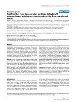
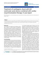
Histologic changes during acute and chronic DSS colitisFigure 4
Histologic changes during acute and chronic DSS colitis. Colon tissues from both control (A) and LMP-420-treated (B,
15 mg/kg LMP-420 i.p.) mice with acute DSS colitis demonstrated similar amounts of edema, acute inflammatory infiltrates, and
focal ulceration. Chronic colitis generated by 3 cycles of 5 days of 3% DSS in drinking water, followed by 16 days of plain water
resulted in development of severe chronic colitis (C) that was not altered by a 16 day treatment with treatment LMP-420 (D,
45 mg/kg i.p.). The cecum is shown in panels A and B and mid-colon is shown in panels C and D. Wild-type and RAG-2
-/-
mice
with chronic DSS colitis developed extensive squamous metaplasia of the rectum (E, F) that in some cases extended proximally
for > 1 cm. The bar equals 100 µm except for panel E, where bar = 1 mm.
Journal of Inflammation 2008, 5:4 />Page 9 of 13
(page number not for citation purposes)
colonic inflammation in response to either acute or
chronic DSS administration. A trend toward decreased
inflammation (p = 0.06) was observed in IL-10
-/-
mice
treated orally with LMP-420, which correlated with
decreased colonic TNF levels. Histologic healing of
colonic inflammatory lesions is known to lag behind clin-
ical remission and endoscopic healing. Thus, it is possible
that LMP-420 treatments of longer durations might result
in beneficial clinical effects that were not observed in this
pilot study using short treatment periods.
The availability of LMP-420, a small-molecule, orally-
active inhibitor of TNF production, provided us an attrac-
Effect of LMP-420 in chronic DSS colitisFigure 5
Effect of LMP-420 in chronic DSS colitis. Chronic colitis
was generated in wild type C57BL/6 mice by 3 cycles of 5
days of 3% DSS in drinking water, followed by 16 days of
plain water. LMP-420 therapy was given parenterally (i.p.) or
orally in food to groups of 5 mice, beginning after completion
of the 3d administration of DSS and continuing throughout
the final 16 day recovery period. All mice had severe colitis
at the termination of the study. Histologic scores were calcu-
lated as described. No significant differences in histologic
scores were observed in mice treated with LMP-420 doses
of 0 – 45 mg/kg given i.p. (panel A) or 0 – 145 mg/kg given
orally (panel B).
Stool levels of TNF and TNF-RII during induction of chronic DSS colitisFigure 6
Stool levels of TNF and TNF-RII during induction of
chronic DSS colitis. Levels of TNF (panel A) and TNF-RII
(panel B) were determined by enzyme immunoassay in stool
samples obtained before DSS exposure began, at the end of
each 5 day cycle of DSS administration, and after 16 days of
recovery prior to beginning the next DSS cycle for wild type
(n = 49–50 mice for days 0, 47, and 62, and n = 9 – 10 for the
remaining time points) and RAG-2
-/-
mice (n = 14). Mean
cytokine concentrations per 100 mg stool are shown. Error
bars are omitted for clarity. * indicates values significantly dif-
ferent from pre-treatment level for a given genotype. # indi-
cates values significantly different (p ≤ 0.02) in wild type vs.
RAG-2
-/-
mice.
Journal of Inflammation 2008, 5:4 />Page 10 of 13
(page number not for citation purposes)
tive opportunity to define the potential role of TNF in the
murine model of DSS-induced colitis, a commonly-used
model of inflammatory bowel disease. Although we were
able to demonstrate significant inhibition of colon TNF
by LMP-420 in this model, we had no effect on the path-
ological inflammation. However, in contrast to the bio-
logical TNF antagonists currently used in humans which
are capable of "neutralizing" essentially all circulating
TNF, LMP-420 allowed ~20% of colon TNF to be pro-
duced in our model. Thus, while our data might be inter-
preted to suggest that TNF does not play a significant role
in this model, we cannot at this time rule out the possibil-
ity that the small amount of TNF produced was sufficient
to induce pathogenesis. Alternatively, prevention of TNF
synthesis without toxicity to TNF-producing cells may not
be sufficient to stop the inflammatory cascade in vivo.
Spohn et al. recently showed that anti-TNF antibodies that
bound to both membrane-bound and soluble forms of
TNF had a greater anti-inflammatory effect in a murine
model of rheumatoid arthritis than antibodies reactive
only with soluble TNF [29]. Furthermore, significant
immunosuppression leading to reactivation of latent
Colonic TNF levels and histologic scores in control and LMP-420-treated IL-10
-/-
mice with chronic colitisFigure 7
Colonic TNF levels and histologic scores in control and LMP-420-treated IL-10
-/-
mice with chronic colitis. A, C.
Levels of TNF in colonic tissues of IL-10
-/-
mice with chronic colitis following 16 days of treatment with i.p. (panel A) or oral
(panel C) LMP-420 at the indicated doses. LMP-420 doses of 5 mg/kg/day i.p. and 41 mg/kg/day oral significantly decreased total
colon tissue TNF content by 44% (p = 0.03) and 39% (p = 0.0003), respectively. B, D. Histologic scoring of colon tissues from
the same mice showed a trend toward decreased histologic evidence of inflammation in mice treated with 41 mg LMP-420/kg/
day orally (panel D), however this change approached (p = 0.06) but did not reach statistical significance. No decreases in his-
tologic scores were seen in mice treated with i.p. LMP-420 (panel B), despite the decreased colonic TNF levels observed in
panel A.
Journal of Inflammation 2008, 5:4 />Page 11 of 13
(page number not for citation purposes)
tuberculosis was observed only in mice with antibodies
reactive with membrane-bound TNF [29]. Thus, it is pos-
sible that additional beneficial effects of TNF antagonists
result from binding to membrane-bound TNF followed
by cytolysis of TNF-producing cells, an activity that is
present with infliximab and other antibodies that bind
membrane-bound TNF, but not with etanercept or LMP-
420. Given the lack of efficacy of etanercept in treating CD
[17] despite its demonstrated efficacy in rheumatoid
arthritis and psoriasis and the inability of LMP-420 to
ameliorate murine DSS-colitis despite lowering colon
TNF levels, the role of direct TNF inhibition in the patho-
genesis and/or treatment of inflammatory bowel disease
remains undetermined.
Levels of TNF excreted in the stool correlated well with
levels of TNF measured in colon tissues harvested at the
time of euthanasia, providing a method to follow colonic
TNF levels non-invasively. Longitudinal measurements of
TNF in the stool of mice subjected to multiple cycles of
DSS demonstrated that, although TNF levels are elevated
in acute DSS colitis, these levels decrease spontaneously
and eventually return to baseline despite ongoing severe
inflammation. Taken together with the lack of efficacy of
LMP-420 in acute or chronic DSS colitis despite its ability
to significantly lower colonic TNF levels, these data sug-
gest that elevated levels of TNF are not required for the ini-
tiation and maintenance of colonic inflammation in this
murine model. Our data thus is similar to that of Olson et
al, who previously reported that TNF was not detectable in
colon tissue or plasma of CBA/J mice with acute DSS col-
itis, and that a polyclonal anti-TNF antiserum had no
effect on disease severity [30]. However, Naito et al
showed that TNF-deficient mice have increased intestinal
inflammation in response to DSS compared with wild
type mice [31]. These contrasting data suggest that
increased investigation will be necessary to clarify the role
of TNF in DSS-induced colitis.
Our studies showed that levels of TNF and TNF-RII in the
stool are highly correlated for RAG-2
-/-
mice, in contrast to
the markedly higher TNF-RII levels observed in stool of
wild type mice subjected to multiple cycles of DSS. The
manufacturer reports that the presence of TNF-RII does
not affect the ability of its TNF ELISA assay to quantitiate
TNF. Thus additional mechanisms beyond simple shed-
ding of receptor that has bound TNF likely account for the
marked and sustained increase in stool TNF-RII during
chronic DSS colitis in wild type mice. The induction of
severe chronic colitis following repeated DSS administra-
tion to RAG-2
-/-
mice that lack T and B cells suggests that
innate immunity is sufficient to drive the development of
chronic colitis in the DSS model. Use of other models that
are TNF-dependent or driven by induced T cell responses
will be necessary to determine if LMP-420 may have effi-
cacy in maintaining remission of chronic colitis driven by
TNF or T cells.
The induction of extensive squamous metaplasia in the
terminal colon and rectum by multiple cycles of DSS
administration is an interesting pathologic observation
that is of uncertain clinical significance. Metaplasia has
been observed in a range of organs and is thought to occur
in response to chronic irritation [32]. T and B cells are
apparently not required for this histopathologic change,
since similar degrees of squamous metaplasia were
observed in both wild type and RAG-2
-/-
mice. The pres-
ence of squamous mucosa is typically limited to the anus
in both humans and mice. The simple columnar epithe-
lium that is normally present in the terminal colon and
rectum functions to absorb fluid from stool as well as to
secrete mucus to lubricate its passage. These functions
would be missing from metaplastic squamous epithe-
lium. By analogy with other organs, it is possible that mice
with extensive squamous metaplasia of the colon might
be at increased long-term risk for development of squa-
mous carcinomas of the colon rather than the adenocarci-
nomas that typically develop at this site. Longer term
studies will be needed to address this possibility.
The effect of LMP-420 was additionally studied in the IL-
10
-/-
model of murine colitis. As we saw for acute DSS col-
itis, LMP-420 treatment (5 mg/kg i.p. and 41 mg/kg oral)
significantly decreased colonic TNF levels in a setting
where TNF was elevated in the colons of mice that did not
receive this drug. However, again we saw no statistically
significant decreases in severity of colitis as measured his-
tologically, although there was a trend to decreased histo-
logic inflammation for mice that received 41 mg/kg oral
LMP-420. Increased potential effect of oral as compared
with systemically administered drug suggests that LMP-
420 may exhibit local anti-inflammatory activity as it
passes through the gastrointestinal tract. Lack of TNF low-
ering in the IL-10
-/-
model at higher LMP-420 doses indi-
cates a complex dose-response profile that may reflect
dual activity of LMP-420 in competing inflammatory/
anti-inflammatory pathways.
Taken together, these studies demonstrate that short-term
treatment with a transcriptional inhibitor of TNF produc-
tion does not decrease the severity of acute and chronic
DSS colitis or piroxicam-accelerated colitis in IL-10
-/-
mice. A detailed dose-response study and longer treat-
ment durations using other colitis models that are more
dependent on TNF elevation should be performed to
more accurately assess the potential of LMP-420 for ther-
apy of inflammatory bowel disease.
Journal of Inflammation 2008, 5:4 />Page 12 of 13
(page number not for citation purposes)
Abbreviations
BSA: bovine serum albumin; CD: Crohn's disease; DSS:
dextran sulfate sodium; FBS: fetal bovine serum; i.p.:
intraperitoneal; IBD: inflammatory bowel disease; IFN:
interferon; IL: interleukin; LPS: lipopolysaccharide; MCP:
monocyte chemoattractant protein; PBS: phosphate buff-
ered saline; SD: standard deviation; SEM: standard error
of the mean; TNF: tumor necrosis factor; TNF-RII: tumor
necrosis factor receptor, type II; UC: ulcerative colitis.
Competing interests
LPH has no competing interests. GJC also has no compet-
ing interests, but discloses that he was a co-discoverer of
LMP-420 and was associated with LeukoMed Inc (the
company that holds the license for LMP-420) until Octo-
ber 2005.
Authors' contributions
LPH conceived of and designed the studies, obtained
funding, performed the pathologic and data analyses, pre-
pared figures, and drafted the manuscript. GJC assisted in
study design, reviewed data, prepared figures, and helped
to draft the manuscript. All authors read and approved the
final manuscript.
Acknowledgements
The authors would like to acknowledge the expert technical assistance of
Chau T. Trinh, Paula K. Greer, and Margaret Kennedy. This work was
funded by the Broad Medical Research Program of The Eli and Edythe L.
Broad Foundation, which had no role in the study design, the collection,
analysis, and interpretation of data, the writing of the manuscript, or in the
decision to submit the manuscript for publication.
References
1. MacDonald TT, Monteleone G: Immunity, inflammation, and
allergy in the gut. Science 2005, 307:1920-1925.
2. Podolsky DK: Inflammatory bowel disease. New Engl J Med 2002,
347:417-429.
3. O'Neil D, Steidler L: Cytokines, chemokines, and growth fac-
tors in the pathogenesis and treatment of inflammatory
bowel disease. Adv Exp Med Biol 2003, 520:252-285.
4. Papadakis KA: Chemokines in inflammatory bowel disease.
Curr Allergy Asthma Reports 2004, 4(1):83-89.
5. Knight DM, Trinh H, Le J, Siegel S, Shealy D, McDonough M, Scallon
B, Moore MA, Vilcek J, Daddona P, Ghrayeb J: Construction and
initial characterization of a mouse-human chimeric anti-
TNF antibody. Molec Immunol 1993, 30:1443-1453.
6. Scallon B, Cai A, Solowski N, Rosenberg A, Song XY, Shealy D, Wag-
ner C: Binding and functional comparison of two types of
tumor necrosis factor antagonists. J Pharmacol Exp Ther 2002,
301:418-426.
7. Van den Brande JM, Braat H, van den Brink GR, Versteeg HH, Bauer
CA, Hoedemaeker I, van Montfrans C, Hommes DW, Peppelenbosch
MP, van Deventer SJ: Infliximab but not etanercept induces
apoptosis in lamina propria T-lymphocytes from patients
with Crohn's disease. Gastroenterol 2003, 124:1774-1785.
8. PDR (Physicians Desk Reference) 61th edition. 2007:584-591. 971–979
9. Wooley PH, Dutcher J, Widmer MB, Gillis S: Influence of a recom-
binant human soluble tumor necrosis factor receptor Fc
fusion protein on type II collagen-induced arthritis in mice. J
Immunol 1993, 151:6602-6607.
10. Graninger W, Smolen J: Treatment of rheumatoid arthritis by
TNF-blocking agents. Int Arch Allergy Immunol 2002, 127:10-14.
11. Leonardi CL, Powers JL, Matheson RT, Goffe BS, Zitnik R, Wang A,
Gottlieb AB: Etanercept as monotherapy in patients with pso-
riasis. New Engl J Med 2003, 349:2014-2022.
12. Present DH, Rutgeerts P, Targan S, Hanauer SB, Mayer L, van Hoge-
zand RA, Podolsky DK, Sands BE, Braakman T, DeWoody KL, Schai-
ble TF, van Deventer SJ: Infliximab for the treatment of fistulas
in patients with Crohn's disease. N Engl J Med 1999,
340:1398-1405.
13. Nahar IK, Shojania K, Marra CA, Alamgir AH, Anis AH: Infliximab
treatment of rheumatoid arthritis and Crohn's disease. Ann
Pharmacol 2003, 37(9):1256-1265.
14. Hanauer SB, Feagan BG, Lichtenstein GR, Mayer LF, Schreiber s,
Colombel JF, Rachmilewicz D, Wolf DC, Olson A, Bao W, Rutgeerts
P: Maintenance infliximab for Crohn's disease: the ACCENT
I randomised trial. Lancet 2002, 359:1541-1549.
15. Sands BE, Anderson FH, Bernstein CN, Chey WY, Feagan BG,
Fedorak RN, Kamm MA, Korzenik JR, Lashner BA, Onken JE, Rach-
milewitz D, Rutgeerts P, Wild G, Wold DC, Marsters PA, Suzanne
Travers, Blank MA, van Deventer SJ: Infliximab maintenance
therapy for fistulizing Crohn's disease. New Engl J Med 2004,
350:876-885.
16. Rutgeerts P, Sandborn WJ, Feagan BG, Reinisch W, Olson A, Johanns
J, Travers S, Rachmilewitz D, Hanauer SB, Lichtenstein GR, de Villiers
WJS, Present D, Sands BE, Colombel JF: Infliximab for induction
and maintenance therapy for ulcerative colitis. New Engl J Med
2005, 353:2462-2476.
17. Sandborn WJ, Hanauer SB, Katz S, Safdi M, Wolf DG, Baerg RD,
Tremaine WJ, Johnson T, Diehl NN, Zinmeister AR: Etanercept for
active Crohn's disease: a randomized, double-blind, placebo-
controlled trial. Gastroenterol 2001, 121(5):1088-1094.
18. Kupper TS: Etanercept for Crohn's disease. New Engl J Med
2004, 350:840.
19. Gottlieb GS, Lesser CF, Holmes KK, Wald A: Disseminated sporo-
trichosis associated with treatment with immunosuppres-
sants and tumor necrosis factor-alpha antagonists. Clin Infect
Dis 2003, 37:838-840.
20. Hage CA, Wood KL, Winer-Muram HT, Wilson SJ, Sarosi G, Knox
KS: Pulmonary cryptococcosis after initiation of anti-tumor
necrosis factor-alpha therapy. Chest 2003, 124:2395-2397.
21. Keane J, Gershon S, Wise RP, Mirabile-Levens E, Kasznica J, Schwiet-
erman WD, Siegel JN, Braun MM: Tuberculosis associated with
infliximab, a tumor necrosis factor alpha-neutralizing agent.
New Engl J Med 2001, 345:1098-1104.
22. Gardam MA, Keystone EC, Menzies R, Manners S, Skamene E, Long
R, Vinh DC: Anti-tumour necrosis factor agents and tubercu-
losis risk: mechanisms of action and clinical management.
Lancet Inf Dis 2003, 3(3):148-155.
23. Wolfe F, Michaud K, Anderson J, Urbansky K: Tuberculosis infec-
tion in patients with rheumatoid arthritis and the effect of
infliximab therapy. Arthritis Rheum 2004, 50:372-379.
24. Benson BJ, Chen X, Cianciolo GJ, Diaz J-L, Ishaq KS, Morris-Natschke
SL, Uhing RJ, Wong H: N-substituted-(Dihydroxyboryl)alkyl
purine, indole and pyrimidine derivatives, useful as inhibitors
of inflammatory cytokines. US Patent 5,643,893 1997.
25. Haraguchi S, Day NK, Kamchaisatian W, Beigier-Pompadre M,
Stenger S, Tangsinmankong N, Sleasman JW, Pizzo SV, Cianciolo GJ:
LMP-420, a small-molecule inhibitor of TNF-alpha transcrip-
tion, reduces replication of HIV-1 and Mycobacterium tuber-
culosis in human cells. AIDS Res & Ther 2006, 3:8.
26. Hale LP, Gottfried MR, Swidsinski A: Piroxicam treatment of IL-
10 deficient mice enhances colon epithelial apoptosis and
mucosal exposure to intestinal bacteria. Inflamm Bowel Dis
2005, 11:1060-1069.
27. Burich A, Hershberg R, Waggie K, Zeng W, Brabb T, Westrich G,
Viney JL, Maggio-Price L: Helicobacter-induced inflammatory
bowel disease in IL-10 and T cell-deficient mice. Am J Physiol
Gastrointest Liver Physiol 2001, 281:G764-G778.
28. Dieleman LA, Ridwan BU, Tennyson GS, Beagley KW, Bucy RP, Elson
CO: Dextran sulfate sodium-induced colitis occurs in severe
combined immunodeficient mice. Gasroenterol 1994,
107(6):1643-1652.
29. Spohn G, Guler R, Johansen P, Keller I, Jacobs M, Bek M, Rohner F,
Bauer M, Dietmeier K, Kundig TM, Jennings GT, Brombacher F, Bach-
mann MF: A virus-like particle-based vaccine selectively tar-
geting soluble TNF-α protects from arthritis without
Publish with Bio Med Central and every
scientist can read your work free of charge
"BioMed Central will be the most significant development for
disseminating the results of biomedical research in our lifetime."
Sir Paul Nurse, Cancer Research UK
Your research papers will be:
available free of charge to the entire biomedical community
peer reviewed and published immediately upon acceptance
cited in PubMed and archived on PubMed Central
yours — you keep the copyright
Submit your manuscript here:
/>BioMedcentral
Journal of Inflammation 2008, 5:4 />Page 13 of 13
(page number not for citation purposes)
inducing reactivation of latent tuberculosis. J Immunol 2007,
178:7450-7457.
30. Olson AD, DelBuono EA, Bitar KN, Remick DG: Antiserum to
tumor necrosis factor and failure to prevent murine colitis.
J Pediatr Gastroenterol Nutr 1995, 21:410.
31. Naito Y, Takagi T, Handa O, Ishikawa T, Nakagawa S, Yamaguchi T,
Yoshida N, Minami M, Kita M, Imanishi J, Yoshikawa T: Enhanced
intestinal inflammation induced by dextran sulfate sodium in
tumor necrosis factor-alpha deficient mice. J Gastroenterol
Hepatol 2003, 18:560-569.
32. Kumar V, Abbas AK, Fausti N: Robbins and Cotran Pathologic
Basis of Disease. 7th edition. Elsevier Saunders. Philadelphia, PA;
2005:10-11.