Báo cáo y học: "Gadolinium decreases inflammation related to myocardial ischemia and reperfusion injury" pdf
Bạn đang xem bản rút gọn của tài liệu. Xem và tải ngay bản đầy đủ của tài liệu tại đây (286.47 KB, 8 trang )
BioMed Central
Page 1 of 8
(page number not for citation purposes)
Journal of Inflammation
Open Access
Research
Gadolinium decreases inflammation related to myocardial ischemia
and reperfusion injury
Jennifer L Strande*
1
, Kasi V Routhu
1
, Anna Hsu
2
, Alfred C Nicolosi
3
and
John E Baker
3,4
Address:
1
Division of Cardiovascular Medicine, Medical College of Wisconsin, Milwaukee, Wisconsin, USA,
2
Department of Pharmacology and
Toxicology, Medical College of Wisconsin, Milwaukee, Wisconsin, USA,
3
Division of Cardiothoracic Surgery, Medical College of Wisconsin,
Milwaukee, Wisconsin, USA and
4
Department of Pharmacology and Toxicology, Medical College of Wisconsin, Milwaukee, Wisconsin, USA
Email: Jennifer L Strande* - ; Kasi V Routhu - ; Anna Hsu - ;
Alfred C Nicolosi - ; John E Baker -
* Corresponding author
Abstract
Background: The lanthanide cation, gadolinium (GdCl
3
) protects the myocardium against
infarction following ischemia and reperfusion. Neutrophils and macrophages are the main
leukocytes responsible for infarct expansion after reperfusion. GdCl
3
interferes with macrophage
and neutrophil function in the liver by decreasing macrophage secretion of inflammatory cytokines
and neutrophil infiltration. We hypothesized that GdCl
3
protects against ischemia and reperfusion
injury by decreasing inflammation. We determined the impact of GdCl
3
treatment for reperfusion
injury on 1) circulating monoctye and neutrophil counts, 2) secretion of inflammatory cytokines,
and 3) influx of monocytes and neutrophils into the myocardium.
Methods: Rats (n = 3-6/gp) were treated with saline or GdCl
3
(20 μmol/kg) 15 min prior to a 30
min period of regional ischemia and 120 min reperfusion. Sham rats were not subject to ischemia.
Blood was collected either after 30 min ischemia or 120 min reperfusion and hearts were harvested
at 120 min reperfusion for tissue analysis. Blood was analyzed for leukocytes counts and cytokines.
Tissue was analyzed for cytokines and markers of neutrophil and monocyte infiltration by
measuring myeloperoxidase (MPO) and α-naphthyl acetate esterase (ANAE).
Results: GdCl
3
did not affect the number of circulating neutrophils prior to ischemia. Two hours
reperfusion resulted in a 2- and 3- fold increase in circulating monocytes and neutrophils,
respectively. GdCl
3
decreased the number of circulating monocytes and neutrophils during
reperfusion to levels below those present prior to ischemia. Furthermore, after 120 min of
reperfusion, GdCl
3
decreased ANAE and MPO activity in the myocardium by 1.9-fold and 6.5-fold
respectively. GdCl
3
decreased MPO activity to levels below those measured in the Sham group.
Serum levels of the major neutrophil chemoattractant cytokine, IL-8 were increased from pre-
ischemic levels during ischemia and reperfusion in both control and GdCl
3
treated rats. Likewise,
IL-8 levels increased throughout the 3 hour time period in the Sham group. There was no difference
in IL-8 detected in the myocardium after 120 min reperfusion between groups. In contrast, after
120 min reperfusion GdCl
3
decreased the myocardial tissue levels of macrophage secreted
cytokines, GM-CSF and IL-1.
Published: 10 December 2009
Journal of Inflammation 2009, 6:34 doi:10.1186/1476-9255-6-34
Received: 3 August 2009
Accepted: 10 December 2009
This article is available from: />© 2009 Strande et al; licensee BioMed Central Ltd.
This is an Open Access article distributed under the terms of the Creative Commons Attribution License ( />),
which permits unrestricted use, distribution, and reproduction in any medium, provided the original work is properly cited.
Journal of Inflammation 2009, 6:34 />Page 2 of 8
(page number not for citation purposes)
Conclusion: GdCl
3
treatment prior to ischemia and reperfusion injury decreased circulating
monocytes and neutrophils, macrophage secreted cytokines, and leukocyte infiltration into injured
myocardium. These results suggest GdCl
3
decreased monoctye and neutrophil migration and
activation and may be a novel treatment for inflammation during ischemia and reperfusion.
Background
The lanthanide cation, gadolinium (GdCl
3
) protects the
myocardium against infarction following ischemia and
reperfusion (IR) in vivo [1], although this preconditioning
is not observed in a buffer perfused, isolated heart model
of acute reperfusion injury (unpublished observation).
This discrepancy suggests that GdCl
3
-induced cardiopro-
tection is dependent upon factors found only in vivo, such
as blood cells, proteins or hormones among others.
Inflammatory cells are important in the pathophysiologi-
cal response to injury associated with IR. While crucial to
healing, the influx of inflammatory cells, specifically mac-
rophages and neutrophils, results in tissue injury beyond
that caused by ischemia alone. Many studies have focused
on the acute myocardial inflammatory reaction as a medi-
ator of ischemia-reperfusion injury [2]. Monocytes and
other leukocytes infiltrate the area at risk soon after the
onset of ischemia. Activated macrophages secrete
cytokines that promote tissue damage and recruit neu-
trophils [3]. Accordingly, the influx of neutrophils into
ischemic tissue increases tissue necrosis by releasing pro-
teolytic enzymes and reactive oxygen species and expands
the zone of infarction [4].
Strategies aimed at reducing the levels of inflammatory
cytokines [5] or the infiltration of leukocytes [6] attenuate
myocardial damage associated with reperfusion. Evidence
suggests that GdCl
3
interferes with macrophage and neu-
trophil function in the liver by decreasing macrophage
secretion of inflammatory cytokines and toxic oxygen rad-
icals [7] and by inhibiting neutrophil infiltration [8]. The
role GdCl
3
plays in monocyte and neutrophil infiltration
during myocardial ischemia and reperfusion is unknown.
Accordingly, this study tests the hypothesis that GdCl
3
modulates leukocyte function either directly by interfer-
ing with migration or indirectly by decreasing the genera-
tion of inflammatory cytokines and chemokines, thereby
decreasing the signal that triggers leukocytes to infiltrate
into the injured tissue.
Methods
Male Sprague Dawley rats at 8 weeks of age (250-300 g)
were used in this study and received humane care in com-
pliance with the "Guide for the Care and Use of Labora-
tory Animals" published by the US National Institutes of
Health (NIH Publication No. 85-23, revised 1996). This
project was granted approval by the local IACUC review
board.
Instrumentation, ischemia-reperfusion protocol and GdCl
3
treatment
Rats were anesthetized with 20-40 mg/kg intraperitoneal
sodium pentobarbital. The right jugular vein was cannu-
lated for delivery of saline solution. A catheter was
inserted in the left femoral artery to measure both blood
pressure and heart rate and to withdraw blood. Pressure
and rate measurements were monitored using a Gould
PE50 or PE23 pressure transducer connected to a Grass
model 7 polygraph. The rats were intubated with a 14-
gauge catheter and ventilated at 38-45 breaths/min (Har-
vard Apparatus, model 683; South Natick, MA) with sup-
plemental oxygen. Atelectasis was prevented by
maintaining a positive end-expiratory pressure of 5-10
mm H
2
O. Arterial pH, pCO2 and pO2 were monitored
with an AVL 995 pH/blood gas analyzer, and normal val-
ues were maintained by adjusting respiratory rate, tidal
volume and/or oxygen flow. Body temperature was main-
tained between 35 and 37°C using a heating pad.
A left thoracotomy was performed, the pericardium was
opened and the left coronary artery was identified. A liga-
ture (6-0 Prolene) was passed around the proximal seg-
ment of the left coronary artery, and the ends of the suture
were threaded through a propylene tube to form a snare.
Regional left ventricular ischemia was induced by tighten-
ing the snare for 30 min. Coronary artery occlusion was
confirmed by epicardial cyanosis and a decrease in blood
pressure. Reperfusion was achieved by releasing the snare
and was confirmed by a marked hyperemic response of
the myocardium. The heart was reperfused for 120 min
then excised and assessed for extent of tissue injury.
Gadolinium chloride hexahydrate (20 μmol/kg dissolved
in 0.9% NaCl solution; Sigma, Milwaukee, WI) was given
as an intravenous (i.v.) bolus 15 min before inducing
myocardial ischemia [1]. Experimental groups are shown
in Figure 1A and included Sham (no treatment, no
ischemia), Sham + GdCl
3
treated (treated, no ischemia),
Control (no treatment but subject to ischemia and reper-
fusion) and GdCl
3
(treated and subject to ischemia and
reperfusion) groups. Infarct size was also measured using
this protocol and serves as a positive control for this study
(Figure 1B).
Myocardial Tissue Myeloperoxidase Activity Assay
Myeloperoxidase (MPO) activity was assayed as a measure
of neutrophil activity in hearts using a modified protocol
[9]. The heart was homogenized in 50 mM potassium
Journal of Inflammation 2009, 6:34 />Page 3 of 8
(page number not for citation purposes)
phosphate buffer (pH 6.0) and centrifuged. The pellet was
washed twice in 5 mM potassium phosphate buffer. After
washing, the pellets were resuspended in extraction buffer
(50 mM potassium phosphate buffer (pH 6.0) containing
0.5% hexadecyltrimethyl ammoniumbromide), followed
by three rounds of freeze-thawing. The suspension was
incubated at 4°C for 20 minutes and then centrifuged at
13,000 rpm at 4°C for 15 minutes. The supernatant (100
μL) was mixed with 100 μL of reaction buffer (50 mM
potassium phosphate buffer (pH 6.0) containing 0.6 mg/
mL O-dianisidine dihydrochloride and 0.03% hydrogen
peroxide). Absorbance was measured at 450 nm after 5
minutes of incubation. After normalization for protein
concentration, the MPO content was expressed as units of
MPO activity per milligram of protein.
Myocardial Tissue
α
-Naphthyl Acetate Esterase Assay
The activity of α-naphthyl acetate esterase (ANAE), a
marker enzyme of monocytes and macrophages was
detected using a previous published protocol [10]. In
brief, frozen specimens from Sham, Sham + GdCl
3
, Con-
trol and GdCl
3
groups were separately homogenized in
ice-cold 0.25 mol/L sucrose (1:5; weight to volume) for 2
× 5 seconds. The samples were centrifuged at 10,000 g for
10 min at 4°C. The supernatant was further sonicated for
90 seconds in ice, centrifuged at 105,000 g for 90 minutes
at 4°C, and assayed for protein. An equivalent of 25 mg of
supernatant protein for each sample and 5 mL of 200
mmol/L α-naphthyl acetate (Sigma-Aldrich, St. Louis,
MO), dissolved in 95% ethanol to give a final concentra-
tion of 0.5 mmol/L, was added to a final volume of 2 mL
of saline solution. Blanks received no substrate. After 10
minutes of incubation at 37°C, the reaction was stopped
by adding 116 mL of 12.5% w/v sodium dodecyl sulfate
solution. Subsequently, 5 mL of 200 mmol/L α-naphthyl
acetate was added to the blanks. Finally, 30 mL of fast red
solution (10 mg/mL distilled water; Fast Red B, Sigma-
Aldrich, St. Louis, MO) was added to the sample, followed
by an incubation period of 15 minutes at room tempera-
ture. Optical density absorption at 490 nm was used to
estimate the metabolism of α-naphthyl acetate to α-naph-
thol. Alpha-naphthyl acetate esterase activity was esti-
mated as absorption at 490 nm per 25 mg of protein.
Cytokine Assays
Blood was collected either 30 min after ischemia or 120
min after reperfusion. Plasma was separated by centrifug-
ing the sample at 14,000 rpm × 10 min at 4°C and frozen
at -80°C until analysis. Left ventricular free wall tissue
homogenates were processed and quantiated using meth-
ods described previously [11]. Interleukin (IL)-8 levels
were determined using an IL-8 (Rat cytokine-induced neu-
trophil chemoattractant (CINC)-1) enzyme-linked immu-
noassay kit from R&D Systems (Minneapolis, MN)
according to the manufacture's instructions. CINC-2,
CINC-3, granulocyte monocyte colony stimulating factor
(GM-CSF), Interferon (INF)-γ, IL-1α, IL-1β, IL-4, IL-6, IL-
10, Monocyte chemotactic protein (MCP)-1, Macrophage
inflammatory protein (MIP)-3A, and Tumor necrosis fac-
tor (TNF)- α tissue levels were determined using RayBio
®
Rat Cytokine Antibody Array 1 (Norcross, GA) according
to the manufacture's instructions. Each dot on the immu-
noblot representing a cytokine was quantitated using
ImageJ 1.37v software.
Statistical Analysis
Data are reported as mean ± SEM. Statistical analyses were
performed by the Student's t test for paired values and a
one-way Analysis of Variance (ANOVA) for differences
between treatment groups. If significant, a Newman-Keuls
multiple comparison test was used to perform pair wise
comparisons. Data were considered significant at a p <
0.05. Statistics were performed using WINKS SDA Soft-
ware (Texasoft, Cedar Hill, TX).
Results
GdCl
3
Attenuates IR-induced Increases in Circulating
Monocytes and Neutrophils
We first determined whether GdCl
3
decreases circulating
monocytes and neutrophils following ischemia and reper-
fusion. We have previously shown that the optimal cardi-
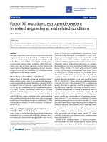
In vivo rat model of ischemia and reperfusion injuryFigure 1
In vivo rat model of ischemia and reperfusion injury.
A) Rats were treated with either saline or GdCl
3
(20 μmol/
kg) 15 minutes prior to a 30 minute period of regional
ischemia and 2 hours reperfusion.↑ Blood collection.↓ Har-
vest the free wall of the left ventricle. B) Measurement of inf-
arct size as a percentage area at risk using this protocol.
A.
Regional
Ischemia
Perfusion
Reperfusion
1. Control
saline
2. GdCl
3
15 min
prior to ischemia
3. Sham
(no ischemia)
Time
(
mins
)
0 200805035
4. Sham + GdCl
3
GdCl
3
GdCl
3
saline
80
Infarct Size (% Area at Risk)
B.
0
40
*
60
20
0 20
GdCl
3
(μmol/kg)
Journal of Inflammation 2009, 6:34 />Page 4 of 8
(page number not for citation purposes)
oprotective dose of GdCl
3
was 20 μmol/kg (Figure 1B) [1].
Therefore, we used this dose for the experiments in this
study. Rats were treated with either saline or GdCl
3
15
minutes prior to a 30 minute period of regional ischemia
followed by 120 min reperfusion. A complete blood
count and differential was performed after 120 min reper-
fusion. GdCl
3
did not affect the number of circulating
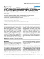
monocytes or neutrophils prior to ischemia. Two hours
reperfusion resulted in a 2-fold increase in circulating
monocytes and a 3-fold increase in circulating neu-
trophils. GdCl
3
decreased the number of circulating
monocytes (Figure 2A) and neutrophils (Figure 2B) dur-
ing reperfusion to levels below those present prior to
ischemia. In addition, GdCl
3
given to the Sham group also
decreased the number of circulating monocytes, but not
neutrophils when compared to the untreated Sham
group.
Macrophage Infiltration in Myocardium
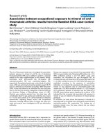
Using α-naphthyl acetate esterase (ANAE) content as an
indicator of myocardial tissue monocyte/macrophage
infiltration, ischemia and reperfusion increased ANAE
activity by 3.6-fold in the IR control group when com-
pared the Sham control group (Figure 3). GdCl
3
partially
reversed increase in ANAE activity after ischemia and
reperfusion injury by 1.9-fold but this was still above
Sham values. GdCl
3
did not change ANAE activity in the
Sham-GdCl
3
group when compared to the Sham control
group.
Neutrophil Infiltration in Myocardium
Using myeloperoxidase (MPO) content as an indicator of
myocardial tissue neutrophil infiltration, ischemia and
reperfusion increased MPO activity by 3.8-fold in control
hearts when compared to a Sham procedure (Figure 4).
GdCl
3
not only reversed this increase in MPO activity after
ischemia and reperfusion injury by 6.5-fold when com-
pared to IR control but also decreased MPO activity below
Sham control values by 1.7-fold. In the Sham groups,
GdCl
3
decreased MPO activity 2.4-fold.
Gadolinium chloride decreases circulating monocytes and neutrophils following ischemia and reperfusionFigure 2
Gadolinium chloride decreases circulating mono-
cytes and neutrophils following ischemia and reper-
fusion. Rats were treated with either saline or GdCl
3
(20
μmol/kg) 15 minutes prior to a Sham procedure or a 30
minute period of regional ischemia and 2 hours reperfusion.
A) Increase in monocytes. B) Increase in neutrophils. Data
mean ± SD, n = 3-6/gp, + = p < 0.05, Sham control vs.
Ischemia-Reperfusion (IR) control,* = p < 0.05, IR Control
vs. IR GdCl
3
, ± = p < 0.05, Sham Control vs. Sham GdCl
3
, § =
p < 0.05, IR GdCl
3
l vs. Sham GdCl
3
A
Increase in Monocytes (%)
400
+
300
200
Increase in Neutrophils (%)
0
100
200
300
400
0
100
*
±
Control GdCl
3
GdCl
3
IR
Control
Sham
B
+
*
,§
GdCl
3
Control
Sham
Control
IR
GdCl
3
Gadolinium chloride decreases Alpha naphthyl acetate este-rase activity in the reperfused myocardiumFigure 3
Gadolinium chloride decreases Alpha naphthyl ace-
tate esterase activity in the reperfused myocardium.
Rats were either treated with vehicle or GdCl
3
either before
30 min ischemia and 120 min reperfusion or a Sham proce-
dure. Alpha naphthyl acetate esterase activity was measured
after 120 min reperfusion in IR Control and IR + GdCl
3
groups and at a comparable time point in Sham Control and
Sham + GdCl
3
groups. Data is mean ± SD, n = 6/group, * = p
< 0.05 vs. IR control.
1.4
Į-naphthyl acetate esterase activity
A490/25 microgram protein
1.2
1
0.8
0
0.2
0.4
0.6
*
*
*
Control GdCl
3
Control GdCl
3
Ischemia-Reperfusion Sham
Journal of Inflammation 2009, 6:34 />Page 5 of 8
(page number not for citation purposes)
GdCl
3
Modulation of Cytokine Levels
Inflammatory cytokines were then measure from either
serum or myocardial tissue at the end of ischemia or
reperfusion periods. There was no difference in IL-8/
CINC-1 levels in the serum or the tissue when measured
after 30 min ischemia (serum) or 120 min reperfusion
(serum and tissue) (Figure 5A and 5B). Interestingly, IL-8/
CINC-1 serum levels increased throughout the study in all
groups when compared to the respective baseline levels.
A macroarray was performed using homogenized tissue
after 120 min reperfusion to look for the tissue levels of
CINC-2, CINC-3, GM-CSF, INF-γ, IL-1α, IL-1β, IL-4, IL-6,
IL-10, MCP-1, MIP-3A, and TNF-α. GdCl3 treatment did
not reduce the major monocyte chemoattactant, MCP-1.
However, GdCl
3
-treatment did reduce the macrophage
secreted cytokines such as GM-CSF and IL-1 after 120 min
reperfusion (Figure 6). In addition, an increase in TNF-α
was observed in the GdCl
3
groups after 120 min reper-
fusion.
Discussion
The important finding in this study is that a single treat-
ment of GdCl
3
prior to ischemia decreased the numbers of
circulating monocytes and neutrophils following reper-
fusion and reduced infiltration of these leukocytes into
the injured myocardium. In addition, GdCl
3
decreased
production of cytokines that are typically secreted by
monocytes. These finding are associated with GdCl
3
-
mediated reduction in infarct size after ischemia and
reperfusion [1].
The early phase of myocardial infarction is associated with
tissue infiltration of circulating leukocytes in response to
chemotactic factors [12]. As the leukocytes infiltration the
tissue and become activated, they release more cytokines
thereby recruiting further leukocytes to the injured area.
Activated leukocytes within the tissue cause further tissue
injury by releasing reactive oxygen species and proteases
and cause capillary plugging leading further tissue
hypoxia. Inhibiting neutrophil infiltration using anti-neu-
trophil antibodies [13], neutrophil depleting antimetabo-
lites [14] or neutrophil filters [15] have been successful in
limiting myocardial infarct size. We have shown that
GdCl
3
is also a potent monocyte and neutrophil depleting
compound as it prevents these leukocytes from circulating
and infiltrating into the myocardium.
Gadolinium chloride decreases myocardial tissue myeloper-oxidase activityFigure 4
Gadolinium chloride decreases myocardial tissue
myeloperoxidase activity. Rats were either treated with
vehicle or GdCl
3
either before 30 min ischemia and 120 min
reperfusion or a Sham procedure. Myeloperoxidase activity
was measured after 120 min reperfusion in IR Control and IR
+ GdCl
3
groups and at a comparable time point in Sham and
Sham + GdCl
3
groups. Data is mean ± SD, n = 3/group, * = p
< 0.05 vs. IR control; § = p < 0.05 vs. Sham control.
0.8
0
0.2
0.4
0.6
Myeloperoxidase activity
milliunits/mg protein
Control
GdCl
3
*
,§
*
*
,§
Control GdCl
3
Sham
Ischemia-Reperfusion
Gadolinium chloride does not decrease IL-8/CINC-1 produc-tion in the myocardium or serum during ischemia and reper-fusionFigure 5
Gadolinium chloride does not decrease IL-8/CINC-1
production in the myocardium or serum during
ischemia and reperfusion. Rats were either treated with
vehicle or GdCl
3
either before 30 min ischemia and 120 min
reperfusion or a Sham procedure. (Ischemia) and (Reper-
fusion) in the Sham groups is equal to the time period that
correlates with Ischemia and Reperfusion in the Control and
GdCl
3
groups. IL-8/CINC-1 was measured from the serum at
30 min ischemia and 120 min reperfusion and from the tissue
at 120 min reperfusion. A) Quantitation of tissue IL-8/CINC-
1 B) Quatitation of serum IL-8/CINC-1. Data mean ± SD, n
= 3-4/gp, * = p < 0.05 vs. perfusion. No significant difference
between IR vs. Sham groups or Control vs. GdCl
3
groups.
A
60
50
40
30
20
10
0
GdCl
3
GdCl
3
Control Control
Ischemia-Reperfusion Sham
0
500
1000
1500
2000
2500
3000
3500
Perfusion
Ischemia
Reperfusion
Perfusion
Ischemia
Reperfusion
Perfusion
(Ischemia)
(Reperfusion)
Perfusion
(Ischemia)
(Reperfusion)
Control GdCl
3
Control
GdCl
3
Serum IL-8/CINC-1 (pg/ml)
B
Ischemia-Reperfusion
Sham
*
*
*
*
*
*
*
*
Tissue IL-8/CINC-1 (pg/ml)
Journal of Inflammation 2009, 6:34 />Page 6 of 8
(page number not for citation purposes)
Gadolinium is known to modulate inflammatory
responses by liver macrophages (Kupfer cells) [7,16].
Gadolinium causes a 70% reduction in the phagocytosis
of radiolabeled bacteria [17], and GdCl
3
has been shown
to prevent the release of both inflammatory cytokines and
toxic oxygen radicals such as superoxide anion produced
by activated Kupffer cells [18,19]. Gadolinium also upreg-
ulates secretion of TNF-α from Kupfer cells in response to
endotoxemia.
In the present study, GdCl
3
treatment pre-ischemia was
associated with attenuation of the IR-induced increases in
circulating monocytes and neutrophils, and it decreased
circulating monocytes in animals not exposed to IR. The
selective action of GdCl
3
on monocytes is based on the
fact that this compound is readily dissolved in normal
saline; however, when it is injected into the bloodstream,
it rapidly aggregates into relatively large colloidal particles
at neutral pH. The particles of GdCl
3
are taken up exclu-
sively by circulating phagocytic mononuclear cells of a
monocyte lineage (CD11b+, CD13+, and CD14+)
[20,21]. Once inside the cell and after exceeding the
threshold concentration, GdCl
3
causes cell apoptosis [22].
The GdCl
3
aggragates are not expected to cross the
endothelial barrier and thus would not be taken up by res-
ident tissue phagocytic cells.
This decrease in circulating leukocytes was not associated
with any changes in the levels of the major monocyte che-
moattractants, MCP-1 and the neutrophil chemoattract-
ants IL-8/CINC-1, CINC-2, and CINC-3 suggesting that
GdCl
3
modulates leukocyte trafficking to the heart via
some other, as yet undefined mechanism following myo-
cardial IR. Interleukin-8/CINC-1 serum levels increased
over baseline levels during ischemia and reperfusion.
Interestingly, there was a similar increase in serum levels
at the corresponding time points in the Sham group.
Although the Sham animals do not undergo LAD occlu-
sion, they still receive a tracheostomy and a thoracotomy.
These two invasive surgical procedures may be enough to
induce an inflammatory response in these rats.
Gadolinium triggered an increase of TNF-α levels in the
myocardium after 120 min reperfusion (Figure 6B). TNF-
α is a potent cytokine which is elevated in a variety of
inflammatory conditions. Endogenous TNF-α has been
correlated with the deterioration of myocardial perform-
ance, while blockade of TNF-α with a neutralizing anti-
body preserves myocardial function after IR [23].
Somewhat paradoxically, exogenously added TNF-α is
protective during ischemia and reperfusion through
mechanisms which involve the JAK/STAT3 pathway, K
ATP
channels and the mitochondrial permeability transition
pore [24,25]. This protective effect of TNF-α may be
related to our previous data, which showed that GdCl
3
-
induced cardioprotection is dependent upon the activa-
tion of JAK-STAT3 and K
ATP
channels [1].
We have previously shown that GdCl
3
given 24-72 hours
prior to the index ischemic event is also able to attenuate
reperfusion injury as determined by infarct size [1]. Inter-
esting, in that study even though GdCl
3
was intravenously
administered to the rats in vivo, infarct size was deter-
mined in isolated buffer perfused hearts subject to IR
injury. Since the isolated heart model of IR does not have
circulating blood components, the infarcts are not likely
solely caused by the inflammatory response. However, it
still could be possible that GdCl
3
-mediated up-regulation
of TNF-α in the myocardium could be part of the mecha-
nism of GdCl
3
-mediated delayed cardioprotection. TNF-α
was not measured in the delayed cardioprotection studies
but this hypothesis forms the basis of further studies.
Gadolinium also attenuated IR-induced expression of the
macrophage-secreted cytokines GM-CSF and IL-1 in this
study, suggesting that attracted monocytes may not be dif-
ferentiating into activated macrophages. Treatment with
GdCl
3
also did not significantly alter levels of INF-γ and
MIP-3 which are activators of macrophage function. In
support, we observed a decrease in ANAE activity in the
myocardium which suggests decreased infiltration and
activation of monocytes into the myocardium. Similarly,
Gadolinium chloride decreases macrophage secreted cytokines after ischemia and reperfusion in ratsFigure 6
Gadolinium chloride decreases macrophage secreted
cytokines after ischemia and reperfusion in rats. (A)
Representative blots from cytokine arrays from control
hearts vs. GdCl
3
-treated hearts. The GM-CSF and IL-1 dots
are indicated. Dots were quantitated and normalized to the
positive control dots (lower left 4 dots) and results displayed
above (B). Data mean ± SD, n = 3-4/gp, * = p < 0.05, GdCl
3
vs. control, += macrophage secreted cytokines, ‡ = cardio-
myocyte secreted cytokines.
GM-CSF
IL-1
GdCl
3
Control
GM-CSF
INF-Ȗ
MCP-1
IL-1Į
MIP-3A
IL-1ȕ
CINC-2
IL-4
CINC-3
IL-6
IL-10
-40
-30
-20
-10
0
10
20
30
TNF-Į
*
*
+
+
+
+
+,‡
+
% Change of cytokine production
A
B
*
+
Journal of Inflammation 2009, 6:34 />Page 7 of 8
(page number not for citation purposes)
we observed decreased MPO activity in the myocardial tis-
sue which suggests a decrease of neutrophil infiltration
into the myocardium although the levels of circulating
and tissue IL-8/CINC-1 are not modified by GdCl
3
treat-
ment. This suggests that GdCl
3
may be inhibiting leuko-
cyte chemotaxis and in essence "paralyzing" the
monocytes and neutrophils at their storage locations.
Swirski et al. have recently identified the splenic subcap-
sular red pulp as the reservoir for monocytes [26]. From
their splenic reservoirs, the monocytes are deployed to
inflammatory sites such as the infarcted myocardium
[26]. The reservoir of neutrophils is yet to be verified but
the neutrophils are thought to reside in the post-capillary
venule sites in the periphery such as the bone marrow
[27]. Although previous studies have shown that GdCl
3
does not affect the migration of neutrophils [28], inhibi-
tion of monocyte migration has not been reported.
One limitation of this study is that we did not study the in
vitro migration of isolated monocytes or neutrophils
treated with GdCl
3
. However, the decrease in monocytes
and neutrophils into the circulation after GdCl
3
treatment
indicates that the leukocytes are either not migrating from
their reservoirs or are being scavenged prior to the 120
min reperfusion time point.
In summary, a single dose of GdCl
3
when given prior to
ischemia and reperfusion decreased circulating mono-
cytes and neutrophils as well as decreased the infiltration
of these leukocytes into the myocardium. Furthermore,
this was not associated with a change in tissue levels of the
major monocyte chemoattractant MCP-1 or neutrophil
chemoattractant IL-8/CINC-1 but was associated with an
up-regulation of TNF-α.
List of abbreviations
(GdCl
3
): Gadolinium; (IR): Ischemia and Reperfusion;
(IL): Interleukin; (CINC): cytokine-induced neutrophil
chemoattractant; (GM-CSF): monocyte colony stimulat-
ing factor; (INF): Interferon; (MCP): monocyte chemotac-
tic protein; (MIP): macrophage inflammatory protein;
(TNF): tumor necrosis factor; (ANOVA): Analysis of Vari-
ance.
Competing interests
The authors declare that they have no competing interests.
Authors' contributions
JLS conceived the study, designed the experiments, per-
formed the cytokine assays, data analysis, and prepared
the manuscript. KVR performed the MPO and ANAE
assays. AH performed the rat studies needed. ACN con-
tributed to the development of the study and revising the
manuscript critically for important intellectual content.
JEB supervised the experiments and oversaw manuscript
construction with JLS. All authors read and approved the
final manuscript.
Acknowledgements
This study was supported in part by National Institutes of Health Grant
HL54075 to J.E.B. J.L.S. was supported by National Institutes of Health
Training Grant HL07792 (awarded to the Medical College of Wisconsin).
References
1. Nicolosi AC, Strande JL, Hsu A, Fu X, Su J, Gross GJ, Baker JE: Gado-
linium limits myocardial infarction in the rat: dose-response,
temporal relations and mechanisms. J Mol Cell Cardiol 2008,
44:345-351.
2. Zahler S, Massoudy P, Hartl H, Hahnel C, Meisner H, Becker BF:
Acute cardiac inflammatory responses to postischemic
reperfusion during cardiopulmonary bypass. Cardiovasc Res
1999, 41:722-730.
3. Colletti LM, Kunkel SL, Walz A, Burdick MD, Kunkel RG, Wilke CA,
Strieter RM: Chemokine expression during hepatic ischemia/
reperfusion-induced lung injury in the rat. The role of epithe-
lial neutrophil activating protein. J Clin Invest 1995, 95:134-141.
4. Frangogiannis NG, Smith CW, Entman ML: The inflammatory
response in myocardial infarction. Cardiovasc Res 2002,
53:31-47.
5. Crawford MH, Grover FL, Kolb WP, McMahan CA, O'Rourke RA,
McManus LM, Pinckard RN: Complement and neutrophil activa-
tion in the pathogenesis of ischemic myocardial injury. Circu-
lation 1988, 78:1449-1458.
6. Altavilla D, Squadrito F, Ioculano M, Canale P, Campo GM, Zingarelli
B, Caputi AP: E-selectin in the pathogenesis of experimental
myocardial ischemia-reperfusion injury. Eur J Pharmacol 1994,
270:45-51.
7. Iimuro Y, Yamamoto M, Kohno H, Itakura J, Fujii H, Matsumoto Y:
Blockade of liver macrophages by gadolinium chloride
reduces lethality in endotoxemic rats analysis of mecha-
nisms of lethality in endotoxemia. J Leukoc Biol 1994,
55:723-728.
8. Chen YX, Sato M, Kawachi K, Abe Y: Neutrophil-mediated liver
injury during hepatic ischemia-reperfusion in rats. Hepatobil-
iary Pancreat Dis Int 2006, 5:436-442.
9. Hillegass LM, Griswold DE, Brickson B, Albrightson-Winslow C:
Assessment of myeloperoxidase activity in whole rat kidney.
J Pharmacol Methods 1990, 24:285-295.
10. Formigli L, Manneschi LI, Nediani C, Marcelli E, Fratini G, Zecchi
Orlandini S, Perna AM: Are macrophages involved in early myo-
cardial reperfusion injury? The Annals of Thoracic Surgery 2001,
71:1596-1602.
11. Strande JL, Hsu A, Su J, Fu X, Gross GJ, Baker JE: Inhibiting pro-
tease-activated receptor 4 limits myocardial ischemia/reper-
fusion injury in rat hearts by unmasking adenosine signaling.
J Pharmacol Exp Ther 2008, 324:1045-1054.
12. Bosnjak JJ, Terata K, Miura H, Sato A, Nicolosi AC, McDonald M,
Manthei SA, Saito T, Hatoum OA, Gutterman DD: Mechanism of
thrombin-induced vasodilation in human coronary arteri-
oles. Am J Physiol Heart Circ Physiol 2003, 284:H1080-1086.
13. Romson JL, Hook BG, Kunkel SL, Abrams GD, Schork MA, Lucchesi
BR: Reduction of the extent of ischemic myocardial injury by
neutrophil depletion in the dog. Circulation 1983, 67:1016-1023.
14. Mullane KM, Read N, Salmon JA, Moncada S: Role of leukocytes in
acute myocardial infarction in anesthetized dogs: relation-
ship to myocardial salvage by anti-inflammatory drugs. J
Pharmacol Exp Ther 1984, 228:510-522.
15. Engler RL, Dahlgren MD, Morris DD, Peterson MA, Schmid-Schon-
bein GW: Role of leukocytes in response to acute myocardial
ischemia and reflow in dogs. Am J Physiol 1986, 251:H314-323.
16. Rai RM, Yang SQ, McClain C, Karp CL, Klein AS, Diehl AM: Kupffer
cell depletion by gadolinium chloride enhances liver regen-
eration after partial hepatectomy in rats. Am J Physiol 1996,
270:G909-918.
17. Husztik E, Lazar G, Parducz A: Electron microscopic study of
Kupffer-cell phagocytosis blockade induced by gadolinium
chloride. Br J Exp Pathol 1980, 61:624-630.
18. Vega VL, Maldonado M, Mardones L, Schulz B, Manriquez V, Vivaldi E,
Roa J, Ward PH: Role of Kupffer cells and PMN leukocytes in
Publish with Bio Med Central and every
scientist can read your work free of charge
"BioMed Central will be the most significant development for
disseminating the results of biomedical research in our lifetime."
Sir Paul Nurse, Cancer Research UK
Your research papers will be:
available free of charge to the entire biomedical community
peer reviewed and published immediately upon acceptance
cited in PubMed and archived on PubMed Central
yours — you keep the copyright
Submit your manuscript here:
/>BioMedcentral
Journal of Inflammation 2009, 6:34 />Page 8 of 8
(page number not for citation purposes)
hepatic and systemic oxidative stress in rats subjected to
tourniquet shock. Shock 1999, 11:403-410.
19. Kono H, Fujii H, Matsuda M, Yamamoto M, Matsumoto Y: Gadolin-
ium chloride prevents mortality in hepatectomized rats
given endotoxin. J Surg Res 2001, 96:204-210.
20. Wasserman AJ, Monticello TM, Feldman RS, Gitlitz PH, Durham SK:
Utilization of Electron Probe Microanalysis in Gadolinium-
Treated Mice. Toxicol Pathol 1996, 24:588-594.
21. Frid MG, Brunetti JA, Burke DL, Carpenter TC, Davie NJ, Reeves JT,
Roedersheimer MT, van Rooijen N, Stenmark KR: Hypoxia-
Induced Pulmonary Vascular Remodeling Requires Recruit-
ment of Circulating Mesenchymal Precursors of a Monocyte/
Macrophage Lineage. Am J Pathol 2006, 168:659-669.
22. Singh B, Pearce JW, Gamage LN, Janardhan K, Caldwell S: Depletion
of pulmonary intravascular macrophages inhibits acute lung
inflammation. Am J Physiol Lung Cell Mol Physiol 2004, 286:L363-372.
23. Gurevitch J, Frolkis I, Yuhas Y, Lifschitz-Mercer B, Berger E, Paz Y,
Matsa M, Kramer A, Mohr R: Anti-tumor necrosis factor-alpha
improves myocardial recovery after ischemia and reper-
fusion. J Am Coll Cardiol 1997, 30:1554-1561.
24. Gao Q, Zhang S-Z, Cao C-M, Bruce IC, Xia Q: The mitochondrial
permeability transition pore and the Ca2+-activated K+
channel contribute to the cardioprotection conferred by
tumor necrosis factor-[alpha]. Cytokine 2005, 32:199-205.
25. Lecour S, Suleman N, Deuchar GA, Somers S, Lacerda L, Huisamen
B, Opie LH: Pharmacological preconditioning with tumor
necrosis factor-alpha activates signal transducer and activa-
tor of transcription-3 at reperfusion without involving classic
prosurvival kinases (Akt and extracellular signal-regulated
kinase). Circulation 2005, 112:3911-3918.
26. Swirski FK, Nahrendorf M, Etzrodt M, Wildgruber M, Cortez-Reta-
mozo V, Panizzi P, Figueiredo JL, Kohler RH, Chudnovskiy A, Water-
man P, et al.: Identification of splenic reservoir monocytes and
their deployment to inflammatory sites. Science 2009,
325:612-616.
27. Babior BM, Golde DW: Production, distribution, and fate of
neutrophils. In Williams Hematology
Edited by: Beutler E, Coller BS,
Lichtman MA, Kipps TJ, Seligsohn U. New York: McGraw-Hill;
2001:753-759.
28. Elferink JGR, de Koster BM: Inhibition of interleukin-8-activated
human neutrophil chemotaxis by thapsigargin in a calcium-
and cyclic AMP-dependent way. Biochemical Pharmacology 2000,
59:369-375.