Báo cáo y học: " Histone deacetylase inhibitors induce apoptosis in human eosinophils and neutrophils" docx
Bạn đang xem bản rút gọn của tài liệu. Xem và tải ngay bản đầy đủ của tài liệu tại đây (1.79 MB, 15 trang )
RESEARC H Open Access
Histone deacetylase inhibitors induce apoptosis
in human eosinophils and neutrophils
Hannu Kankaanranta
1,2*
, Mirkka Janka-Junttila
1
, Pinja Ilmarinen-Salo
1
, Kazuhiro Ito
3
, Ulla Jalonen
1
, Misako Ito
3
,
Ian M Adcock
3
, Eeva Moilanen
1
, Xianzhi Zhang
1
Abstract
Background: Granulocytes are important in the pathogenesis of several inflammatory diseases. Apoptosis is pivotal
in the resolution of inflammation. Apoptosis in malignant cells is induced by histone deacetylase (HDAC) inhibitors,
whereas HDAC inhibitors do not usually induce apoptosis in non-malignant cells. The aim of the present study was
to explore the effects of HDAC inhibitors on apoptosis in human eosinophils and neutrophils.
Methods: Apoptosis was assessed by relative DNA fragmentation assay, annexin-V binding, and morphologic
analysis. HDAC activity in nuclear extracts was measured with a nonisotopic assay. HDAC expression was measured
by real-ti me PCR.
Results: A HDAC inhibitor Trichostatin A (TSA) induced apoptosis in the presence of survival-prolonging cytokines
interleukin-5 and granulocyte-macrophage colony stimulating factor (GM-CSF) in eosinophils and neutrophils. TSA
enhanced constitutive eosino phil and neutrophil apoptosis. Similar effects were seen with a structurally diss imilar
HDAC inhibitor apicidin. TSA showed additive effect on the glucocorticoid -induced eosinophil apoptosis, but
antagonized glucocorticoid-induced neutrop hil survival. Eosinophils and neutrophils expressed all HDACs at the
mRNA level except that HDAC5 and HDAC11 mRNA expression was very low in both cell types, HDAC8 mRNA was
very low in neutrophils and HDAC9 mRNA low in eosinophils. TSA reduced eosinophil and neutrophil nuclear
HDAC activities by ~50-60%, suggesting a non-histone target. However, TSA did not increase the acetylation of a
non-histone target NF-B p65. c-jun-N-terminal kinase and caspases 3 and 6 may be involved in the mechanism of
TSA-induced apoptosis, whereas PI3-kinase and caspase 8 are not.
Conclusions: HDAC inhibitors enhance apoptosis in human eosinophils and neutrophils in the absence and
presence of survival-prolonging cytokines and glucocorticoids.
Background
Eosinophils are important inflammatory cells involved in
the pathogenesis of asthma and exacerbations of chronic
obstructive pulmonary disease (COPD) [1]. Accumula-
tion and activation of neutrophils at the inflamed site is
involved in the pathogenesis of COPD, severe asthma
and asthma exacerbations [1]. The process of apoptosis
of granulocytes is believed to be pivotal in the resolution
of inflammation, since it determine s the rapid clearance
of intact senescent eosinophils and neutrophils, thus
providing an injury-limiting granulocyte clearance
mechanism [2,3]. Eosinophil and neutrophil apoptosis
can be modulated by glucocorticoids and death recep-
tors i.e. Fas and inhibited by survival-prolonging cyto-
kines such as interleukin-5 (IL-5) and granulocyte-
macrophage colony-stimulating f actor (GM-CSF) [2,3].
We, and others, have previously shown that eosinophil
apoptosis is delayed in patients with asthma or inhalant
allergy [4-6]. However, the mechanisms of apoptosis in
these cells remain largely unknown. In fact, it is not
even known whether the main event controlling eosino-
phil apoptosis is upregulation or downregulation of
genes [3].
Histone acetylation regulates inflammatory gene expres-
sion and also plays a role in diverse functions such as
DNA repair and cell proliferation and apoptosi s [7,8]. In
the resting cell, DNA is tightly compacted around core
histones. Specific residues within the N-terminal tails of
* Correspondence:
1
The Immunopharmacology Research Group, Medical School, FIN-33014,
University of Tampere and Research Unit, Tampere University Hospital,
Tampere, Finland
Kankaanranta et al. Journal of Inflammation 2010, 7:9
/>© 2010 Kankaanranta et al; licensee BioMed Central Ltd. This is an Open Acces s article distributed under the terms of the Creative
Commons Attribution License ( which permits unrestricted use, distribution, and
reproductio n in any medium, provide d the original work is properly cited.
histones can be posttranslationally modified by acetylation,
leading to release of the tightly wound DNA. Conversely,
histone deacetylation is thought to re-establish the tight
nucleosomal structure [7,8]. Histone acetylation is regu-
lated by a d ynamic balance between histone acetyltrans-
ferases (HAT) and histone deacetylases (HDAC). Changes
in histone acetylation patterns have been reported in
many human diseases, particularly cancer, and investiga-
tors have used HDAC inhibitors against many malignan-
cies. HDAC inhibitors induce apoptotic cell death in a
number of tumor cell types [9,10]. In contrast, normal
cells are usually resistant to cell death caused by HDAC
inhibitors [9,10].
However, recent in vivo data in animal models suggest
that HDAC inhibitors may have potential to act as a nti-
inflammatory and anti-allergic agents. For example, evi-
dence from an adjuvant-induced arthritis-model suggests
that HDAC inhibitors may be useful in rheumatoid
arthritis [11]. Recently, Choi and cowork ers [12] demon-
stratedthattrichostatinA(TSA) blocked ovalbumin
(OVA) -in duced airway hyper-responsive ness, as well as
reduced the numbers of eosinophils in lavage fluid. Even
though HDAC inhibitors do not usually induce apoptosis
in non-malignant cells, t he promising in vivo findings
prompted us to test the effects of HDAC inhibitors on
apoptosis of terminally differentiated primary cells such
as human eosinophils and neutrophils.
Methods
Blood donors
For neutrophil experiments blood was obta ined from
healthy donors. For eosinophil experiments, blood (50-
100 ml) was obtained from eosinophilic individuals.
However, patients with hypereosinophilic syndrome
were exclude d. All subjects gave informed c onsent to a
study protocol approved by the ethical committee of
Tampere University Hospital (Tampere, Finland).
Neutrophil and eosinophil isolation
Neutrophils from venous blood were isolated under
sterile conditions as previously reported [13,14]. Neutro-
phil populations with purity of >98% were accepted for
the experiments. The neutrophils were resuspended at 2
×10
6
cells/ml, cultured for 16 h (37°C; 5% CO
2
)in
RPMI 1640 (Dutch modification) with 10% fetal calf
serum plus antibiotics. Eosinophils were purified by
using immunomagnetic anti-CD16 antibody conjugated
beads as prev iously described [5,15-17]. The purity of
eosinophil population was > 99%. The eosinophils were
resuspended at 1 × 10
6
cells/ml, cultured (37°C, 5%
CO
2
) for 18 h (morphological and Annexin -V assays) or
40 h (relative DNA fragmentation assay) in the absence
or presence of cytokines, glucocorticoids and HDAC
inhibitors in RPMI 1640 (Dutch modification) with 10%
fetal calf serum plus antibiotics in 96-well plates.
Macrophage cultures
J774.2 macrophages (The European Collection of Cell
Cultures, Porton Down, Wiltshire, UK) were cultured at
37°C, 5% CO2 atmosphere, in Dulbecco’ sModified
Eagle’s Medium with Ultraglutamine 1 (DMEM/U1) sup-
plemented with 5% of heat inactivated foetal bo vine
serum, penicillin (100 U/ml), streptomycin ( 100 μg/ml)
and amphotericin B (250 ng/ml). Cells were seeded on 24
well plates and grown to confluence prior to experiments.
Cells were cultured for 24 h in the presence or absence of
various concentrations of TSA or lipopolysaccharide
(LPS; 10 ng/ml) and ammonium pyrrolidinedithiocarba-
mate (PDTC; 100 μM), whereafter medi um was removed,
cells were washed once with phosphate-buffered saline
(PBS) and double-stained with Annexin-V and PI.
Apoptosis assays
Apoptosis was determined by propidium iodide staining
of DNA fragmentation and flow cytometry (FACScan,
Becton Dickinson, San Jose, CA) as previously described
[15-17]. The cells showing decrease d relative DNA con-
tent were considered apoptotic [15,16]. Annexin V-bind-
ing assay was performed as p reviously described [14,1 6]
and cells showing positive staining with Annexin-V (i. e.
both early apoptotic Annexin V
+ve
/PI
-ve
and late apopto-
tic/secondary necrotic cells: Annexin V
+ve
/PI
+ve
)were
considered to be apoptotic. For morphological analysis,
eosinophils or neutrophils were centrifuged onto cytos-
pin slides (1000 rpm, 7 min) and stained w ith May-
Grünwald-Giemsa after fixation in methanol. The cells
showing typical features of apoptosis such as cell shrink-
age, nuclear coalescence and nuclear chromatin conden-
sation were considered as apoptotic [5,15,16].
Western blotting
Eosinophils were suspended at 10
6
cells/ml and cultured
at +37°C for 1 h in the absence and presence of DMSO
(solvent control), TSA (330 nM) or GM-CSF (0.1 ng/
ml). Thereafter the samples were ce ntrifuged at 1000 g
for 1 min. The cell pellet w as lysed by incubati ng for
15-30 min in 40 μl of ice-co ld RIPA buffer with pro-
tease inhibitors. The sample was centrifuged at 12000 g
for 5 min and the d ebris was carefully removed. Sam-
ples were mixed into SDS (sodium dodecyl sulfate)-con-
tainingloadingbufferandstoredat-20°Cuntilthe
Western blot analysis. The protein sample (25-30 μg)
was loaded o nto 10% SDS-polyacrylamide electrophor-
esis gel and electrophoresed for 2 h at 120 V. The sepa-
rated proteins wer e transferred to Hybond enhanced
chemiluminescence nitrocellulose membrane
Kankaanranta et al. Journal of Inflammation 2010, 7:9
/>Page 2 of 15
(Amersham Biosciences UK, Ltd., Little Chalfont, Buck-
inghamshire, UK) with a semidry blotter at 2 mA cm
-2
for 60 min. After transfer, the membranes were blocked
by 5% bovine serum albumin (BSA) in TBST (20 mM
Tris base pH 7.6, 150 mM NaCl, 0.1% Tween-20) for 1
h at room temperature and incubated with the specific
primary ant ibody overnight at +4°C in the blocking
solution. The membrane was thereafter washed 3× with
TBST for 5 min, incubated for 30 min at room tem-
perature with the secondary antibody in the blocking
solution and washed 3× with TBST for 5 min. Bound
antibody was detected by using SuperSignal West Dura
chemiluminescent substrate (Pierce, Cheshire, UK) and
FluorChem 8800 imaging system (Alpha Innotech Cor-
poration, San Leandro, CA, USA). The chemilumines-
cent signal was quantified by using the FluorChem
software version 3.1.
HDAC colorimetric activity assay
Nuclear extracts were prepared from 5 × 10
6
cells using
a modification of method of D ignam et al [18]. Briefly,
isolated cells were washed with cold PBS and suspended
in hypotonic buffer A (20 mM HEPES-KOH, pH 7.9,
3.0 mM MgCl
2
, 20 mM KCl and protease inhibitor mix-
ture). After incubation for 30 min on ice, 0 .2 volumes
of 10% igepal CA-30 (v/v) was added, and the cells
were vortexed for 30 s. Eosinophils were further pro-
cessed by Dounce tissue homogenize r. Following centri-
fugation at 12,000 g for 10 s, the supernatant was
discarded and the pellet was washed in 100 μl of buffer
A without Igepal and re-centrifuged. The pelleted nuclei
were resuspended in buffer C ( 40 mM HEPES-KOH,
pH 7.9, 50% glycerol, 840 mM NaCl, 3 mM MgCl
2
,0.2
mM EDTA and protease inhibitor cocktail tablet solu-
tion) and incubated for 20 min on ice. Nuclei were vor-
texed for 1 min an d nuclear extracts were obtained by
centrifugation at 12,000 g for 2 min, 4°C and stored at
-76°C until use.
HDAC colorimetric activity assay was carried out
according to the manufacturer’s instructions. HDAC
inhibitors and assay buffer were mixed to the wells of
the microtiter plate. Nuclear extracts were added to
appropriate wells and equilibrated to assay temperature
(37°C). Color de Lys™ substrate was added and mixed in
each well to initiate HDAC reactions an d incubated at
37°C for 30 min. Color de Lys™ developer was added to
stop HDAC reaction. The mixture was incubated at 37°
C f or 15 min and read in microtiter-plate reader (W al-
lac, Turku, Finland) at 405 nm.
Real-time PCR
To isolate mRNA from human eosinophils and neu-
trophils, the cells were first sedimented whereafter
TRI REAGENT (1.0 ml/5 × 10
6
eosinophils) was
added. mRNA was isolated according to the manu-
facturer’ s instructions and reverse transcription of
RNA to cDNA was performed as described pre-
viously [19].
Gene transcript levels of HDAC1 to 11 and the
housekeeping genes glyce raldehydes-3 phosphate dehy-
drogenase (GAPDH) and GLB2L1 were quantified by
real-time PCR using a Taqman master mix (Applied
Biosystems, Foster City, CA) on a Rotor-Gene 3000
PCR apparatus (Corbett Research, N.S.W., Australia).
The primer pairs were purchased fr om Applied Biosys-
tems. Variations in cDNA concentration between differ-
ent samples were corrected using the housekeeping
gene. The rela tive amount of gen e transcript present
was calculated and normalized by dividing the calcu-
lated value for the gene of i nterest by the housekeeping
gene value.
Materials
Reagents were obtained as follows: apicidin, MC-1293
and MS-275 (Alexis, Lausen, Switze rland), CD95 mono-
clonal antibody (clone CH-11; Immunotech, Marseille,
France), NF-kB p65 and acetyl-NF-kB p65 (Lys310) anti-
bodies (Cell Signaling Technology, Inc., Danvers, USA),
fluticasone, igepal CA-630, LPS, PDTC and trichostatin
A(SigmaChemicalCo.,St.Louis,MO,USA),Z-VE
(OMe)ID(OMe)-FMK, Z-D(OMe)QMD(OMe)-FMK,
IETD-CHO, Q-VD-OPh and LY294002 (Calbiochem,
San Diego, USA), HDAC colorimetric activity kit (Bio-
mol, Plymouth Meeting, USA), mometasone (Schering-
Plough, Kenilworth, NJ), DMEM/U1 (Lonza Verviers
SPRL, Verviers, Belgium), penicillin, streptomycin and
amphotericin (Invitrogen, Paisley UK), wortmanni n
(Merck, Darmstadt, Germany) and TRI REAGENT
(Molecular Research Center, Inc., Cincinnati, OH).
Other reagents were obtained as previously described
[5,13-17,19]. Stock solutions of budesonide (50 mM)
were prepared in ethanol. The final concentration of
ethanol in the culture was 0.2%. Stock solutions of
HDAC inhibitors were prepared in DMSO. The final
concentration of DMS O in the culture was 0.5%. A
similar concentration of DMSO was used in control
experiments.
Statistics
Results are expressed as Mean ± SEM. The EC
50
was
defined as the concentration of drug producing 50% of
its maximal effect. Statistical signifi cance was calculated
by analysis of variance for repeated measures supported
by Student-Newman-Keuls multiple comparisons test or
Dunnett test. HDAC expression levels obtained by
quantitative PCR were compared using Mann-Whitney
U-test. Differences were considered significant when P
< 0.05.
Kankaanranta et al. Journal of Inflammation 2010, 7:9
/>Page 3 of 15
Results
HDAC inhibitors enhance eosinophil apoptosis in the
presence of survival-prolonging cytokines
IL-5 inhibited human eosinophil apoptosis in a concen-
tration-dependent manner and maximal inhibition of
apoptosis was obtained at 0.3 ng/ml concentration (per-
centage of apoptotic cells 41 ± 3 and 8 ± 1 in the
absence a nd presence of IL-5, respectively, n = 5, P <
0.001). TSA (330 nM) enhanced apoptosis in the pre-
sence of IL-5 as evidenced by an increase in the number
of cells showing decreased relative DNA content (Figure
1A-C). The effect of TSA was concentration-dependent
and the EC
50
value for the enhancement of apopt osis in
thepresenceofIL-5was92±8nM,n=6;Figure1D).
This increase in the number of apoptotic cells was con-
fir med by showing increased phosphatidylserine expres-
sion on the outer leaflet of cell membrane of IL-5-
treated cells, i.e. the percentage of Annexin-V-positive
cells (Figure 1E-H). Furthermore, an increase in the
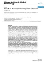
Figure 1 The effect of TSA (330 nM in C, G, J, K) on eosinophil apoptosis in the presence of IL-5 (0.3 ng/ml) as measured by relative
DNA fragmentation assay (A-D), Annexin V binding assay (E-H; Annexin V-FITC: FL1-H and propidium iodide: FL2-H)) and
morphological analysis (I-K). Figures in top right hand corner represent the percentage of eosinophils showing decreased relative DNA
content (A-C) or total percentage of apoptotic eosinophils (all Annexin V-FITC
+ve
cells) (E-G). In A-C, E-G and I-J a representative of 6 similar
experiments is shown. Mean ± SEM, n = 6 (D, H, K). ***P < 0.001 vs. solvent control in the presence of IL-5 and ### P < 0.001 vs. the control in
the absence of IL-5 and TSA.
Table 1 The EC
50
Values for the effects of trichostatin A
on apoptosis in eosinophils and neutrophils.
EC
50
(nM)
Apoptosis Eosinophils neutrophils P value
GM-CSF
0.01 ng/ml 79 ± 2
0.1 ng/ml 102 ± 1
10 ng/ml 93 ± 1 123 ± 9 0.0042
IL-5 92 ± 8
Constitutive 34 ± 10 97 ± 22 0.0007
Budesonide 32 ± 17 99 ± 7 0.026
Fluticasone 47 ± 15 100 ± 11 0.017
Mometasone 20 ± 5 87 ± 9 < 0.0001
Fas 31 ± 10
Values are the mean ± S.E.M. of six duplicate experiments with cells isolated
from different donors.
Kankaanranta et al. Journal of Inflammation 2010, 7:9
/>Page 4 of 15
number of eosinophils showing the typical morphologi-
cal features of apoptosis such as nuclear coalescense,
chromatin condensation and cell shrinkage was found
with TSA (Figure 1-K).
To evaluate whether the effect of TSA is specifically
related t o IL-5, we employed another eosinophil s urvi-
val-prolonging cytokine, i. e. GM-CSF. GM-CSF (0.01 -
10 ng/ml) promoted eosinophil survival in a concentra-
tion-dependent manner (Figure 2A). TSA (3.3-330 nM)
enhanced apoptosis in the presence of GM-CSF (0.01 -
10 ng/ml) (Figure 2A, Table 1).
Glucocorticoids are known to partially antagonize the
survival-prolonging action of IL-5 or GM-CSF on eosi-
nophils. However, this effect of glucocorticoids is abol-
ishedwhenthecytokineisusedathigher
concentrations [14,20-22]. For example, recently, we
reported that budesonide (1 μM) partly antagonizes
cytokine-afforded survival in the presence of low but
not in the presence of high concentrations of IL-5 [16].
The maximal response and the EC
50
values (Table 1) of
TSA were almost similar independently of the concen-
tration o f GM-CSF, suggesting that the cellular targets
of TSA are different from that of glucocorticoids.
To evaluate whether the ability to an tagonize cyto-
kine-afforded eosinophil survival is not related to TSA
only, we employed other pharmacological inhibitors of
HDACs. Another general HDAC inhibitor, apicidin
(0.1 - 10 μM) antagonized GM-CSF-mediated eosino-
phil survival by inducing apoptosis with an EC
50
of
427 ± 42 nM (Figure 2B). MC-1293, a commercially
available HDAC1 inhibitor, antagonized GM-CSF-
mediated eosinophil survival only partially at high (10
μM) drug concentrations (Figure 2C). Another HDAC
inhibitor, MS-275 (0.1-1 μM), at concentrations known
to inhibit HDAC1 [23] did not affect GM-CSF-afforded
eosinophil survival. In contrast, at higher concentra-
tions (10-100 μM) known to inhibit HDAC3 [ 23], MS-
275 enhanced apoptosis in GM-CSF-treated eosino-
phils (Figure 2D).
HDAC inhibitors enhance constitutive eosinophil
apoptosis
In the absence of life -supporting cytokines, TSA
increased the number of cells showing decreased relative
DNA content suggesti ng apoptosis (Figure 2A, Table 1).
Similarly, an increase in the number of cells presenting
with the typical morphological features of apopt osis was
found with TSA (percentage of apoptotic cells 11 ± 3
and 62 ± 8 in the absence and presence of 330 nM
TSA, respectively, n = 5, P < 0.001). This was confirmed
by showing an increase in the percentage of Annexin-V-
positive cells in the absence and presence o f TSA (330
nM)(15±3%and68±8%,respectively,n=6,P<
0.001).
Apicidin enhanced spontaneous eosinophil apoptosis
(Figure 3A). The selective HDAC1 inhibitor, MC1293,
did no t enhance eosinophil apoptosis (Figure 3B). MS-
275 (0.1-1 μM) inhibited constitutive eosinophil apopto-
sis slightly, but at higher concentrations (10-100 μM),
known to inhibit HDAC3 [23], MS-275 enhanced con-
stitutive eosinophil apoptosis (Figure 3C).
HDAC inhibitors have additive effect on glucocorticoid-
induced eosinophil apoptosis
Glucocorticoids increase apoptosis of human eosinophils
at clinically relevant drug concentrations [3,14,20].
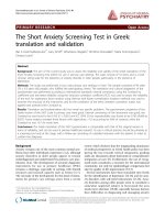
Figure 2 The effect of HDAC inhibitors Trichostatin A (TSA; A), apicidin (B), MC1293 (C) and MS-275 (D) on eosinophil apoptosis in the
presence of GM-CSF (in B-D: 0.1 ng/ml). In (A) the black colums indicate the effect of TSA in the absence of GM-CSF. Apoptosis was assessed
by flow cytometry measuring the relative DNA fragmentation. *P < 0.05, **P < 0.01 and ***P < 0.001 as compared with the respective control.
Mean ± S.E.M., n = 5-6.
Kankaanranta et al. Journal of Inflammation 2010, 7:9
/>Page 5 of 15
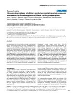
Figure 3 The effect of HDAC inhibitors apicidin (A), MC1293
(B) and MS-275 (C) on apoptosis in eosinophils in the absence
of survival-prolonging cytokines (ie. spontaneous apoptosis).
Apoptosis was assessed by flow cytometry measuring the relative
DNA fragmentation in propidium iodide-stained cells. **P < 0.01
and ***P < 0.001 as compared with the respective control in the
absence of HDAC inhibitors. Mean ± S.E.M. of 5-6 independent
determinations using cells from different donors.
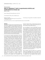
Figure 4 The effect of trichostatin A (A-C) on human
eosinophil apoptosis in the presence of budesonide (1 μM; A),
fluticasone (1 μM; B) or mometasone (1 μM; C). In (D-F) is
shown the effects of HDAC inhibitors apicidin (D), MC1293 (E) and
MS-275 (F) on eosinophil apoptosis in the presence of budesonide
(1 μM). Apoptosis was assessed by flow cytometry measuring the
relative DNA fragmentation in propidium iodide-stained cells. **
indicates P < 0.01 and *** P < 0.001 as compared with the
respective control in the absence of HDAC inhibitors. Mean ± S.E.M.
of 5-6 independent determinations using cells from different
donors. The corresponding percentage of apoptotic cells in the
absence of glucocorticoids and HDAC-inhibitors was 49 ± 3 (n =
25).
Kankaanranta et al. Journal of Inflammation 2010, 7:9
/>Page 6 of 15
Budesonide, f luticasone and mometasone (all at 1 μM)
enhanced constitutive eosinophil apoptosis (Figure 4A-C
and figure legend). A general HDAC inhibitor, TSA
(3.3-330 nM), had an additive effect in t he presence of
glucocorticoids (Figure 4A-C) on eosinophil apoptosis.
The EC
50
values of TSA for the enhancement of eosino-
phil apoptosis in the presence of glucocorticoids ranged
from 20 ± 5 nM to 47 ± 15 nM (Table 1). The additive
effect of TSA (3.3-330 nM) on budesonide-induced eosi-
nophil apoptosis was confirmed by using morphological
analysis and Annexin-V binding assay ( n = 5-6, P <
0.05; data not shown).
Apicidin (1 nM-10 μM) also had an additive effect on
budesonide-induced eosinophil apoptosis (Figure 4D). In
contrast, MC-1293 (1 nM-10 μM, Figure 4E) failed to
enhance budesonide-enhance d eosinophil apoptosis.
MS-275 at higher concentrations (10-100 μM) had an
additive effect on budesonide-induced eosinophil apop-
tosis (Figure 4F).
HDAC-inhibitors have an additive effect on Fas-induced
eosinophil apoptosis
Activation of Fas enhanced c onstitutive apoptosis of
eosinophils (percentage of apoptot ic cells 47 ± 4 and 65
± 2 in the absence and presence of 100 ng/ml activating
CD95 monoclonal antibody, respective ly, n = 6, P <
0.01). TSA (3.3-330 nM) had an additive effect on Fas-
induced eosinophils apoptosis (Table 1 and Table 2).
This was confirmed by measuring the percentage of
Annexin-V-positive cells in the absence and presence of
TSA (330 nM) (36 ± 6% vs 74 ± 8%, n = 6, P < 0.001).
Furthermore, an increase in the number of eosinophils
showing the typical morphological features of apoptosis
was found with TSA (percentage of apoptotic cells 26 ±
7 and 78 ± 7 in the absence and presence of 330 nM
TSA, respectively, n = 6, P < 0.001).
Effect of HDAC inhibitors on neutrophil apoptosis
Neutrophils rapidly undergo apoptosis when cultured in
the absence of survival-prolonging factors. GM-CSF
inhibited constitutive apoptosis in neutrophils (percen-
tage of apoptotic cells 60 ± 5 and 34 ± 4 in the absence
and pre sence of 10 ng/ml GM-CSF, respectively, n = 6,
P < 0.001). TSA (3.3-330 nM) antagonized the the survi-
val promoting action of GM-CSF (Figure 5A) with an
EC
50
of 123 ± 9 nM. The enhancement of neutrophil
apoptosis by TSA in the presence of GM-CSF was con-
firmed by annexin-V binding analysis (47 ± 5% vs 60 ±
8%,n=4,P<0.05).TSAalsoenhancedspontaneous
neutrophil apoptosis 1.5-fold (Figure 5B).
In contrast to t he enhancing effect on eosinphil apop-
tosis, glucocorticoids inhibit apoptosis in human neutro-
phils [13,14,24]. For example, budesonide inhibited
neutrophil apoptosis, the percentages of apoptotic cells
were 60 ± 5 and 42 ± 5 in the absence and presence of
budesonide (1 μM), respectively (n = 6, P < 0.001, Fig-
ure 5C). TSA (3.3-330 nM) antagonize d the inhibito ry
effect of budesonide (Figure 5C) on neutrophil apopto-
sis. This was confirmed by Annexin-V binding analysis
(55 ± 4% vs 91 ± 1% Annexin V-positive cells, n = 6, P
< 0.001). Furthermore, TSA antagonized fluticasone-
(Figure 5D) and mometasone- (Figure 5E)-induced sur-
vival of neutrophils by inducing apoptosis. The EC
50
values of TSA for antagonizing glucocorticoid-afforded
survival in neutrophils were not different between the
glucocorticoids (Table 1).
Pharmacological nature of the effect of HDAC inhibitors
To further evaluate whether the effects of HDAC inhibi-
tors on eosinophil and neutrophil apoptosis in the pre-
sence of glucocorticoids or Fas are additive or
synergistic, dose-response curves of TSA in the absence
or presence of survival-prolonging cytokines, glucocorti-
coids and Fas are compared (Figure 6A and 6B). In eosi-
nophils, the maximal percentage of apoptotic cells is
similar in the presence of TSA (330 nM) alone and in
thepresenceofbudesonideandTSA(330nM)(Figure
6A). This indicates that the effect is additive, but not
synergistic. The same can be seen with the combination
of TSA and Fas. Similarly, in neutrophils, the maximal
percentage of apoptotic cells is similar in the presence
ofTSA(330nM)aloneandinthepresenceofFasand
TSA (330 nM) (Figure 6B). In neutrophils, TSA
enhanced apoptosis in the presence of GM-CSF and
budesonide in a similar manner within the same con-
centration range ( Figure 6B). Similarly, in eosinophils
TSA enhanced apoptosis in the presence of IL-5 (Figure
6A). This suggests that the antagonism of the actions of
survival-prolonging cytokines IL-5 and GM-CSF in both
cell types and the antagonism of the actions of
Table 2 The effects of trichostatin A on Fas-induced
eosinophil apoptosis.
Percentage of apoptotic cells
Control 47 ± 4
Fas 65 ± 2##
Fas +trichostatin A 3.3 nM 67 ± 3
Fas +trichostatin A 33 nM 79 ± 2***
Fas +trichostatin A 330 nM 89 ± 1***
Shown is the percentage of apoptotic cells after 40 h incubation as analyzed
by relative DNA fragmentation assay
Values are the mean ± SEM of independent determinations using cells from
different donors. n = 6. *** indicates P < 0.001 as compared with the
respective solvent control in the absence of trichostatin A and ## P < 0.01 as
compared with the respective control in the absence of Fas. The
concentration of Fas activating antibody was 100 ng/ml.
Kankaanranta et al. Journal of Inflammation 2010, 7:9
/>Page 7 of 15
glucocorticoids does not occur at the level of IL-5, GM-
CSF or glucocorticoid receptors.
HDAC expression in human eosinophils and neutrophils
To evaluate whether granulocytes express HDACs, we
isolated mRNA from human eosinophils and neutrophils
and measured the expression of different HDACs using
real-time PCR. To confirm the accuracy of the results,
the expression of different HDACs was normalized
against two different housekeeping genes, namely
GAPDH and GLB2L1. This analysis gave almost identi-
cal results. Expression of HDAC5, 9 and 11 was very
low in eosinophils and expression of HDAC5, 8 and 11
was very low in neutrophils (Figure 7). The expression
of HDAC2 and HDAC9 was higher in neutrophils than
in eosinophils and the expression of HDAC8 was signifi-
cantly higher in eosinophils (Figure 7).
HDAC activity in eosinophils and neutrophils
The HDAC activity in eosinophil nuclear extracts was
somewhat higher (0.37 ± 0.05 OD/mg/min; n = 6) than
in neutrophil nuclear extracts (0.22 ± 0.05 OD/m g/min;
n = 5, P < 0.05). For comparison, we included HeLa-
cell nuclear extracts which had clea rly higher HDAC
activity (0.70 ± 0.04 OD/mg/min, n = 6, P < 0.001 ver-
sus eosinophil and neutrophil nuclear extracts). TSA
inhibited substrate (1.25 mM) deacetylation by eosino-
phil and neutrophil nuclear extracts only partially. The
maximal inhibition of HDAC activ ity by TSA (1000
nM) in eosinophil nucle ar extracts was 59 ± 13% (n =
6, P < 0.05) and in neutrophil nuclear extracts it was 50
± 4% (n = 5, P < 0.001), whereas in HeLa nuclear
extracts HDAC activity was inhibited almost completely
(93 ± 1% inhibition, n = 6, P < 0.001) by 1000 nM TSA
(Figure 8).
Acetylation of NF-B p65 does not explain the apoptosis-
inducing effect of TSA in human eosinophils
The above data suggest that the effects of HDAC inhibi-
tors in eosinophils or neutrophils may not be mediated
via regulation of acetylation status of histones, but
rather might be mediated via so me non-histone targets.
NF-B has been shown to be involved in the regulation
of eosinophil apoptosis [3]. NF-B assembly with IB, as
Figure 5 The effect of Trichostatin A on apoptosis in human neutrophils in the presence (A) or absence (B) of the survival-prolonging
cytokine GM-CSF (10 ng/ml). In (C-E) is shown the effect of trichostatin A on human neutrophil apoptosis in the presence of budesonide (1
μM; C), fluticasone (1 μM; D) or mometasone (1 μM; E). Apoptosis was assessed by flow cytometry measuring the relative DNA fragmentation
assay. *** P < 0.001 as compared with the respective control in the absence of HDAC inhibitors. ### P < 0.001 as compared with the respective
control in the absence of HDAC inhibitors and GM-CSF or glucocorticoids. Mean ± S.E.M. of 6 independent determinations using cells from
different donors.
Figure 6 Concentration-response curves of TSA in eosinophils
(A) and neutrophils (B) in the absence (black circle) and
presence of survival-prolonging cytokines (black up-pointing
triangle; IL-5 0.3 ng/ml in eosinophils and GM-CSF 10 ng/ml in
neutrophils), budesonide (black down-pointing triangle; 1 μM)
or Fas (black square; 100 ng/ml). Apoptosis was assessed by flow
cytometry measuring the relative DNA fragmentation (A) or Annexin
V-binding (B). Eosinophils or neutrophils were isolated and
concentration-response curves in the absence or presence of
cytokines, budesonide or Fas were prepared simultaneously from the
cells of the same donor. Mean ± S.E.M. of 6 independent
determinations using cells from different donors.
Kankaanranta et al. Journal of Inflammation 2010, 7:9
/>Page 8 of 15
well as its DNA binding and transcriptional activ ity, are
regulated by p300/CBP acetyltransferases that principally
target Lys218, Lys221 and Lys310 [25-27]. This process
is reciprocally regulated by HDACs and several HDAC
inhibitors have bee n shown to activate NF-B[25-27].
To evaluate whether the effects of HDAC inhibitors
could be mediated via acetylation of a non-histone tar-
get such as NF-B, we evaluated the effect of TSA on
the acetylation status of NF-B p65. However, TSA (330
nM) did not enhance acetyl-p65 expression in human
eosinophils either in the absence (n = 5; Figure 9) or
presence of GM-CSF (n = 2) (data not shown).
Effect of c-jun-N-terminal kinase and PI3K-Akt pathway
inhibitors on TSA-induced apoptosis in human
eosinophils
c-jun-N-terminal kinase (JNK) and PI3K-Akt pathways
have been proposed to be involved in the modulation of
human eosinophil longevity [3,28,29]. To test the invol-
vement of these pathways in HDAC-inhibitor-induced
apoptosis, we employd pharmacological inhibitors of
JNK and PI3K. Inhibition of JNK activity by the cell
permeable inhibitory peptide L-JNKI1 almost completely
abolished TSA (330 nM)-enha nced DNA breakdo wn. In
contrast, the negative control peptide L-TAT had no
effect (Figure 10).
Inhibition of PI3K-Akt pathway by two chemically dis-
tinct inhibitors, namely wortmannin (10-100 nM) and
LY294002 (5-50 μM) did not affect TSA-induced apop-
tosis in human eosinophils (n = 6, data not shown).
Involvement of caspases in TSA-induced apoptosis in
human eosinophils
Even though the involvement of caspases in apoptosis in
general is well established, surprisingly little is known of
the role caspases in human eosinophils [3,30] and the
actual caspases mediating apoptosis in human eosino-
phils remain largely unknown [3,30]. Gene ral caspase
inhibitors Q-Vd-OPh and Z-Asp-CH2-DCB completely
antagonized the effect of TSA on apoptosis in human
eosinophils (Figure 11). Inhibitors of caspase 6 (Z-VE
(OMe)ID(OMe)-FMK) and 3 (Z-D(OMe)QMD(OMe)-
FMK) compeletely and partly antagonized TSA-induced
DNA breakdown in human eosinophils, respectively
Figure 7 The expression of histone deacetylases (HDAC) 1-11 in human eosinophils (black circle; E) and neutrophils (black up-pointing
triangle; N). HDAC mRNA levels were normalized against GLB2L1 mRNA. Total mRNA from eosinophils (n = 4) and neutrophils (n = 5) was
extracted and subjected to RT-PCR. *P < 0.05 for the difference between eosinophils and neutrophils.
Kankaanranta et al. Journal of Inflammation 2010, 7:9
/>Page 9 of 15
Figure 8 The effect of HDAC inhibitor Trichostatin A (TSA) on HDAC activity in nuclear ext racts isolated from human eosinophils (n =
6) and neutrophils (n = 5). For comparison is shown the effect of TSA on HDAC activity in HeLa nuclear extracts (n = 6). Nuclear extracts were
prepared and HDAC activity was measured as described in materials and methods. HDAC activity in the absence of TSA was set as 100%. *P <
0.05 and *** P < 0.001 as compared with the respective control in the absence of TSA. Mean ± S.E.M.
Figure 9 The effect of TSA (330 nM) on the expression of
acetyl-NF-kB p65 (Lys310). Human eosinophils were treated with
solvent or TSA for 1 h and immunoblots were run using antibodies
against acetyl-NF-kB p65 and total NF-kB p65. The
chemiluminescent signal was quantified as described under
Materials and Methods. Acetyl-NF-kB p65 values were normalized to
NF-kB p65 values and the value in the absence of TSA was set as
100%. Results are expressed as mean ± S.E.M., n = 5.
Figure 10 The effect of the c-jun-N-terminal kinase inhibitor L-
JNKI1 (10 μM) on TSA (330 nM)-induced human eosinophil
apoptosis. Apoptosis was assessed by the relative DNA
fragmentation assay. Each data point represents the mean ± SEM of
6 independent determinations using eosinophils from different
donors. *** indicates p < 0.001 as compared with the respective
control (10 μM L-TAT or solvent).
Kankaanranta et al. Journal of Inflammation 2010, 7:9
/>Page 10 of 15
(Figure 11). In cont rast, inhibition of caspase 8 (IETD-
CHO) had no effect (Fig ure 11). These results suggest a
role for caspases 3 and 6, but not 8, in the mechanism
of action of TSA in human eosinophils.
HDAC inhibitors enhance apoptosis in J774 macrophages
Macrophages are considered to be important in the
removal of apoptotic cells. To evaluate whether HDAC
inhibitors could affect macrophage survival, we evalu-
ated the effects of TSA on apoptosis in J774.2 macro-
phages. TSA increased the percentage of Annexin V-
positive cells in J774.2 macrophages in a concentra tion-
dependent manner, although to a lesser extent than a
combination of LPS and an inhibitor of NF-BPDTC
(100 μM), previously known to induce apoptosis in
macrophages (Figure 12).
Discussion
In the present study we show that HDAC inhibitors
inhibit HDAC acitivity and induce apoptosis in human
eosinophils and neutrophils in the absence and presence
of survival-prolonging cytokines and g lucocorticoids.
Furthermo re, we report that eosinophils and neutrophils
express a different pattern of HDACs, namely the
expression of HDAC2 and HDAC9 is higher in neutro-
phils than in eosinophils and the expression of HDAC8
Figure 11 The effect of caspase inhibitors on TSA (330 nM)-induced human eosinophil apoptosis. The concentrations used were: Q-Vd-
Oph (20 μM), Z-Asp-CH2-DCB and IETD-CHO (100 μM) and Z-D(OMe)QMD(OMe)-FMK and Z-VE(OMe)ID(OMe)-FMK (200 μM). Apoptosis was
assessed by the relative DNA fragmentation assay. Each data point represents the mean ± SEM of 6-7 independent determinations using
eosinophils from different donors. * indicates p < 0.05 and *** p < 0.001 as compared with the respective control in the absence of caspase
inhibitors.
Figure 12 The effect of HDAC inhibitor Trichostatin A (TSA) on
apoptosis in J774.2 macrophages during culture for 24 h. For
comparison, is shown the effect of a known inducer of apoptosis in
J774 macrophages, i.e. combination of LPS (10 ng/ml) and PDTC
(100 μM). Apoptosis was assessed by flow cytometry measuring the
percentage of Annexin V-positive cells. *P < 0.05 and **P < 0.01 as
compared with the respective control. Mean ± S.E.M., n = 12.
Kankaanranta et al. Journal of Inflammation 2010, 7:9
/>Page 11 of 15
is higher in eosinophils than in neutrophils. T he
mechanism of apoptosis-enhancing action of HDAC
inhibitors in human eosinophils seems to involve JNK
and caspases 3 and 6.
HDAC inhibitors have been reported to cause apopto-
tic cell death in a variety of cultured transformed cells,
including human bladder, breast, prostate, lung, ovary
and colon cancers and acute myelogenous leukemia
[31]. For example, HDAC inhibitors such as apicidin,
sodium butyrate, suberoylanilide hydroxamic acid
(SAHA) and TSA have been reported to reduce viability
or induce apoptosis in HeLa cells [32,33]. In contrast,
normal cells are usually resistant to cell death caused by
HDAC inhibitors [9,10] and there is no previous data to
describe the effects of HDAC inhibitors on apoptosis in
human eosinophils or neutrophils. Supporting our
results on the possible anti-inflammatory effects of
HDAC inhibitors on granulocytes, recent in vivo data in
animals suggest that HDAC inhibitors may have poten-
tial to act as anti-inflammatory agents. Choi and cowor-
kers [12] demonstrated that TSA given prophylactically
blocked OVA-induced airway hyper-responsiveness, as
well as reduced the numbers of eosinophils in lavage
fluid [12]. Interestingly, HDAC inhibitors seem not to
block the production of eosinophil life-supporting cyto-
kines such as I L-5, but rather may enhance the activity
of IL-5 promoter [34]. Thus, it is tempting to speculate
that as HDAC inhibitors may not reduce the concentra-
tions of eosinophil survival-prolonging cytokines. The
finding that TSA enhances apoptosis in the presence of
IL-5- and GM-CSF, may, at least partly, explain the ben-
eficial effects of TSA in m odels of eosinophilic
inflammation.
Structurally distinct HDAC inhibitors were used.
Unfortunately, the inhibitory profiles of HDAC inhibi-
tors against all HDAC isoforms have not been thor-
oughly characterized. T SA has been reported to be a
general HDAC inhibitor [31,35-37]. HDAC1 selective
inhibitors, MC-1293 [38] and MS-275 at low concentra-
tions [23] did not affect eosinophil apoptosis to a similar
extent than TSA or apicidin. This probably excludes
HDAC1 as a target of HDAC inhibitors. However, given
that the effect of TSA in the HDAC activity assay
experiments using nuclea r extracts obtained from eosi-
nophils or neutrophils revealed that the HDAC activity
was reduced only by 50-60% even at 1 μMsuggests
either that granulocytes possess a TSA-insensitive
HDAC e.g. HDAC4 or 7 or that HDACs are not the
major target for HDAC inhibitors in these cells. The
EC
50
values for TSA in enhancing apoptosis in the pre-
sence or absence of glucocorticoids were different
between eosinophils and neutrop hils, whereas no differ-
ence was found in the EC
50
values for TSA in the pre-
sence of GM-CSF. This suggests that there may be two
or more HDACs responsible mediating these effects or
that the effect may reflect the combined effect of two or
more HDACs. The expression of HDAC2, HDAC8 and
HDAC9 were different between eosinophils and neutro-
phils. This suggests that one or more of these HDACs
may also be involved.
In malignant cell lines activation of caspase cascades
as well as changes in the expression of Bcl-2 family
members have been described [9,10]. The exact mechan-
isms how the survival-prolonging cytokines IL-5 and
GM-CSF induce eosinophil survival or glucocorticoids
induce eosinophil death are not known in detail
[3,22,30]. In fact, it is not even known whether gluco-
corticoid-induced apoptosis involves mainly transcrip-
tional activation or repression [39]. Mechanistically,
inhibition of HDAC activity should lead to increased
transcription. Treatment with HDAC inhibitors in an in
vitro situation leads almost u p to 10% of transcription-
ally active genes having altered expression [9]. Surpris-
ingly, nearly an equal number of genes are repressed in
their expression as those that are activated [9]. Treat-
ment with HDAC inhibitors in vitro causes an increase
in the acetylation levels of histones in both normal and
tumor cells, including melano cytes and melanoma cell
lines [9]. However, normal melanocytes are resistant to
cell death caused by HDAC inhibitors, whereas most
melanoma cell lines undergo apoptosis [9]. This suggests
that the difference between survival and death between
normal and malignant cells may be due to acetylation of
non-histone proteins rather than histones themselves
[9,10]. In eosinophils, NF-B has been shown to be
involved in the regulation of apoptosis [3]. NF-B
assembly with IB, as well as its DNA binding and tran-
scriptional activity, are regulated by p300/CBP acetyl-
transferases that principally target Lys218, Lys221 and
Lys310 [25-27]. This proc ess is reciprocally regulated by
HDACs and several HDAC inhibitors have been shown
to activate NF-B [25-27]. In fact, ineffectiveness of
HDAC inhibitors to induce apoptosis in certain cell
lines has been proposed to involve the transcriptional
activation by acetylation of RelA/p65 subunit of NF-kB
through the Akt pathway [26]. However, we were not
able to detect any increased acetylation of NF-kB p65 in
response to TSA in human eosinophils. Similarly, inhibi-
tion of the PI3K-Akt pathway by pharmacological inhi-
bitors did not modulate TSA-induced apoptosis. These
results suggest that NF-kB p65 or PI3K-Akt pathway are
not involved, but we cannot exclude other non-histone
targets.
c-jun-N-terminal kinase (JNK) pathway has been pro-
posed to be involved in spontaneous and nitric oxide-
and orazipone-induced apoptosis of human eosinophils
[3,16,17,28]. Inhibition of JNK activity by the cell perme-
able inhibitory peptide L-JNKI1 almost completely
Kankaanranta et al. Journal of Inflammation 2010, 7:9
/>Page 12 of 15
abolished TSA-enhanced DNA breakdown, suggesting a
role for JNK. E ven though the involvement of caspases
in apoptosis in general is well established, surprisingly
little is known of the role caspases in human eosinophils
[3,30] and the actual c aspases mediating apoptosis in
human eosinophils remain largely unknown [3,30]. Gen-
eral caspase inhibitors Q-Vd-OPh and Z-Asp-CH2-DCB
completely antagonized the effect of TSA on apoptosis
in human eosinophils similarly to inhibitors of caspases
6 an d 3, whereas inhibition of caspase 8 had no effect.
However, caspase inhibition also reduced spontaneous
apoptosis as previously described [16]. These results
suggest a role for JNK and caspa ses 3 and 6, but not 8,
in the mechanism of action of TSA in human eosino-
phils. This interpretation may be complicated by the
fact that the specificity of these inhibitors for caspases 3,
6 and 8 has not been completely characterized. How-
ever, neither JNK nor caspases 3 and 6 appear specific
for HDAC-inhibitor-induced apoptosis as they have
been reported to affect spontaneous or induced apopto-
sis in human eosinophils [3,16,17,28].
In contrast to the potentiation of glucocorticoid effects
in eosinophils, in neutrophils TS A antagonized the sur-
vival-prolonging eff ect of glucocorticoids on neutrophil
survival. In addition, the EC
50
value for TSA for antag-
onism of glucocorticoid-induced survival in neutrophils
was higher t han that in eosinophils for enhancement of
glucocorticoid-induced apoptosis. One might argue that
the effect of HDAC inhibitors is non-specific in that
they override the effects of any survival-prolonging fac-
tor in granulocytes.
Accumulation, activation and delayed death of neutro-
phils at the inflamed site has recently been implicated in
the pathogenesis of COPD, severe asthma and asthma
exacerbations [1]. We found that TSA antagonized GM-
CSF-afforded neutrophil survival by inducing apoptosis.
In addition, TSA enhanced apoptosis in the absence and
presence of glucocorticoids in neutrophils. We were not
able to identify any studies exploring the effects of TSA
on neutrophilic inflammation in the lung and based on
our results such studies are warranted.
HDACinhibitorsareuniqueinthesensethatthey
antagonize cytoki ne-afforde d survival of eosinophils and
neutrophils despite the vast amount of literature that
indicates that they are not toxic towards several types of
normal non-malignant cell lines [9,10]. In fact, the pub-
lished phase I-II clinical trials suggest that HDAC inhi-
bitors: 1. inhibit HDAC activity in vivo in humans and
2. show moderate to good tolerability in humans
[40-44]. Thus, it is tempting to speculate that HDAC
inhibitors might be used to treat also eosinophilic and/
or neutrophilic inflammation.
Macrophages are considered to be important in the
removal of apoptotic cells. The finding that TSA at
similar concentrations induced apoptosis also in a
macrophage cell-line suggests that removal of apoptotic
cells in the lungs could be impaired. However, in addi-
tion to macrophages, lung epithelial cells have been
implicated in the removal of apoptotic eosinophils [45]
and A54 9 lung epithelial cells have been reported to be
insensitive to apoptosis induced by HDAC inhibitors
[26].
Conclusions
Taken together, our results suggest that HDAC inhibi-
tors such as TSA enhance apoptosis both in the pre-
sence and absence of survival-prolonging cytokines in
eosinophils and neutrophils. In addition, TSA has an
additive effect on apoptosis in the presence of glucocor-
ticoids in eosinophils and antagonizes glucocorticoid-
induced neutrophil survival. The mechanism of action
in eosinophils involves c-jun-N-terminal kinase and cas-
pases 3 and 6. Thus, HDAC inhibitors have anti- eosino-
philic and anti-neutrophilic properties and are possible
drug candidates to treat eosinophil ic or neutrophili c
inflammation.
List of abbreviations
(COPD): Chronic obstructive pulmonary disease; (GM-
CSF): Granulocyte-macrophage colony-stimulating fac-
tor; (HAT): Histo ne acetyltransferase; (HDAC): Histone
deacetylase; (IL): Interleukin; (L-JNKI1):
GRKKRRQRRR-PP-RPKRPTTLNLFPQVPRSQD-amide;
(LPS): Lipopolysaccharid e; (L-TAT): RKKRRQRRR-
amide, negative control for L-JNKI1; (MC1293): 3-(4-
toluoyl-1-methyl-1H-2-pyrrolyl)-N-hydroxy-2-propena-
mide; ( MS-275): [N-(2-aminophenyl)-4-[N-(pyridine-3-
ylmethoxy-carbonyl)aminomethyl]benzamide]; (PDTC):
ammo nium pyrrolidinedithiocarbamate; (TSA): Trichos-
tatin A.
Acknowledgements
Supported by Tampere Tuberculosis Foundation (Finland), the Academy of
Finland, the Finnish Anti-Tuberculosis Association Foundation (Finland), and
the Medical Research Funds of Tampere University Hospital (Finland) and
Seinäjoki Central Hospital (Finland). The skilful technical help of Ms. Elina
Heiskanen is gratefully acknowledged.
Author details
1
The Immunopharmacology Research Group, Medical School, FIN-33014,
University of Tampere and Research Unit, Tampere University Hospital,
Tampere, Finland.
2
Department of Respiratory Medicine, Seinäjoki Central
Hospital, Seinäjoki, Finland.
3
Airway Disease, Imperial College School of
Medicine at the National Heart and Lung Institute, London, UK.
Authors’ contributions
HK, MJ-J, PI and XZ carried out the eosinophil and neutrophil isolation, flow
cytometric assays, morphological analyses, HDAC assays and western blot
analysis. UJ gave valuable collaboration in laboratory studies. KI and MI
performed the HDAC mRNA assays. KI, IMA and EM participated in the
design of the study and helped to draft the manuscript. HK conceived the
Kankaanranta et al. Journal of Inflammation 2010, 7:9
/>Page 13 of 15
study, and participated in its design and coordination and drafted the
manuscript with XZ. All authors read and approved the final manuscript.
Competing interests
The authors declare that they have no competing interests.
Received: 8 April 2009
Accepted: 4 February 2010 Published: 4 February 2010
References
1. Watt AP, Schock BC, Ennis M: Neutrophils and eosinophils: clinical
implications of their appearance, presence and disappearance in asthma
and COPD. Curr Drug Targets-Inflammation Allergy 2005, 4:415-423.
2. Walker A, Ward C, Taylor EL, Dransfield I, Hart SP, Haslett C, Rossi AG:
Regulation of neutrophil apoptosis and removal of apoptotic cells. Curr
Drug Targets-Inflammation Allergy 2005, 4:447-454.
3. Kankaanranta H, Moilanen E, Zhang X: Pharmacological regulation of
human eosinophil apoptosis. Curr Drug Targets-Inflammation Allergy 2005,
4:433-445.
4. Wedi B, Raap U, Lewrick H, Kapp A: Delayed eosinophil programmed cell
death in vitro: a common feature of inhalant allergy and extrinsic and
intrinsic atopic dermatitis. J Allergy Clin Immunol 1997, 100:536-43.
5. Kankaanranta H, Lindsay MA, Giembycz MA, Zhang X, Moilanen E,
Barnes PJ: Delayed eosinophil apoptosis in asthma. J Allergy Clin Immunol
2000, 106:77-83.
6. Vignola AM, Chanez P, Chiappara G, Siena L, Merendino A, Reina C:
Evaluation of apoptosis of eosinophils, macrophages, and T lymphocytes
in mucosal biopsy specimens of patients with asthma and chronic
bronchitis. J Allergy Clin Immunol 1999, 103:563-73.
7. Adcock IM: Histone deacetylase inhibitors as novel anti-inflammatory
agents. Curr Opin Invest Drugs 2006, 7:966-73.
8. Barnes PJ: How corticosteroids control inflammation: Quintiles Prize
Lecture 2005. Br J Pharmacol 2006, 148:245-54.
9. Boyle GM, Martyn AC, Parsons PG: Histone deacetylase inhibitors and
malignant melanoma. Pigment Cell Res 2005, 18:160-6.
10. Dokmanovic M, Marks PA: Prospects: histone deacetylase inhibitors. J Cell
Biochem 2005, 96:293-304.
11. Chung YL, Lee MY, Wang AJ, Yao LF: A therapeutic strategy uses histone
deacetylase inhibitors to modulate the expression of genes involved in
the pathogenesis of rheumatoid arthritis. Mol Ther 2003, 8:707-17.
12. Choi JH, Oh SW, Kang MS, Kwon HJ, Oh GT, Kim DY: Trichostatin A
attenuates airway inflammation in mouse asthma model. Clin Exp Allergy
2005, 35:89-96.
13. Zhang X, Moilanen E, Kankaanranta H: Beclomethasone, budesonide and
fluticasone propionate inhibit human neutrophil apoptosis. Eur J
Pharmacol 2001, 431:365-371.
14. Zhang X, Moilanen E, Adcock IM, Lindsay MA, Kankaanranta H: Divergent
effect of mometasone on human eosinophil and neutrophil apoptosis.
Life Sci 2002, 71:1523-1534.
15. Kankaanranta H, De Souza PM, Barnes PJ, Salmon M, Giembycz MA,
Lindsay MA: SB 203580, an inhibitor of p38 mitogen-activated protein
kinase, enhances constitutive apoptosis of cytokine-deprived human
eosinophils. J Pharm Exp Ther 1999, 290:621-628.
16. Kankaanranta H, Ilmarinen P, Zhang X, Nissinen E, Moilanen E:
Antieosinophilic activity of orazipone. Mol Pharmacol 2006, 96:1861-70.
17. Zhang X, Moilanen E, Lahti A, Hämäläinen M, Giembycz MA, Barnes PJ,
Lindsay MA, Kankaanranta H: Regulation of eosinophil apoptosis by nitric
oxide: Role of c-Jun-N-terminal kinase and signal transducer and
activator of transcription 5. J Allergy Clin Immunol 2003, 112:93-101.
18. Dignam JD, Lebovitz RM, Roeder RG: Accurate transcription initiation by
RNA polymerase II in a soluble extract from isolated mammalian nuclei.
Nucleic Acids Res 1983, 11:1475-1489.
19. Lahti A, Jalonen U, Kankaanranta H, Moilanen E: c-Jun NH
2
-Terminal Kinase
Inhibitor anthra(1,9-cd)pyrazol-6(2H)-one Reduces Inducible Nitric-Oxide
Synthase Expression by Destabilizing mRNA in Activated Macrophages.
Mol Pharmacol 2003, 64:308-315.
20. Zhang X, Moilanen E, Kankaanranta H: Enhancement of human eosinophil
apoptosis by fluticasone propionate, budesonide, and beclomethasone.
Eur J Pharmacol 2000, 406:325-332.
21. Hagan JB, Kita H, Gleich GJ: Inhibition of interleukin-5 mediated
eosinophil viability by fluticasone 17-propionate: comparison with other
glucocorticoids. Clin Exp Allergy 1998, 28:999-1006.
22. Druilhe A, Letuve S, Pretolani M: Glucocorticoid-induced apoptosis in
human eosinophils: mechanisms of action. Apoptosis 2003, 8:481-495.
23. Hu E, Dul E, Sung C-M, Chen Z, Kirkpatrick R, Zhang G-F, Johanson K, Liu R,
Lago A, Hofmann G, Macarron R, de los Frailes M, Perez P, Krawiec J,
Winkler J, Jaye M: Identification of novel isoform-selective inhibitors
within class I histone deacetylases. J Pharm Exp Ther 2003, 307:720-728.
24. Meagher LC, Cousin JM, Seckl JR, Haslett C: Opposing effects of
glucocorticoids on the rate of apoptosis in neutrophilic and eosinophilic
granulocytes. J Immunol 1996, 156:4422-4428.
25. Ashburner BP, Westerheide SD, Baldwin AS: The p65 (RelA) subunit of NF-
kB interacts with the histone deacetylase (HDAC) corepressors HDAC1
and HDAC2 to negatively regulate gene expression. Mol Cell Biol 2001,
21:7065-7077.
26. Mayo MW, Denlinger CE, Broad RM, Yeung F, Reilly ET, Shi Y, Jones DR:
Ineffectiveness of histone deacetylase inhibitors to induce apoptosis
involves the transcriptional activation of NF-kB through the Akt
pathway. J Biol Chem 2003, 278:18980-18989.
27. Chen LF, Mu Y, Greene WC: Acetylation of RelA at discrete sites regulates
distinct nuclear functions of NF-kB. EMBO J 2002, 21:6539-6548.
28. Hasala H, Zhang X, Saarelainen S, Moilanen E, Kankaanranta H: c-jun-N-
terminal kinase mediates constitutive human eosinophil apoptosis. Pulm
Pharmacol Ther 2007, 20:580-587.
29. Machida K, Inoue H, Matsumoto K, Tsuda M, Fukuyama S, Koto H, Aizawa H,
Kureishi Y, Hara N, Nakanishi Y: Activation of PI3K-Akt pathway mediates
antiapoptotic effects of b-adrenergic agonist in airway eosinophils. Am J
Physiol Lung Cell Mol Physiol 2005, 288:L860-L867.
30. Simon HU: Cell death in allergic diseases. Apoptosis 2009, 14:439-446.
31. Marks PA, Richon VM, Rifkind RA: Histone deacetylase inhibitors: inducers
of differentiation or apoptosis of transformed cells. J Natl Cancer Inst
2000, 92:1210-1216.
32. Shao Y, Gao Z, Marks PA, Jiang X: Apoptotic and autophagic cell death
induced by histone deacetylase inhibitors. Proc Natl Acad Sci USA 2004,
101:18030-18035.
33. Ishihara K, Hong J, Zee O, Ohuchi K: Possible mechanism of action of the
histone deacetylase inhibitors for the induction of differentiation of HL-
60 clone 15 cells into eosinophils. Br J Pharmacol 2004, 142:1020-1030.
34. Han S, Lu J, Zhang Y, Cheng C, Li L, Han L, Huang B: HDAC inhibitors TSA
and sodium butyrate enhanced the human IL-5 expression by altering
histone acetylation status at its promoter region. Immunol Lett 2007,
108:143-50.
35. Furumai R, Komatsu Y, Nishino N, Khochbin S, Yoshida M, Horinouchi S:
Potent histone deacetylase inhibitors built from trichostatin A and cyclic
tetrapeptide antibiotics including trapoxin. Proc Natl Acad Sci USA 2001,
98:87-92.
36. Grozinger CM, Hassig CA, Schreiber SL: Three proteins define a class of
human histone deacetylases related to yeast Hda1p. Proc Natl Acad Sci
USA 1999, 96:4868-4873.
37. Tong JJ, Liu J, Bertos NR, Yang X-J: Identification of HDAC10, a novel class
II human histone deacetylase containing a leucine-rich domain. Nucleic
Acids Res 2002, 30:1114-1123.
38. Mai A, Massa S, Ragno R, Esposito M, Sbardella G, Nocca G, Scatena R,
Jesacher F, Loidl P, Brosch G: Binding mode analysis of 3-(4-benzoyl-1-
methyl-1H-2-pyrrolyl)-N-hydroxy-2-propenamide: a new synthetic
histone deacetylase inhibitor inducing histone hyperacetylation, growth
inhibition, and terminal cell differentiation [abstract]. J Med Chem 2002,
45:1778.
39. Janka-Junttila M, Moilanen E, Hasala H, Zhang X, Adcock I, Kankaanranta H:
The glucocorticoid RU24858 does not distinguish between
transrepression and transactivation in primary human eosinophils. J
Inflammation 2006, 3:10.
40. Kuendgen A, Knipp S, Fox F, Strupp C, Hildebrandt B, Steidl C, Germing U,
Haas R, Gattermann N: Results of a phase 2 study of valproic acid alone
or in combination with all-trans retinoic acid in 75 patients with
myelodysplastic syndrome and relapsed or refractory acute myeloid
leukaemia. Ann Hematol 2005, 84(suppl 1):61-66.
41. Hauschild A, Trefzer U, Garbe C, Kaehler K, Ugurel S, Kiecker F, Eigentler T,
Krissel H, Schadendorf D: A phase II multicenter study on the histone
Kankaanranta et al. Journal of Inflammation 2010, 7:9
/>Page 14 of 15
deacetylase (HDAC) inhibitor MS-275, comparing two dosage schedules
[abstract]. J Clin Oncol 2006, 24(suppl):8044.
42. Gojo I, Jiemjit A, Trepel JB, Sparreboom A, Figg WD, Rollins S, Tidwell ML,
Greer J, Chung EJ, Gore SD, Sausville EA, Zwiebel J, Karp JE: Phase 1 and
pharmacological study of MS-275, a histone deacetylase inhibitor, in
adults with refractory and relapsed acute leukemias. Blood 2007,
109:2781-2790.
43. Garcia-Manero G, Assouline S, Cortes J, Kantarjian H, Yang H,
Newsome WM, Miller WH Jr, Rousseau C, Kalita A, Liu J, Dubay M,
Patterson TA, Li Z, Besterman JM, Reid G, Laille E, Martell RE, Minden MD:
Phase I study of the oral isotype specific histone deacetylase inhibitor
MGCD0103 in leukemia. Blood 2008, 112:981-989.
44. Siu LL, Pili R, Duran I, Messersmith WA, Chen EX, Sullivan R, MacLean M,
King S, Brown S, Reid GK, Li Z, Kalita AM, Laille EJ, Besterman JM, Martell RE,
Carducci MA: Phase I study of MGCD0103 given as three-times-per-week
oral dose in patients with advanced solid tumors. J Clin Oncol 2008,
26:1940-1947.
45. Walsh GM, Sexton DW, Blaylock MG: Mechanism of steroid action and
resistance in inflammation: Corticoisteroids, eosinophils and bronchial
epithelial cells: new insights into the resolution of inflammation in
asthma. J Endocrinol 2003, 178:37-43.
doi:10.1186/1476-9255-7-9
Cite this article as: Kankaanranta et al.: Histone deacetylase inhibitors
induce apoptosis in human eosinophils and neutrophils. Journal of
Inflammation 2010 7:9.
Submit your next manuscript to BioMed Central
and take full advantage of:
• Convenient online submission
• Thorough peer review
• No space constraints or color figure charges
• Immediate publication on acceptance
• Inclusion in PubMed, CAS, Scopus and Google Scholar
• Research which is freely available for redistribution
Submit your manuscript at
www.biomedcentral.com/submit
Kankaanranta et al. Journal of Inflammation 2010, 7:9
/>Page 15 of 15