Geochemical and Hydrological Reactivity of Heavy Metals in Soils - Chapter 11 ppt
Bạn đang xem bản rút gọn của tài liệu. Xem và tải ngay bản đầy đủ của tài liệu tại đây (822.19 KB, 39 trang )
11
Analytical Techniques for
Characterizing Complex
Mineral Assemblages:
Mobile Soil and
Groundwater Colloids
John C. Seaman, M. Guerin, B.P. Jackson,
P.M. Bertsch, and J.F. Ranville
CONTENTS
11.1 Introduction
11.2 Light-Scattering Techniques for Colloid Characterization
11.2.1 Turbidimetric Methods
11.2.2 Dynamic Light Scattering
11.2.3 Laser Doppler Velocimetry and Particle Charge
11.3 Acoustic Spectroscopy
11.3.1 Acoustic Attenuation and Particle Sizing
11.3.2 Electroacoustics
11.4 Field Flow Fractionation
11.4.1 Sedimentation (Sd-FFF) and Flow-Field Flow
Fractionation (Fl-FFF)
11.4.2 FFF Applications
11.5 Electron-Based Analysis Techniques
11.5.1 Scanning Electron Microscopy
11.5.2 Automated SEM Techniques: Removing Instrument and
Operator Biases
11.5.3 Transmission Electron Microscopy
11.6 Other Analytical Methods
11.7 Conclusions
11.8 Acknowledgments
11.9 List of Symbols
11.10 List of Greek Symbols
References
© 2003 by CRC Press LLC
11.1 INTRODUCTION
Surface chemical reactions play a major role in controlling contaminant fate and
transport in the environment. To better understand such processes, one often resorts
to well-defined laboratory studies using mineral and organic standards or synthetic
analogs as surrogates for the more complicated natural systems, either focusing on
homogeneous systems or assuming the additivity of the major system components.
In reality, such mixtures may display changes in particle size, surface area, and
reactivity that differ from the individual surrogate components or the natural diage-
netic environment that the investigator wishes to emulate.
1–5
For example, natural
colloids observed in the electron microscope often appear irregularly eroded or
coated with other mineral or organic phases and rarely resemble synthetic or pure
mineral particles.
6,7
Complex mixtures and the presence of “surface coatings” or
surface heterogeneities, often representing only a small fraction of the total suspen-
sion or matrix composition, can alter the reactivity of the more abundant components
in ways that are difficult to quantify or predict based on the idealized systems.
8–11
Even common lab practices, such as homogenization and air-drying of soil materials
can alter surface reactivity more than generally recognized.
12–14
In recent years the study of mobile soil and groundwater colloids has received
considerable attention because of concerns that such a vector may enhance the
mobility of strongly sorbing contaminants, a process that is often referred to as
“facilitated transport.”
15,16
However, our ability to predict colloid movement and
deposition is often confounded by the complexities of surface interactions in such
dynamic, unstable systems. The lack of universally accepted analytical techniques
and failure to realize instrumental limitations have made it difficult to compare and
critically evaluate the results of different studies. Artifacts associated with ground-
water sampling, filtration, and storage, and the dilute nature of most soil and ground-
water suspensions further hamper characterization efforts.
17–21
Not surprisingly, elevated concentrations of mobile groundwater colloids are
generally associated with a disruption in the native hydrogeochemical environ-
ment, including those induced by artificial recharge, groundwater contamination,
and even elevated flow rates associated with conventional sampling practices.
22,23
When precautions are taken to ensure that groundwater samples are representative
of actual geohydrologic conditions within an aquifer, background or control wells
outside the influence of the contamination source generally yield few mobile
colloids.
24,25
However, artifactual colloids can be introduced during well construc-
tion or development (drilling fluids, bentonite, etc.),
26
result from changes in
chemistry or redox due to inadequate sample preservation,
22,24,25
or become sus-
pended from the immobile matrix by the shear forces associated with pump-
ing.
17,22,23
Aggregation after sampling and membrane clogging can increase the
efficiency of phase separation and reduce the average size and percentage of total
suspended solids passing through the filter
18,19
; thus, the relative percentage of
colloid-associated metals in filtered samples may not vary systematically with
turbidity, that is, with colloid mass or concentration. In addition, larger size
particles may contribute much of the colloidal mass, but reflect a smaller portion
of the surface area available for contaminant sorption.
20
L1623_FrameBook.book Page 272 Thursday, February 20, 2003 9:36 AM
© 2003 by CRC Press LLC
Common colloidal materials found in subsurface environments include phyllo-
silicate clays, Al, Fe, and other metal oxyhydroxides, CaCO
3
, microorganisms, and
other biological debris. Field and laboratory studies have identified several mecha-
nisms by which such materials can be mobilized in the environment: (1) clay
dispersion due to changes in groundwater pH, ionic strength, and/or Na
+
/Ca
2+
ratios
27–32
; (2) manipulation of surface charge using a chemical dispersing agent
33,34
;
(3) dissolution of carbonate or Fe-cementing agents resulting in the release and
transport of silicate clays
35–38
; and (4) precipitation of colloidal particulates resulting
from a change in groundwater chemistry.
24
In some instances, more than one of
these mechanisms may be operative,
38
but essential in the development of such
hypotheses is a thorough characterization of the composition and chemical nature
of the colloidal suspension, including the inherent associations between various
colloidal components, and their reactivity with respect to aggregation/filtration pro-
cesses, as well as contaminant sorption properties.
Bulk quantification and characterization techniques, such as turbidity and
chemical digestion/extraction methods, and certain commonly used instrumental
characterization techniques [i.e., photon correlation spectroscopy (PCS), x-ray
diffraction, electron microscopy, etc.] are extremely sensitive to the presence of
artifactual colloids that are not inherently mobile within the soil or aquifer. Small
sample sizes and the presence of organics and poorly ordered mineral phases can
confound identification by x-ray diffraction, the primary method used by many in
identifying clay minerals. Furthermore, discrete particle analysis techniques have
confirmed that contaminants tend to be associated with specific colloidal types
within a complex suspension and not generally distributed on all surfaces.
39
Chem-
ical digestion may result in the overestimation of contaminant metals due to the
dissolution of particulates that are not truly mobile, making it difficult to correlate
elevated contaminant levels with a specific solution chemistry or sorptive colloidal
fraction. Even nondestructive surface characterization methods, such as streaming
potential, can yield macroscopic information about the surface charge of the
immobile matrix that may not be indicative of surface chemical processes regu-
lating colloidal deposition.
10,30
This chapter will focus on a few key instrumental analysis methods that have
wide application to the study of mobile colloids, including light-scattering methods
(i.e., PCS), acoustic/electroacoustic methods, field flow fractionation (FFF), and
electron microscopy (scanning electron microscope, SEM, and transmission electron
microscope, TEM). Although the current chapter focuses on mobile colloids, the
discussion is of general utility to any discipline in which the physicochemical
characteristics of a suspension are of interest. The objective is to improve the
quantitative nature of colloid characterization and description within an environ-
mental context, and to ensure that the limitations of analytical techniques are fully
recognized by environmental practitioners when the results are interpreted and
reported in the literature. When appropriate, specific examples will be given illus-
trating the biases associated with certain widely applied analytical techniques. This
text is not meant, however, to serve as a comprehensive discussion of the colloid
transport literature, for which several excellent reviews have been published.
15,16,23
L1623_FrameBook.book Page 273 Thursday, February 20, 2003 9:36 AM
© 2003 by CRC Press LLC
11.2 LIGHT-SCATTERING TECHNIQUES FOR COLLOID
CHARACTERIZATION
Various methods including sedimentation, centrifugation, zone-sensing and sequen-
tial filtration have been used to determine the concentration and particle size of
submicron colloidal suspensions.
40–42
Such time-consuming methods may be sensi-
tive to changes in particle size due to aggregation or the unforeseen alteration of
solution chemistry during the sampling and analysis process. In contrast, light
scattering provides a rapid noninvasive method of estimating particle size and con-
centration for dilute environmental suspensions. An extensive review of light scat-
tering is beyond the scope of this chapter; thus, only a few qualitative aspects with
respect to the characterization of environmental colloids are discussed. Those inter-
ested in an in-depth treatment of light-scattering methods and their application to
the study of environmental colloids are directed to an excellent review by Schurten-
berger and Newman.
43
11.2.1 T
URBIDIMETRIC
M
ETHODS
When a light beam passes through a suspension, the dispersed particles scatter light
away from the forward direction, thus reducing the intensity of the transmitted beam.
Turbidity, the reduction in light intensity due to such scattering, is directly analogous
to the Beer–Lambert relationship used in absorption spectrophotometry,
44,45
I
l
= I
o
e
−τ
l
where
τ
is the turbidity or turbidimetric coefficient, analogous to the absorption
coefficient
, I
o
is the incident beam intensity, and
I
l
is the remaining transmitted
intensity after the beam passes through a sample of path length,
l
.
46
Turbidimetric methods are often used to estimate the relative mass of suspended
solids generated in laboratory column studies or present in surface- and ground-
water samples.
7,17,29,38,47–50
In fact, turbidity is commonly used as an indicator when
the chemistry within a monitoring well has stabilized during pumping so that a
representative groundwater sample can be taken.
In many instances, researchers have simply used a UV/Vis spectrophotometer
to estimate the colloid concentration, rather than a dedicated turbidimeter. For dilute
suspensions, a linear relationship
for particle concentration,
c
, is usually observed where
k
turb
is a turbidimetric pro-
portionality constant.
Nephelometric turbidimeters measure the radiant power,
I
sc
, of the scattered
radiation at 90
°
from the incident light path, a scattering angle that is least sensitive
to the presence of relatively few large particles. A calibration curve is obtained by
−=log
I
I
klc
l
o
turb
L1623_FrameBook.book Page 274 Thursday, February 20, 2003 9:36 AM
© 2003 by CRC Press LLC
simply relating the concentration of a given standard, usually a formazin suspension,
to the sample scattering under carefully controlled conditions with results reported
in nephelometric turbidity units:
I
sc
=
I
o
I
sc
c
Nephelometric turbidimeters are more accurate for measuring dilute suspensions
and less sensitive to minor changes in instrumental design. Sensitivity increases with
path length; however, linearity is sacrificed at higher suspension concentrations and
self-quenching can result in anomalously low turbidity levels.
9
Obviously, dirty,
scratched, or etched glassware, air bubbles, and vibration can all interfere with the
accurate determination of turbidity.
Correlating turbidity with the actual mass of suspended particulates is often
difficult because, in addition to the concentration of suspended solids, the size,
shape, relative refractive index of the suspended particulates, and the wavelength
of the incident radiation affect the light-scattering properties of the suspension.
42,46
To account for variations in scattering efficiency associated with different minerals,
researchers often use reference minerals that are deemed to be representative of
the mobile colloidal phase to calibrate instrumental response and estimate the
mass of suspended colloids generated in column and groundwater studies.
17,29,37,51
In one case, Ryan and Gschwend
29
observed that the mass of suspended colloids
generated in a laboratory column study was 5.1 to 11% less than calibration
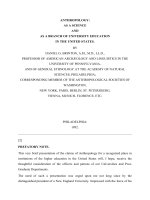
FIGURE 11.1
Turbidity of suspensions containing one of three synthetic Fe oxides display-
ing different particle sizes and morphologies: goethite (acicular, needle-like crystals 200+ nm
in length); Al-substituted goethite (somatoidal crystals ∼∼
∼∼
100 nm in length); and hematite
(diamond-shaped crystals ∼∼
∼∼
30 to 50 nm at the longest dimension), or the <2.0 µµ
µµ
m fraction
of kaolinite or montmorillonite.
50403020100
0
20
40
60
80
100
mg/L
Turbidity vs. Colloid Mass
NTU
Al-sub. goethite
goethite
hematite
montmorillonite
kaolinite
L1623_FrameBook.book Page 275 Thursday, February 20, 2003 9:36 AM
© 2003 by CRC Press LLC
estimates using kaolinite, possibly resulting from the presence of more efficient
scatterers. To illustrate the impact of such differences, the turbidity of three
synthetic Fe oxides/oxyhydroxides and the <2
µ
m fraction of kaolinite and mont-
morillonite suspended in deionized water was determined as a function of suspen-
sion concentration (Figure 11.1). Linear turbidity relationships with colloid mass
were observed for each of the mineral suspensions. However, dramatic differences
in the turbidity were observed for the two phyllosilicate clays compared to the Fe
oxides, despite their large variation in size and morphology. Such differences likely
reflect the higher refractive index for the Fe oxides (2.3 to 3.2) when compared
to the phyllosilicates (
∼
1.5).
11.2.2 D
YNAMIC
L
IGHT
S
CATTERING
The determination of particle size distributions for environmentally relevant suspen-
sions is difficult due to their dilute nature, wide distribution of particle sizes (poly-
dispersivity), and the large variation in particle morphologies.
22,37,43,52
Rayleigh light
scattering occurs for particles much smaller than the wavelength of the light, the
intensity of which is dependent on the wavelength (1/
λ
4
) and scattering angle (
θ
)
with short wavelength radiation being scattered more than longer wavelengths.
42
For
particle sizes comparable to the wavelength of light, multiple scattering events occur
at different sites within a given particle, and the resulting scattering pattern becomes
more complicated with an emphasis on forward scattering as particle size
increases.
42,43
As Hunter
42
notes, even though a suspension of colloidal particulates
is beyond the scope of Rayleigh theory, it demonstrates the strong dependence of
scattering intensity (
I
) on particle mass (
m
2
), that is, particle size:
where
N
p
is number of particles per unit volume, and
λ
is the wavelength of light.
42
For a spherical particle, the scattering intensity (
I
sc
) is dependent on the polarizability
(
α
) of the particle:
where
The radius of the spherical particle is
r
, and the relative refractive index,
n,
is the
ratio (
n
p
/
n
o
) of the refractive index for the particle (
n
p
) to that of the suspending
media (
n
o
). As seen in Figure 11.1, particles with higher refractive indices scatter
light more efficiently. The refractive index of polystyrene beads (1.6), generally used
I
mN
sc
p
()
θ
λ
∝
2
4
I
sc
∝
α
2
απε
=
−
+
4
1
2
3
2
2
o
r
n
n
L1623_FrameBook.book Page 276 Thursday, February 20, 2003 9:36 AM
© 2003 by CRC Press LLC
to standardize light-scattering instruments, is similar to the refractive index of phyl-
losilicate clays.
Light scattering from an intense, coherent, monochromatic beam, usually a He-
Ne or Ar laser in most instruments, can be used to estimate colloidal particle size.
Colloidal suspensions, due to the small particle size, are subject to Brownian motion
resulting in local fluctuations in particle concentration and light scattering, the rate
of which depends on the size (dispersion coefficient) of the particles. Thus, small
particles that are more subject to Brownian motion will induce rapid transient
concentration fluctuations while large, slowly moving particles will produce slow
fluctuations in the scattering intensity.
PCS, also known as dynamic light scattering or quasi-elastic light scattering,
quantifies the particle motion (Brownian motion) by measuring the signal intensity
at a given instance and comparing that with signals obtained at successively longer
time intervals, which is integrated over time [Figure 11.2(a)].
41–43
With little or no
motion, the product of the signal will vary little with time; however, subsequent
signals at different time intervals will vary dramatically if particles are moving
rapidly. Scattering intensity measurements are taken at short time intervals relative
to the diffusive motion of colloidal particles, resulting in high initial correlation that
gradually disappears with longer time intervals.
21
Analysis of the decay function for
signal correlation can yield the diffusion coefficient (
D
) for the suspended particu-
lates:
where
k
is the Boltzmann constant,
T
is the absolute temperature,
η
is the viscosity,
and
d
is the hydrodynamic diameter. The noninvasive nature of dynamic light
scattering eliminates artifacts associated with particle isolation, such as centrifuga-
tion, filtration, or sample drying. However, PCS is very sensitive to contamination
of larger particles, provides nondetailed size information, and determines the “effec-
tive” hydrodynamic diameter for nonspherical particles.
41,43
The detection limit (mg
l
−
1
) for PCS is dependent on the particle size and scattering angle, as well as
measurement duration, instrument sensitivity, and laser source.
43
Average size esti-
mates may be heavily weighted in favor of larger particles; thus, fractionation, such
as filtration, sedimentation, or centrifugation, designed to remove extremely large
particles (>1
µ
m) prior to PCS analysis may be necessary to resolve the size of
smaller, more abundant colloids.
43,53
Schurtenberger and Newman
43
emphasized that researchers must be more
explicit in describing the data analysis techniques relating particle size to the mea-
sured autocorrelation function. To date, few studies have been successful at resolving
multimodal size distributions for environmental suspensions.
9
Multi-angle PCS is
critical for sizing environmental suspensions where no
a
priori
knowledge of the
particle size distribution is available to confirm multimodal size distributions, resolve
artifacts associated with particle anisotropy and particle-particle interaction at high
concentrations, and account for variations in scattering intensity as a function of
D
kT
d
=
3
πη
L1623_FrameBook.book Page 277 Thursday, February 20, 2003 9:36 AM
© 2003 by CRC Press LLC
particle size that are manifested as local scattering minima at specific angles.
43
Electron microscopy can be used to confirm PCS results; however, qualitative agree-
ment between the two techniques is not surprising because of the limited resolution
inherent to PCS, with an applicable size range that is consistent with the imaging
and sizing capabilities of the SEM. This, combined with the qualitative nature of
most SEM particle surveys, ensures that most investigators will observe results that
are consistent with PCS. Care must be taken to ensure that particle associations
observed in the electron microscope are typical of the particle state in suspension
and do not reflect changes in aggregation induced by filtration or other sample
preparation artifacts.
Changes in particle size with even limited storage suggest that timely analysis
of environmental suspensions reduces artifacts associated with aggregation, changes
in chemistry and biological activity.
20,21,43,53
The development of
in situ
PCS systems
offers the ability to monitor particle size distribution and concentration of ground-
water colloids without the necessity of altering the system during the sampling and
handling processes before analysis.
25
11.2.3 L
ASER
D
OPPLER
V
ELOCIMETRY
AND PARTICLE CHARGE
Evaluating particle surface charge is critical to understanding the mechanisms of
mobile colloid formation, stabilization, and physicochemical filtration in the envi-
ronment, as well as other sorption phenomena. Without a significant electrostatic
barrier to particle approach, smaller colloidal particles with higher diffusion coeffi-
cients collide more frequently and aggregate faster than larger particles, with discrete
colloids or colloidal aggregates in the size range of 0.1 to 1.0 µm being most stable.
20
In addition, surface charge may be inconsistent with bulk suspension mineralogy
due to the presence of organic or oxide surface modifiers.
8,9,34,54
For example, highly
negative electrophoretic mobilities commonly observed for Fe/Al oxide-rich suspen-
sions under pH conditions well below the reported point of zero charge (PZC; pH
at which net charge is zero) have generally been attributed to organic coatings on
the mobile oxide fractions.
9,37,49,54
Colloidal particulates develop surface charge in one of two ways: either through
isomorphic substitution within the mineral structure, which is insensitive to the
external solution conditions (permanent charge), or from reactions of surface func-
tional groups (e.g., surface hydroxyls associated with organics, edge sites on alu-
minosilicates, and metal oxyhydroxides) with adsorptive ions at the mineral/partic-
ulate-solution interface, which is subject to changes in the aqueous environment
surrounding the particle (i.e., pH, ionic strength, etc.), and therefore considered
“variable charge.”
55,56
The dilute nature of most environmental suspensions does not lend itself to
conventional wet-chemical techniques for evaluating the surface charge of particu-
lates, such as potentiometric titration or ion exchange methods. Potentiometric
titrations, especially when applied to mixed, constant/variable-charge suspensions,
are complicated by the presence of species other than H
+
and OH
−
that act as potential
determining ions (PDI) and various reactions that consume H
+
and OH
−
without
generating equivalent surface charge, such as exchangeable Al.
8,28,57,58
Ion
L1623_FrameBook.book Page 278 Thursday, February 20, 2003 9:36 AM
© 2003 by CRC Press LLC
exchange/extraction methods depend on the identity and concentration of the probe
ion used to extract surface-associated species and yield little information related to
overall colloidal stability. In contrast, electrophoretic methods can be used to evaluate
the surface charge properties of dilute colloidal suspensions under the specific
chemical conditions to which the colloids are subjected, thus reducing the errors
and biases associated with altering the suspending solution or quantifying various
poorly defined surface/solute reactions (ion exchange, mineral dissolution, Al hydrol-
ysis, etc).
When an electrical field is applied to a suspension of charged particles, the
particles migrate toward the electrode of opposite sign, reaching a terminal velocity
in a matter of microseconds. The electrophoretic mobility (EM), u (µm cm s
–1
V
–1
),
for a particle is defined as:
where v
e
is the terminal velocity of the particle at a specified unit field strength, E
(V cm
−1
), with the sign being positive if the particles migrate from a region of high
electrical potential to a region of low electrical potential.
55
A boundary is established
between the strongly sorbed species and solvent that remains associated with the
charged particle as it moves through the solution and the loosely sorbed diffuse
species. The inner potential at the shear plane, known as the zeta potential,
ζ
, depends
on the surface charge density of the particle at the shear plane and is indicative of
the “effective charge” that particles and surfaces experience as they approach each
other, that is, colloid stability.
42,55
Analysis of the solution chemistry (i.e., pH, ionic
strength, solution composition, etc.) is critical to understanding the system, since
the EM (i.e., zeta potential) is a function of the colloidal material and aqueous
chemical environment. Such information can then be used to predict the effect of
various solution–particle and particle–particle interactions on aggregation, flow,
sedimentation, and filtration behavior.
Various equations have been derived for relating EM, u, to the zeta potential,
ζ
.
Traditionally, the Smoluchowski equation,
has been used for soil clays where
ε
o
is the permitivity of a vacuum, D is the dielectric
constant for water, and
η
is the viscosity of the solution. However, the validity of
such an expression depends on a number of assumptions and the choice of molecular
models used to represent the “plane of shear.”
58,59
In many instances it may be more
appropriate to simply report the measured mobility, u.
Electrophoretic instruments for analysis of colloidal suspensions can be divided
into two basic classes: optical instruments for which the operator observes the
migration of particles in a field using a microscope; and laser-based instruments that
measure the Doppler shift in the frequency of the scattered light from particles
u
v
E
e
=
ζ
ε
η
=
o
Du
L1623_FrameBook.book Page 279 Thursday, February 20, 2003 9:36 AM
© 2003 by CRC Press LLC
moving in response to an electric field, that is, laser Doppler velocimetry (LDV).
Analysis using optical instruments is slow and tedious, making it difficult to analyze
unstable suspensions. Particle detection is limited by the resolving power of the light
microscope and possibly biased by differences in particle size and the refractive
index of various colloidal constituents of multicomponent suspensions. Therefore,
electrophoretic results can be biased by the analysis of a relatively few discrete
particles that may display the expected behavior.
60
Design improvements, such as
laser illumination to improve particle resolution and rotating prism systems that
measure the mobility of a field of particles, have addressed some of the inherent
limitations of microscope-based systems.
In many respects, LDV instruments are superior to optical-based instruments,
especially for polydisperse samples with a range of surface properties. Doppler
broadening is generally evaluated at lower scattering angles to reduce the impact of
inherent Brownian motion on the frequency shift, thus increasing instrument resolu-
tion.
60
Information about the particle size of the suspension can be obtained by
measuring frequency broadening due to Brownian motion in the absence of the
electric field. Verification of LDV results generally involves comparing frequency
shifts at different scattering angles or different electric field strengths at a fixed angle;
an alternating electrical field is used to avoid electrode polarization. However, Bertsch
and Seaman
8
observed the disaggregation of colloidal particles that could impact
charge characterization when repeatedly subjected to the alternating field without
sufficient relaxation time between electrophoretic analyses. Operator bias associated
with particle selection is eliminated and the mobility of a much larger population of
particles can be rapidly determined, thus facilitating the analysis of colloidal samples
that are inherently unstable. Typically, greater standard deviations in the measured
mobilities are observed for LDV instruments, but this may reflect a more statistically
relevant sample population that better accounts for actual mobility distributions.
Care must be taken to evaluate the influence of other electrokinetic phenomena
occurring within the EM sample cell. When an electrical field is applied to the
capillary containing a colloidal suspension, migration is observed for the suspending
solution due to the osmotic flow of the counterions (electro-osmosis), as well as the
particles (electrophoresis), resulting in a parabolic velocity distribution in colloid
migration across the capillary that is the sum of the electrophoretic velocity and
electro-osmotic flow (Figure 11.2(b)). Electro-osmosis is a consequence of the
surface potential associated with the capillary walls, which induces a nonuniform
distribution of solution ions within the tube. For example, cations associated with
the capillary wall are attracted to the negative potential of the electrical field in a
manner similar to a positively charged particle. However, there is a location within
the tube, known as the stationary layer, where the net osmotic flow in either direction
is zero.
42,61
Absolute measurements of EM should be taken in the stationary layer
of the capillary tube. Unfortunately, the change in mobility as a function of minor
changes in cell position is quite great in the region surrounding the stationary layer.
Taking numerous measurements across the capillary (i.e., EM fingerprinting) may
be an effective means of evaluating relative changes in surface charge under various
solution conditions (i.e., pH, ionic strength, solution composition, etc.). Such an
approach is also recommended to determine if significant particle settling has
L1623_FrameBook.book Page 280 Thursday, February 20, 2003 9:36 AM
© 2003 by CRC Press LLC
occurred during EM analysis, which would tend to alter the symmetric nature of the
parabolic velocity function as larger particles and aggregates settle away from the
stationary layer located at the top of the capillary and accumulate on the bottom of
the analysis cell. Particle segregation due to aggregation and settling may signifi-
cantly skew results in favor of the smaller, more-stable fraction. Unfortunately, one
is often interested in the EM of a suspension under conditions that are not conducive
to colloidal stability, such as the pH region near the PZC for a given suspension. In
such a case, electroacoustic methods discussed in a subsequent section may be
appropriate. Recent advances in LDV instrumentation have focused on reduc-
ing/eliminating the need for optical alignment, and automating certain time-consum-
ing aspects of analysis, such as incorporating autotitration systems to evaluate
changes in charge as a function of pH.
Despite the poorly defined nature of the shear plane, EM has been commonly
used as a noninvasive technique for evaluating various surface–surface and sur-
face–solute reactions, such as evaluating the PZC for amphoteric minerals; specific
sorption reactions for inorganics and organic compounds on mineral surfaces;
FIGURE 11.2 (a) Schematic representation of dynamic light scattering, and (b) laser Doppler
velocimetry. (Part (a) modified from Buffle, J. and Leppard, G.G., Environ. Sci. Technol., 29,
2176, 1995. With permission.)
+
-
Detector
0
20
40
60
80
100
0-2 -4-6-8-10
Relative Position
Mobility (µm cm/V s)
Carboxylate-Modified
Polystyrene Latex
Beads
Electro-Osmotic Flow
Stationary Layers
Reference beam
Scattering beam
I
= 0
incident
light (l
0
)
time
<I
0
>
I
0
fluctuations due
to brownian motion
scattered
light (I
0
)
Laser
Detector
A.
B.
L1623_FrameBook.book Page 281 Thursday, February 20, 2003 9:36 AM
© 2003 by CRC Press LLC
charge reversal for surfactant modified clays; changes in microbial cell membrane
properties in response to contaminated environments; and cation demixing on 2:1
phyllosilicate clay minerals.
62–66
Electrophoretic behavior, however, is not necessarily indicative of a colloidal
sorption affinity for particular ionic species. For example, zeolite suspended in
HDTMA, a commonly used cationic surfactant, will undergo charge reversal at high
surface coverages resulting from the head-to-tail arrangement of the surfactant once
the external cation exchange sites are filled, yielding a relatively stable suspension
that displays a positive electrophoretic mobility (Figure 11.3).
111
However, the zeolite
retains a high affinity for cations, especially Cs, due to the presence of internal
exchange sites that are inaccessible to HDTMA and contribute little to the electro-
phoretic behavior. Internal exchange sites located within the open zeolite structure
are analogous to surface heterogeneity and similar high affinity sorption sites asso-
ciated with many soil minerals.
11.3 ACOUSTIC SPECTROSCOPY
11.3.1 A
COUSTIC ATTENUATION AND PARTICLE SIZING
Recent advances have been made in the theory and application of acoustic and
electroacoustic spectroscopies for measuring the particle size distribution (PSD) and
ζ
-potential of colloidal suspensions, respectively.
67–69
To date, the use of acoustics
has been confined mainly to industrial applications, despite the clear potential for
the technique to characterize colloids with environmental or agricultural significance.
Acoustic spectroscopy measures the speed and attenuation of sound waves
interacting with a colloidal suspension. When a sound wave in the range of 1 to 100
MHz interacts with a colloidal suspension, the measured acoustic attenuation and
FIGURE 11.3 Electrophoretic mobility of zeolite as a function of HDTMA loading indicating
external charge reversal. (Adapted from Sullivan, E.J., Hunter, D.B., and Bowman, R.S.,
Environ. Sci. Technol., 32, 1948, 1998. With permission.)
806040200
-6
-4
-2
0
2
4
6
HDTMA-Induced Charge Reversal
meq/100g zeolite
Electrophoretic Mobility (µm cm/V s)
L1623_FrameBook.book Page 282 Thursday, February 20, 2003 9:36 AM
© 2003 by CRC Press LLC
sound speed can be theoretically related to PSD by accounting for viscous, thermal,
scattering, intrinsic, electrokinetic, and structural losses.
70,71
The first two losses are
the most significant as particles mainly interact with sound waves hydrodynamically
through viscous losses and thermodynamically through temperature losses. Several
different theoretical approaches and acoustically based instruments are available,
such as the DT-1200, ESA-8000, Malvern Ultrasizer, and AcoustoSizer.
67,69,72
When either of the main attenuation mechanisms (viscous or thermal) predom-
inates, the other may be neglected. A quantity called the “viscous depth,”
δ
V
(L),
characterizes the decay distance of the shear wave from a particle’s surface, while
the “thermal depth,”
δ
t
(L), is the penetration depth of the temperature wave into the
liquid.
70
Each depth has a “critical frequency,” comparable to the particle radius,
corresponding to maximum attenuation. Thermal losses dominate in emulsions and
low-density dispersions, so viscous losses may be neglected. Viscous losses are more
sensitive to suspension concentration, while thermal losses may not become impor-
tant until 30%-volume fractions. When viscous losses predominate, the so-called
“long wavelength requirement” sets a lower limit of 10 nm on detectable particle
size. The formulas for viscous depth and thermal depth, respectively, are
where
υ
(l t
−1
) is the kinematic velocity;
ω
is the sound frequency (radian t
−1
);
κ
(t l
2
mol
−1
) is the thermal conductivity;
ρ
0
(m l
−1
) is the liquid density; and C
0
p
(J T
−1
mol
−1
m
−1
) is the specific heat of the liquid at constant pressure.
70
Two models are available for interpreting attenuation spectra as a PSD in sus-
pensions with chemically distinct, dispersed phases using the extended coupled phase
theory.
68
Both models assume that the attenuation spectrum of a mixture is composed
of a superposition of component spectra. In the “multiphase model,” the PSD is
represented as the sum of two log-normal distributions with the same standard
deviation, that is, a bimodal distribution. The appearance of multiple solutions is
avoided by setting a common standard deviation to the mean size of each distribution.
This may be a poor assumption for the PSD (see section 11.3.2). The “effective
medium” model assumes that only one “target phase” of a multidisperse system
needs to be determined, while all other phases contribute to a homogeneous system,
the so-called “effective medium.” Although not complicated by the possibility of
multiple solutions, this model requires additional measurements to determine the
density, viscosity, and acoustic attenuation of the effective medium. The attenuation
spectrum of the effective medium is modeled via a polynomial fit, while the target
phase is assumed to have a log-normal PSD.
68
This model allows the PSD for
mixtures of more than two phases to be determined.
Acoustic spectroscopy has several characteristics that make it useful. One clear
advantage over light-scattering techniques is the ability to stir, pump, or otherwise
physically agitate the sample during analysis, making the technique well suited to
potentiometric titration and analysis of unstable suspensions. When the acoustic
signal is measured as a function of the transmitter–receiver gap, it requires no
δ
υ
ω
δ
κ
ωρ
Vt
p
C
==
22
0
0
,
L1623_FrameBook.book Page 283 Thursday, February 20, 2003 9:36 AM
© 2003 by CRC Press LLC
calibration. A PSD can be calculated even when there is little density contrast
between sample and fluid. Attenuation spectra are independent of electrical proper-
ties of the particle surface, and supply independent information about PSD, even in
concentrated systems with several dispersed phases.
72
Thus, attenuation spectra can
characterize PSD in uncharged dispersed systems, in highly conducting systems, as
well as in systems with conducting particles. The theoretical success of character-
izing viscous losses in concentrated dispersions with large density contrast gives
acoustics an advantage over light-scattering methods in measuring PSD. Using well-
characterized samples and commercially available instruments, acoustic spectros-
copy can measure the mean of the PSD with a precision and accuracy of up to 1%,
and the width of the PSD with an accuracy of up to 5%.
71
There are several shortfalls in acoustic spectroscopy. Information about particle
shape is lacking in the spectrum, and a substantial amount of physical and thermo-
dynamic information may be needed to interpret acoustic spectra, including particle
density, liquid density and viscosity, and the weight or volume fraction of the
suspension.
73
Such information may not always be available for complex environ-
mental suspensions. Also, relatively large sample requirements may restrict the use
of acoustics to idealized laboratory systems.
Acoustic spectroscopy shows promise for distinguishing particle–particle inter-
actions in concentrated suspensions (up to 30% by volume), as well as in polydis-
perse suspensions with chemically distinct phases. Although acoustic spectroscopy
does not provide information regarding particle shape, it has an advantage over SEM
for determining PSD as in situ measurements are made, so that the colloids are not
subject to changes in particle–particle interactions during filtration and drying.
11.3.2 ELECTROACOUSTICS
Electroacoustic spectroscopy measures either colloid vibration potential/current
(CVP/CVI) or electrokinetic sonic amplitude (ESA), each of which is quantitatively
related to mean
ζ
-potential. In response to an acoustic wave, the density contrast
between the particle and the medium causes a displacement, or polarization, of the
electrical double layer, creating a dipole moment (Figure 11.4) whose magnitude
varies with the sound wave amplitude.
67
In superposition, the individual dipole
moments give rise to the macroscopic alternating electric field measured as the
CVP.
70
Conversely, the application of an alternating electric field produces an oscil-
lating electrophoretic motion for particles with a nonzero
ζ
-potential, which gener-
ates a sound wave, the ESA effect, and the resulting acoustic field is measured.
67,69
The CVI is analogous to a sedimentation current, the current arising when the
potential generated as charged particles settle under gravity is short-circuited
between vertically placed electrodes, while in CVP, the alternating acoustic field
supplies the acceleration instead of gravity.
74
In electrokinetic phenomena such as electroacoustics, theoretical models need
to consider the induced movement of charge within the electrical double layer (EDL),
the “surface current”, I
s
, as well as the interaction of the outer portion of the double
layer with the applied signal (acoustic or electric field) and with the liquid medium.
Hydrodynamic flows generate surface current as liquid moving relative to the particle
L1623_FrameBook.book Page 284 Thursday, February 20, 2003 9:36 AM
© 2003 by CRC Press LLC
surface causes movement of charge in the outer portion of the electric double layer
(EDL). A tangential electric field, E, is generated by the surface current, I
s
, and the
compensating current, I
n
, is measured in CVI electroacoustics.
70
When particle size
and/or electrolyte concentrations are small, surface conduction may be significant.
67
Electroacoustic measurement of
ζ
-potential can be made even in concentrated
suspensions (up to 40% by weight) where optical techniques fail. The interpretation
of electroacoustic measurements is questionable when the EDL is thick and over-
laps for neighboring particles, when the
ζ
-potential is large, or when anomalous
conduction in the inner portion of the EDL is present. However, similar problems
are encountered in theoretical analyses for all available techniques for measuring
ζ
-potential. Excluding electroacoustics,
ζ
-potential has typically been measured in
extremely dilute colloidal suspensions by micro-electrophoresis, although suspen-
sion dilution may affect both particle size distribution and
ζ
-potential. Sedimen-
tation potential measurements are also restricted to dilute suspensions in order to
ensure free settling and uniform particle flow. Conversely, high solid concentrations
of very large particles (e.g., sand) are required for electro-osmosis and streaming
potential measurements due to the requirement of a tightly packed, immobile
porous plug. In contrast, electroacoustic spectroscopy is applicable to a range of
suspension concentrations.
Acoustic methods offer several advantages when compared to other comparable
techniques: (1) applicable to concentrated suspensions; (2) less sensitive to partic-
ulate contamination; (3) better suited to polydisperse suspensions; (4) applicable to
a wide size range; (5) well suited to automated potentiometric titrations and analysis
FIGURE 11.4 Dipole moments generated on charged particles in response to an applied field.
Inset: location of CVI current, I
n
, external to the electric double layer.
+
++
+
+
+
+
+
+
+
+
+
+
+
+
+
+
+
+
+
+
+
+
+
+
+
+
+
+
+
+
+
+
+
+
+
+
+
+
+
+
+
+
+
+
+
+
+
+
+
+
+
+
+
+
+
+
+
+
+
+
++
+
+
+
+
+
+
+
+
+
Electric Field
Acoustic Field
Particle Motion
Is
In
In
L1623_FrameBook.book Page 285 Thursday, February 20, 2003 9:36 AM
© 2003 by CRC Press LLC
of suspensions near their PZC and critical coagulation concentration (CCC) because
of the ability to stir or pump the sample during analysis.
From an experimental standpoint, the ability to use concentrated suspensions
offers obvious advantages when compared to other potentiometric and micro-elec-
trophoretic techniques. Such conditions are more analogous to the solid/solution
ratio encountered in a typical soil/sediment environment, but with less kinetic restric-
tions and system heterogeneity than encountered in the field. In addition, small
aliquot suspensions can be removed during analysis for characterization using
another technique. The large sample volumes and high colloidal loads, however,
may restrict the use of acoustic techniques to idealized laboratory systems where
sufficient sample is available.
11.4 FIELD FLOW FRACTIONATION
FFF is a separation technique that encompasses a range of procedures based on
theory and subsequent instrumentation originally developed and advanced by Gid-
dings.
75,76
FFF is a high-resolution chromatography-like technique applicable to the
separation of macromolecules, colloids and particles encompassing a size range of
1 nm to 100 µm. In practice, a small volume of colloidal suspension (10–20 µL) is
injected into the FFF channel, the concentration of which depends on the sensitivity
of the online detector. The FFF channel is a thin (∼0.02–0.05 cm), open rectangular
channel through which a carrier solution is pumped by means of a peristaltic or
HPLC pump. The breadth and length of the channel are generally on the order of
2 cm by 20 to 50 cm, respectively. The thinness of the channel ensures a laminar
flow profile with the fastest flow vectors in the center and slowest flow at the channel
walls. A force is applied across the thin dimension of the channel, and perpendicular
to the direction of flow, driving molecules and colloidal particles against one of the
channel walls (accumulation wall). Particles also diffuse back into the channel
through Brownian motion; the extent of this diffusion being related to the molecular
weight (M
w
) or hydrodynamic diameter (d) of the particle. After sample injection
the main channel flow is stopped for a period of time (relaxation time) to allow the
particles to reach their equilibrium distribution from the channel wall without migrat-
ing down the channel. The equilibrium positions above the wall are based on the
balance achieved between migration caused by the applied force and the back-
diffusion of the particle (Figure 11.5(a)). After the relaxation time, the channel flow
is resumed and particles whose equilibrium distance is higher in the channel (smaller
particles) are carried in faster moving flow vectors, and, hence, are eluted first from
the channel.
11.4.1 SEDIMENTATION (SD-FFF) AND FLOW-FIELD FLOW
F
RACTIONATION (FL-FFF)
The various FFF techniques arise through the different fields that are employed,
including sedimentation, flow, electrical, and thermal fields. Of these techniques,
sedimentation (Sd-FFF) (Figure 11.5(b)) and flow (Fl-FFF) (Figure 11.5(c)) have
found the widest application in environmental studies. In Sd-FFF, the channel is
L1623_FrameBook.book Page 286 Thursday, February 20, 2003 9:36 AM
© 2003 by CRC Press LLC
created by clamping a mylar spacer, with the channel cut out, between two con-
centric rings, which are then positioned within a centrifuge basket. The centrifugal
forces used make Sd-FFF applicable to the separation of particles from about 30
nm to 100 µm, the retention volume of a particle being a function of the spherical
diameter (d) and density (
ρ
p
). When operated in normal mode (see later description),
that is, particles in the size range 30 nm to 1 µm, the theory of Sd-FFF is well
defined mathematically and, when the gravitational field applied to effect the
FIGURE 11.5 Schematic representation of the FFF. Schematic diagram of (a) Sd-FFF and
(b) Fl-FFF systems coupled to ICP-MS and UV/Vis detector, respectively.
HPLC
PUMP
INJECTION
PORT
MOTOR
CONTROL
ICP DATA
ICP-MS
CARRIER
RESERVOIR
UV DATA
UV-VIS
DETECTOR
ICP-MS
PUMP
ROTATION
FRACTION
COLLECTOR
GFAAS
A.
B.
C.
Carrier
Fluid
Parabolic
Velocity
Vectors
Applied Field
W
High velocity
Low velocity
l
T
2
T
1
PUMPS
FLOW FFF CHANNEL
TO WASTE
TO FRACTION COLLECTOR
DETECTOR
PC BASED
WORK STATION
DATA ACQUISITION
PUMP CONTROL INTERFACE
VALVE CONTROL INTERFACE
L1623_FrameBook.book Page 287 Thursday, February 20, 2003 9:36 AM
© 2003 by CRC Press LLC
separation is constant, the retention ratio R (V
0
/V
r
) can be calculated from the
following equation:
The relationship between R and d
3
affords Sd-FFF a high resolving power;
however, it also means that the retention time of large particles can become unac-
ceptably long when a constant field is employed. To overcome this, power program-
ming is commonly used, where the gravitational field is decayed exponentially over
time after an initial hold time at constant field strength.
77
Computer programs exist
for optimizing run parameters and converting detector response and field strength
as a function of analysis time into particle size distributions.
In Fl-FFF, the channel is created by placing a mylar spacer with the channel cut
out between two porous frits. A membrane filter of a specific molecular weight cutoff
is placed on one of the frits and acts as the accumulation wall to permit flow, without
loss of particles. The applied force is then a perpendicular flow of the carrier solution
across the porous frits. Fl-FFF is a versatile technique capable of separating mac-
romolecules as small as roughly 1000 Da, in which case it is comparable to gel
permeation (size exclusion) chromatography. However, Fl-FFF can also be applied
to the separation of colloidal particles. In this case the hydrodynamic diameter of
the colloidal particle is related to the retention volume, V
r
, by the equation
where w is the channel thickness, v
c
is the cross-flow velocity, and d
s
is the hydro-
dynamic diameter. For macromolecules, the usual practice is to create a calibration
curve relating peak retention time to molecular weight, M
w
. The diffusion coefficient,
D, and can then be calculated from the equations below.
A recent advance in Fl-FFF has been the introduction of asymmetric-flow FFF
instrumentation. In asymmetric Fl-FFF, the upper channel wall is impermeable and
the cross-flow rate is achieved by flow control of the cross-flow and channel flow.
Upon sample injection channel flow is directed through both the channel inlet and
outlet that allows for focusing of the sample and for preconcentration. For elution,
channel flow is just introduced at the channel inlet.
R
kT
wrd
p
=
πρϖ
∆
23
V
wvd
kT
r
cs
=
πη
2
2
dAM
sw
b
=
'
D
kT
d
s
=
3
πη
log log logDAbM
w
=−
"
L1623_FrameBook.book Page 288 Thursday, February 20, 2003 9:36 AM
© 2003 by CRC Press LLC
The study of environmental colloids by FFF is complicated by a change in the
main counter force opposing the applied force as particle size increases above about
0.8–1 µm. In normal mode FFF, the method previously described, the main force
opposing the applied field is Brownian motion. Smaller particles have higher diffu-
sion coefficients and therefore attain higher equilibrium distances from the accumu-
lation wall and thus emerge first from the channel. However, as particle diameter
increases, diffusion becomes less important compared to hydrodynamic lift forces.
These forces are not well understood, but the net effect is to lift larger particles away
from the accumulation wall. Also, for larger particles the actual particle diameter
becomes comparable to the equilibrium cloud thickness for smaller particles. This
is termed the steric effect and results in the opposite elution pattern to normal mode
with larger particles eluting from the column first. The point at which the elution
mode changes from normal to hyperlayer/steric mode is termed the inversion point
and occurs around 1 µm. Steric-mode FFF has found numerous applications in the
separation of particles >1 µm, such as in the separation of cells.
78
However, the
steric effect complicates analysis of environmental suspensions and necessitates
preseparation of particles >1 µm before FFF analysis in either normal or steric mode.
11.4.2 FFF APPLICATIONS
Environmental applications of FFF fall into two broad groups. First, studies utilizing
FFF with a nonspecific detector (i.e., UV spectrophotometer) to determine the
particle size or molecular weight distribution of the sample. This approach has been
applied extensively for the study of humic and fulvic acids in natural waters.
79–82
Fl-FFF is used exclusively for this application, in which it is essentially equivalent
to gel permeation chromatography (GPC). As in GPC, the main analytical consid-
eration is minimizing interactions between organic acids and the accumulation wall
by the judicious choice of carrier solutions and membrane materials. Thang et al.
80
reported optimal recovery of humic substances using a 0.005-M Tris-buffer (tris-
hydroxymethylaminomethane) as the carrier solution at pH 9.1 and utilizing a
regenerated cellulose membrane. For molecular weight calculations, the system is
first calibrated by running polystyrenesulfonate molecular weight standards to create
a calibration curve. Caution is advised when calibrating because differences in
structure between calibration standards and samples may introduce errors in molec-
ular weight calculations of unknowns. UV detectors have been commonly used for
larger colloids based on light scattering by the particles. Multi-angle light scattering
has also been used for accurate molecular weight determinations in FFF studies of
humic substances.
83,84
The second major environmental application of FFF has been the use of an
element-specific detector, usually in series with a UV detector, to provide elemental
composition data along with the PSD. Graphite-furnace atomic absorption spectrom-
etry has been used off-line on fractions collected from the FFF run. However, the
multi-element detection, low detection limits and capability to function as an on-
line detector have made inductively coupled plasma mass spectrometry (ICP-MS)
the ideal detector for FFF.
85,86
The sample introduction system of the ICP-MS is
able to efficiently transport micron-sized particles into the high-temperature plasma,
L1623_FrameBook.book Page 289 Thursday, February 20, 2003 9:36 AM
© 2003 by CRC Press LLC
where the particles are completely decomposed, atomized, and ionized. The advan-
tage of this technique is that it allows for the major element mineralogy of the
colloidal particles to be determined simultaneously with the UV-based PSD to
provide element-based size distributions. Although the light-scattering response of
a UV detector can be a complex function of particle size, agreement between the
UV response and the element-specific response for environmental colloids has been
generally observed. In most cases the sensitivity of the ICP-MS allows multiple
elemental signals to be continuously monitored to determine minor and trace ele-
ments and, therefore, assess potential correlations between element distributions.
Sd-FFF-ICP-MS has been used to determine major mineral constituents and
distributions of soil colloids to study trace metal adsorption to colloids and the effect
of colloidal surface coatings on phosphate sorption.
87–90
Examples of the UV-based
and ICP-MS–based fractograms for soil colloids are shown in Figure 11.6(a). The
original surface soil was prefractionated by centrifugation in order to obtain a 0.2-
0.8-µm size fraction. The example shows the element-based fractogram for a minor
and trace element, manganese (Mn) and uranium (U), respectively. The fractogram
was converted into an element-based particle size distribution (Figure 11.6(b)) and
an element ratio distribution (Figure 11.6(c)) using a major element (Si). The element
ratio shows changes in mineralogy that occur across the size range. In this case, it
appears that U is uniformly distributed across the particle size range whereas a larger
Mn-rich colloid, perhaps Mn oxide, appears to be present. Interpretations of element-
based size distributions may prove useful for understanding trace metal behavior in
soils.
Fl-FFF has also been coupled to ICP-MS for the determination of element size
distributions of 28 elements, including C, in natural waters.
91
Hassellov et al.
91
further
developed methodology for on-channel preconcentration that enables up to 50 ml
of sample to be introduced onto the channel. This significantly enhances the effective
detection limits of the technique, which can otherwise be problematic due to the
low concentration of trace elements in natural waters, the dilution inherent in FFF
analysis, and the small injection volume, typically 10 to 50 µl.
Clearly, FFF techniques are potentially useful in environmental analysis, as they
can provide PSD analysis and, when coupled to a suitable detector, element sized
distribution analysis. However, FFF theory for particle size determination does not
account for particle–particle or particle–wall interactions, both of which will cause
errors in the calculated particle size. Particle–wall interaction forces can be incor-
porated into FFF theory to allow for accurate particle sizing in low-ionic-strength
carrier solutions if standards of the same material and of known size are available.
92
Particle interactions are minimized when the ionic strength of the carrier solution
is near 10
–
3
M. Common carrier solutions employed in previous studies include
dilute surfactant solutions (0.1 mM) sodium pyrophosphate, and 0.01 M sodium
bicarbonate usually at high pH (>7). These conditions might be expected to change
the primary PSD of the natural sample and confound interpretation of trace ele-
ment–colloidal interactions and sorption behavior. Hence, interpretation of FFF
results requires a consideration of the potential changes to surface charge that may
result from the carrier solution.
L1623_FrameBook.book Page 290 Thursday, February 20, 2003 9:36 AM
© 2003 by CRC Press LLC
FIGURE 11.6 UV and ICP-MS-based Sd-FFF fractogram of a colloidal (0.2 to 0.8 µµ
µµ
m)
fraction: detector responses versus time (a); particle size distribution (UV response) and
element-based size distribution for Mn and U (b); and element ratios for Mn/Si and U/Si (c).
010203040
Mass Mn
Mass U*10,000
UV
Time (min)
UV Response
UV
Mn
U
0.0 0.1 0.2 0.3 0.4 0.5 0.6
0.0000
0.0002
0.0004
0.0006
0.0008
0.0010
0.0012
0.0014
Mass Mn and U (µg)
Relative amount
Elemental/Si ratios
0
1
2
3
4
5
0.0
0.1
0.2 0.3
0.4 0.5
Diameter (µm)
Mn/Si
U/Si
A
B
C
Diameter (µm)
L1623_FrameBook.book Page 291 Thursday, February 20, 2003 9:36 AM
© 2003 by CRC Press LLC
11.5 ELECTRON-BASED ANALYSIS TECHNIQUES
11.5.1 S
CANNING ELECTRON MICROSCOPY
A major advantage of the scanning electron microscope (SEM) when compared to
other microscopic analysis methods is that it produces topographical images that
are easily interpreted without an understanding of the theory behind the instrumen-
tation, that is, colloids look as we think they should. SEM combined with energy
dispersive spectrometry (EDS), that is, x-ray microanalysis, also commonly known
as energy dispersive x-ray analysis, has been used as the primary discrete-particle
analysis technique to characterize mobile groundwater colloids generated in field
and laboratory studies, and colloidal particulates suspended in surface water as
well.
7,24,29,30,35,37,39,51,93
A curious feature in many published micrographs from colloid
transport studies is the common presence of particles that are much larger (>1 µm)
than one would expect to be mobile in the subsurface environment. In many cases,
the large particles may be artifacts associated with sampling, but their presence may
also reflect resolution biases inherent in SEM analysis, and perhaps the tendency to
fixate on interesting visual anomalies rather than regions of the sample that reflect
the more mundane common particle morphology (i.e., aesthetic bias).
Typically, colloidal suspensions are deposited on polycarbonate filters immedi-
ately following sampling to reduce post-collection precipitation or aggregation arti-
facts and mimic the operational procedures used to define dissolved and particulate
sample components. Obviously, a well-dispersed dilute sample should be used to
avoid artifactual particle overlap. Filter sections are then mounted on SEM stubs
and carbon or metal coated (Au/Pd) for compositional analysis and micrographic
imaging, respectively. The tedious nature of operator controlled analysis of partic-
ulate samples in the SEM and the TEM has often precluded the acquisition of
statistically valid information regarding suspension composition.
SEM images are generated by rastering a focused electron probe across the
surface of the specimen while measuring various signals as a function of beam
position. Elastic and inelastic electron scattering combine to limit the penetration
of the beam within the sample, resulting in a region known as the “interaction
volume,” the dimensions of which (∼1 µm
3
) are greater than that of the focused
probe.
94
Various signals responsible for image generation, that is, backscattered
(BSE) and secondary electrons (SE), and “characteristic” x-rays indicative of sample
composition are generated within this interaction volume (Figure 11.7(a)). An under-
standing of the size and shape of the interaction volume for a given specimen as
influenced by the beam parameters (i.e., instrumentation) is critical in properly
interpreting SEM images and the resolution capabilities of x-ray microanalysis.
For a given initial probe diameter, the energy of the electron beam (keV) has a
strong influence on the relative size of the interaction volume, with greater electron
penetration occurring as the beam energy increases. Electrons may travel significant
lateral distances from the impact point before generating SE signals or escaping as
BSEs, thus reducing image resolution by contributing to noise. Electron backscat-
tering increases with atomic number (Z) to a degree, and thus, provides additional
information about sample composition, as well as local topography.
94
L1623_FrameBook.book Page 292 Thursday, February 20, 2003 9:36 AM
© 2003 by CRC Press LLC
Imaging at low electron voltages reduces beam penetration and ensures that the
BSE and SE signals are more indicative of local surface features. However, a
reduction in probe size necessary to achieve high resolution reduces the beam current
within that probe, limiting the intensity of the BSE and SE signal (contrast) and the
effective spot size that can be achieved for a given electron source. The brightness
of an electron source, current density per solid angle, is an indicator of its ability
to maintain current within a finely focused probe (Table 11.1); therefore, resolving
power differs for various electron sources, a factor that most studies of environmental
colloids fail to recognize when reporting instrumental operating conditions.
To illustrate the importance of emission source, synthetic goethite particles
deposited on 0.1-µm pore-size polycarbonate filters (150,000 × mag.) were imaged
using both a JEOL 6310 and a JEOL 6320F microscope equipped with thermoionic
LaB
6
and field-emission (FE) electron sources, respectively (Figure 11.8). The
FIGURE 11.7 Electron/sample interactions for (a) conventional and (b) thin-foil mounting
techniques in the SEM. (Adapted from Seaman, J.C., Environ. Sci. Technol., 34, 187, 2000.
With permission.)
TABLE 11.1
Brightness and Stability of Electron Sources at 20 keV
Source Type Brightness Energy Spread (eV)
Current Stability
(%)
Thermoionic
Tungsten 10
5
1–3 1
LaB
6
10
6
1–2 1
Field Emission 10
8
0.3–1 2–5
Source: From Goldstein, J.I. et al., Scanning Electron Microscopy and X-ray Microanalysis, 2nd ed.,
New York, 1992. Reprinted with permission of Kluwer Academic/Plenum Publishers.
Incident Beam
absorption
Charateristic
X Rays
Backscattered
Electrons
Secondary
Electrons
Transmitted
Electrons
Charateristic
X Rays
Thin Foil
Thick Specimen
Auger Electrons
Incident Beam
Backscattered
Electrons
fluorescence
L1623_FrameBook.book Page 293 Thursday, February 20, 2003 9:36 AM
© 2003 by CRC Press LLC
enhanced resolving power of the FE instrument is derived from both the finer spot
size and the ability to image the sample at lower excitation voltages, 3 compared to
10 keV for the FE and LaB
6
instruments, respectively, with image signals originating
from surficial regions of the sample closer to the initial probe location.
The influence of sample coating on SEM imaging and EDS analysis has fre-
quently been neglected. A fraction of the beam electrons lose their initial high energy
and are captured within the specimen. If the sample is a conductor, the charge will
flow to ground; however, if the sample is a poor conductor, such as silicate clay
minerals and polycarbonate filters, or if the path to ground is broken or inadequate
due to poor contact between irregular-shaped particulates and the underlying sub-
strate, excess charge accumulates. Sample charging can result in artifactual contrast
features, beam aberrations that contribute to noise, and particle loss due to electro-
static repulsion with the incident beam.
94,95
Operation at low-beam energies reduces
the potential for charging, but soil clays generally require a thin coating of a
conductive substance to eliminate charging artifacts. Metal coating (e.g., Au/Pd) can
increase both the SE and BSE signals for high-resolution imaging, but may interfere
with performing microanalysis by increasing electron backscattering at the expense
of specimen x-rays while producing x-rays characteristic of the metal coating.
Therefore, a carbon coating that does not significantly interfere with x-ray generation
is typically applied to nonconducting samples for EDS analysis. To illustrate this
effect, mobile colloids generated in a series of column studies were deposited on a
0.1-µm pore size polycarbonate filter, a portion of which was mounted to two
separate SEM stubs.
7,93
One stub was coated with evaporated carbon and the other
was sputter-coated with Au/Pd prior to imaging with a Hitachi S 800 SEM equipped
with a field emission electron source. The poor resolution observed for the carbon-
coated sample illustrates an important limitation to the use of SEM for the analysis
of colloids, with submicron particles becoming less distinct and appearing somewhat
amorphous (Figure 11.9(a) and (b)).
Although the impact of sample coating and beam intensity on image resolution
is quite obvious, there are more subtle implications to the x-ray microanalysis of
FIGURE 11.8 Electron micrographs of synthetic goethite imaged using two different electron
sources, (a) field emission, and (b) LaB
6
, at the same magnification level.
A. FE Electron Source B. LaB
6
Electron Source
L1623_FrameBook.book Page 294 Thursday, February 20, 2003 9:36 AM
© 2003 by CRC Press LLC
submicron-sized particulates.
93,96
X-rays are produced in the SEM as a consequence
of inelastic scattering events, with the resulting spectra consisting of two distinct
components: “characteristic” x-rays indicative of the atomic composition of the
beam–specimen interaction volume with sharply defined energy values, and a non-
specific background continuum from zero to the beam energy, known as Bremsstrahl-
ung x-rays (Figure 11.9(c)). To generate an x-ray signal, the electron beam voltage
must be greater than the x-ray energy for that sample element of interest (i.e., over-
voltage). For example, x-ray lines indicative of the elements beryllium to uranium
are found in the energy range of 0.1 to 14 keV.
94
Typically, a beam energy of 20
keV is used for EDS, which results in greater beam penetration and the generation
of x-rays from other regions of the bulk sample or instrument chamber, yielding less
x-ray intensity for smaller particles than would be predicted for a bulk material with
the same composition and an increase in the Bremsstrahlung background with
decreasing particle size.
Using a smaller electron probe does not improve resolution during microanalysis
because electron scattering and penetration prior to x-ray generation is more a
function of beam current.
94,97
Carbon sample coating necessary to perform x-ray
analysis because of the higher potential for charging at 20 keV further limits visual
and photomicrographic resolution, making it difficult to resolve small particles
against the substrate or distinguish primary particles and aggregates. In essence, the
resolution displayed in published micrographs of metal-coated samples is usually
far superior to the resolution achieved during microanalysis. EDS patterns collected
FIGURE 11.9 Electron micrograph images of mobile colloids collected in a series of column
studies, deposited on polycarbonate filters, and then (a) metal (AuPd) or (b) carbon coated
prior to imaging and EDS analysis with (c) a field-emission SEM.
A. Au/Pd B. Carbon
L1623_FrameBook.book Page 295 Thursday, February 20, 2003 9:36 AM
© 2003 by CRC Press LLC