Báo cáo y học: " Acute compartment syndrome of the hand in Henoch-Schonlein Purpura" ppt
Bạn đang xem bản rút gọn của tài liệu. Xem và tải ngay bản đầy đủ của tài liệu tại đây (819.65 KB, 5 trang )
BioMed Central
Page 1 of 5
(page number not for citation purposes)
Journal of Medical Case Reports
Open Access
Case report
Acute compartment syndrome of the hand in Henoch-Schonlein
Purpura
Guntur E Luis* and Eng-Seng Ng
Address: Department of Orthopedics Surgery, Faculty of Medicine, University of Malaya, Kuala Lumpur, Malaysia
Email: Guntur E Luis* - ; Eng-Seng Ng -
* Corresponding author
Abstract
An eight year old boy with Henoch-Schonlein Purpura (HSP) presented with acute compartment
syndrome (ACS) of his left hand following arterial cannulation of his radial artery in intensive care
unit. Emergency decompression and fasciotomy were performed. The authors report this first case
in literature and discuss how HSP can be complicated by ACS and ways to prevent the latter from
happening.
Background
Henoch-Schonlein Purpura is one of the most common
vasculitides of childhood and is considered to be self-lim-
iting. One manifestation of HSP that can continue to
cause lifelong problems is renal involvement [1].
In 1990, the American College of Rheumatology pub-
lished diagnostic criteria for HSP. These included (1) Pal-
pable purpura-slightly raised "palpable" hemorrhagic
skin lesions, not related to thrombocytopenia; (2) Age less
than 20 at disease onset-patient 20 years or younger at
onset of first symptoms; (3) Bowel angina-diffuse abdom-
inal pain, worse after meals, or the diagnosis of bowel
ischemia, usually including bloody diarrhea; and (4) Wall
granulocytes on biopsy-histologic changes showing gran-
ulocytes in the walls of arterioles or venules. The classifi-
cation further states, "For purposes of classification, a
patient shall be said to have Henoch-Schonlein purpura if
at least 2 of these 4 criteria are present. The presence of any
2 or more criteria yields a sensitivity of 87.1% and a spe-
cificity of 87.7%" [2].
Case report
An eight year old Malay boy, with a history of Henoch-
Schonlein Purpura and G-6-PD deficiency, presented with
left hand swelling and punctate rashes on the dorsum of
his left hand, four hours following his transfer out from
intensive care unit (ICU).
The history dated back to two weeks prior to admission
when he noted rashes on the dorsum of his feet and had
intermittent, diffuse abdominal pain. He then developed
migratory pain in his ankle joints, knee, elbow and small
finger joints in that order. He refused treatment.
He had no drug allergies. Developmental milestones were
normal and immunization was complete.
A week later, he was admitted to the general pediatrics
ward for intermittent vomiting, severe generalized
abdominal pain and passing red-current stools. The pain
was constant, unrelenting and aggravated by solid food
intake despite intravenous omeprazole (16 mg bd), meto-
clopromide (3 mg tds) and tramadol (30 mg 4 hourly).
Rashes now developed on his left flank, buttock and
Published: 2 March 2007
Journal of Medical Case Reports 2007, 1:6 doi:10.1186/1752-1947-1-6
Received: 15 December 2006
Accepted: 2 March 2007
This article is available from: />© 2007 Luis and Ng; licensee BioMed Central Ltd.
This is an Open Access article distributed under the terms of the Creative Commons Attribution License ( />),
which permits unrestricted use, distribution, and reproduction in any medium, provided the original work is properly cited.
Journal of Medical Case Reports 2007, 1:6 />Page 2 of 5
(page number not for citation purposes)
medial aspects of both feet. He was febrile and mictura-
tion was normal.
His vital parameters were as follows: BP 127/96, PR 99/
min, RR 24/min and SpO2 97% on room air. Clinical
examination revealed non-blanching purpuric rashes on
the dorsum of his feet, left flank and buttock [Fig. 3].
There was also localized tenderness on deep palpation of
his left iliac fossa. Hematological, coagulation and renal
profiles were within normal limits.
Urgent transabdominal ultrasound did not show any
"pseudokidney" or "doughnut sign" to suggest intussus-
ception." Peristalsis was normal. All intraabdominal
organs were normal and there was no free fluid. There
was, in the left iliac fossa, a bowel loop filled with echo-
genic material likely to be a stool."
Thirty-six hours later, he was admitted to the pediatric
ICU owing to poor intake, severe per rectal bleeding and
deteriorating general conditions. Fluid challenge with 300
mls of Ringer's Lactate solution and 2 units of packed red
cell transfusion were given. Intra-arterial cannulation of
the left radial artery was performed for continuous blood-
pressure monitoring. Intravenous fluids, methylpred-
nisolone (16 mg od), PCA morphine (bolus 0.5 mg, lock-
out at 15 mg), intravenous ceftriaxone (800 mg od) and
metronidazole (120 mg tds) were administered.
Following two days of stabilization, he was transferred
out from ICU after the removal of his arterial line and
urine catheter. Clinical examination in the morning
showed diffuse, oedematous swelling on the dorsum of
the left hand and all fingers with non-blanching purpuric
rash restricted to an area 4 cm in diameter. There were no
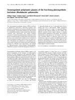
Decompression and fasciotomy of the dorsal interosseous compartmentsFigure 1
Decompression and fasciotomy of the dorsal interosseous compartments.
Journal of Medical Case Reports 2007, 1:6 />Page 3 of 5
(page number not for citation purposes)
signs of fluid extravasations, inflammation or infection at
the site of cannulation.
An urgent Doppler ultrasound of the left forearm was per-
formed to exclude a thrombo-embolic event. Both radial
and ulnar arteries and corresponding veins were patent.
Normal Doppler flow pattern was obtained within these
vessels up to the level of cubital fossa.
In the late afternoon, the whole left hand was swollen
intensely, with extension of the purpura from the hand,
up the wrist into the forearm. The metacarpophalangeal
joints of all the left fingers were extended with flexion of
all the distal interphalangeal joints. Capillary refill was
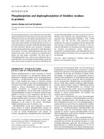
normal. Emergency fasciotomy decompression of the dor-
sal [Fig 1], thenar and hypothenar compartments [Fig 2]
of the left hand were performed. Serous fluid accumula-
tion was noted in all compartments with marked tissue
oedema. Additional hematoma was noted in the hypoth-
enar compartment. The rashes and swelling subsided
quite quickly and he was discharged uneventfully on the
fifth post-operative day.
Discussion
This is the first case of acute compartment syndrome of
the hand in HSP to be reported in the literature. Compli-
cations such as gastrointestinal bleeding [3], intracranial
bleeding [4], pulmonary hemorrhages [5], bilateral cen-
tral artery occlusion [6], orbital hematomas [7], nephritis
and penile involvement [8] have been well documented.
While nephritis is the most important determinant of HSP
survival outcome, acute compartment syndrome (ACS) of
the hand cannot be overlooked since all these patients
will need arterial or venous cannulation at some point in
their treatments. ACS can lead to ischemia, necrosis and
Decompression and fasciotomy of the thenar and hypothenar compartmentsFigure 2
Decompression and fasciotomy of the thenar and hypothenar compartments.
Journal of Medical Case Reports 2007, 1:6 />Page 4 of 5
(page number not for citation purposes)
lost of the hand. It is a dire emergency requiring immedi-
ate surgical decompression.
Clinical astuteness in the early diagnosis of ACS is
extremely important and cannot be overemphasized.
In this case, the severity of purpura correlated well with
the severity in hand swelling. There was immediate regres-
sion of the purpura following surgical decompression.
It can be hypothesized that in HSP, the initial inflamma-
tion and thrombosis of the post-capillary venules in the
dermis caused increased exudation, red blood cells
extravasation and edema fluid accumulation in the sur-
rounding tissues. The subsequent increase in interstitial
pressure exacerbated the vasculitic process, worsened the
exudation and purpura. This vicious cycle will eventually
culminate in acute compartment pressure.
In the later stage, the inflamed arterioles caused ischemia
by compromising the circulation to muscles distal to the
cannulation site. This further exacerbated the compart-
ment syndrome.
In HSP, the inflammation and damage occurred primarily
in small vessels, especially the post-capillary venules. The
following are noted to play major roles in the disease
process: 1) complement pathway activation, 2) lym-
phokines and 3) hemodynamic factors [9].
Arterial cannulation breached the endothelium and
caused complement system activation. This resulted in
chemotaxis of neutrophils and vessel wall injury with exu-
dation of serum, erythrocytes and fibrin.
Lymphokines such as TNF and IL-1 stimulated the
endothelium to activate the intrinsic and extrinsic coagu-
Non-blanching purpura of the left flank and buttockFigure 3
Non-blanching purpura of the left flank and buttock.
Publish with BioMed Central and every
scientist can read your work free of charge
"BioMed Central will be the most significant development for
disseminating the results of biomedical research in our lifetime."
Sir Paul Nurse, Cancer Research UK
Your research papers will be:
available free of charge to the entire biomedical community
peer reviewed and published immediately upon acceptance
cited in PubMed and archived on PubMed Central
yours — you keep the copyright
Submit your manuscript here:
/>BioMedcentral
Journal of Medical Case Reports 2007, 1:6 />Page 5 of 5
(page number not for citation purposes)
lation pathways and reduce its fibrinolytic activity. This
resulted vessel thrombosis.
Hemodynamic factors like turbulence, ischemia and
increased venous pressure, as well as reduced fibrinolytic
activity that occurred in the legs and flanks, may explain
why localisation of the lesions and hence purpura to these
sites commonly occur. In this case, the pronounced sub-
epidermal edema also resulted in vesicobullous lesion
and skin infarction on the dorsum of his hand.
The high incidence of arterial thrombosis in ICU patients
with radial artery cannulation, as shown by Martin et al,
must be highlighted to all clinicians. His study (1983)
found that in 134 ICU patients with radial cannulation for
more than 4 days, no thrombosis was observed in 31
patients (24%), a partial thrombosis was found in 73
patients (57%), and a total thrombosis with vessel
obstruction was found in 25 patients (19%) [10].
To prevent the problem of ACS in similar patients, coagu-
lation profiles and bleeding times must be normalized
prior to cannulation. Hypoxia and hypotension, if
present, must be corrected. There should only be a single
attempt at cannulation to prevent hematoma formation.
One should avoid the dorsum of the hand or foot, and
away from the wrist and ankle joints, so as to minimize
flow turbulence. An open cut-down procedure for cannu-
lation under direct vision is recommended if the intended
artery for cannulation proved too small to manipulate.
Finally, a high index of suspicion for acute compartment
syndrome is required since the diagnosis is still based on
clinical findings. All of us should be alerted to similar
cases of ACS in patients requiring arteriovenous fistulae
creation, abdominal compartment syndrome in patients
requiring CPPD or thoracic outlet syndrome in those with
central lines.
Competing interest declaration
The author(s) declare that they have no competing inter-
ests.
Authors' contributions
Dr ES Ng contributed to the content of this article.
Acknowledgements
Consent for publication of this article has been given by the patient's parent.
References
1. Ronkainen J, Nuutinen M, Koskimies O: The adult kidney 24 years
after childhood Henoch-Schonlein purpura: a retrospective
cohort study. Lancet 2002, 360:666-670.
2. Mills JA, Michel BA, Bloch DA, et al.: The American College of
Rheumatology 1990 criteria for the classification of Henoch-
Schonlein purpura. Arthritis Rheum 1990, 33:1114-1121.
3. Lawes D, Wood J: Acute abdomen in Henoch-Schonlein pur-
pura. Journal of the Royal Society of Medicine 2002, 95:505-506.
4. Imai T, Okada H, Nanba M, et al.: Henoch-Schonlein purpura
with intracerebral hemorrhage. Brain Dev 2002, 24:115-117.
5. Al-Harbi NN: Henoch-Schonlein nephritis complicated with
pulmonary hemorrhage but treated successfully. Pediatr
Nephrol 2002, 17:762-764.
6. Wu TT, Sheu SJ, Chou LC, et al.: Henoch-Schonlein purpura with
bilateral central retinal artery occlusion. Br J Ophthalmol 2002,
86:351-352.
7. Ma'luf RN, Zein WM, El Dairi MA, et al.: Bilateral subperiosteal
orbital hematomas and Henoch-Schonlein purpura. Arch
Ophthalmol 2002, 120:1398-1399.
8. Sandell J, Ramanan R, Shah D: Penile involvement in Henoch-
Schonlein purpura. Indian J Pediatr 2002, 69:529-530.
9. David Weeden Skin Pathology. In Churchill Livingstone Harcourt
Publisher Ltd; 1998:194-7.
10. Martin C, Saux P, Papazian L, et al.: Long-term Arterial Cannula-
tion in ICU Patients Using the Radial Artery or Dorsalis
Pedis Artery. Chest 2001, 119:901-906.