Báo cáo y học: "Primary malignant melanoma of the oesophagus: a case report" docx
Bạn đang xem bản rút gọn của tài liệu. Xem và tải ngay bản đầy đủ của tài liệu tại đây (826.11 KB, 3 trang )
BioMed Central
Page 1 of 3
(page number not for citation purposes)
Journal of Medical Case Reports
Open Access
Case report
Primary malignant melanoma of the oesophagus: a case report
Justin Kelly*
1
, Mary Leader
2
and Patrick Broe
1
Address:
1
Department of General Surgery, Beaumont Hospital, Dublin 9, Ireland and
2
Department of Pathology, Beaumont Hospital, Dublin 9,
Ireland
Email: Justin Kelly* - ; Mary Leader - ; Patrick Broe -
* Corresponding author
Abstract
Primary malignant melanoma of the oesophagus is a rare neoplasm comprising less than 0.2% of all
primary oesophageal neoplasms. There are fewer than 250 reported cases in worldwide literature.
Several reports suggest that it has a mean survival rate of 2.2% at 5 years and a median survival rate
of 10 months. A 48 year old male presented to our surgical service complaining of a three month
history of progressively worsening dysphagia with associated regurgitation and unintentional weight
loss of 14 kg. There was no prior history of cutaneous or ocular melanoma. He was treated with
a combination of subtotal oesophageal resection and immunomodulatory therapy. We present
herein a case of primary malignant melanoma of the oesophagus including the associated clinical,
pathological and radiological findings.
Case presentation
A previously healthy 48 year old male presented to our
surgical out-patient service complaining of a 3 month his-
tory of progressively worsening dysphagia for solids with
associated regurgitation and unintentional weight loss of
14 kg. Physical examination was unremarkable and there
was no evidence of organomegaly or lymphadenopathy.
Subsequent oesophagoscopy revealed a large polypoid
pigmented lesion at 30 cm. The lesion did not impede the
passage of the scope. Multiple biopsies were taken. Two
pigmented cutaneous lesions (no sinister features present
in either lesion) were also excised – histology showed
benign lesions. No evidence of melanoma was found.
A staging CT scan of his thorax, abdomen and pelvis
showed a well-defined eccentric mass in the mid-lower
oesophagus. There was no apparent local invasion or
regional lymphadenopathy.
Whole body FDG PET/CT body scan confirmed the mass
in the oesophagus with increased uptake in a high abdom-
inal pre-aortic and high right paratracheal node consistent
with metastasis. (see figure 1).
Biopsy results from the oesophagoscopy showed an infil-
trating malignant tumour with prominent nucleoli and
cells prominent in the submucosa and also in the basal
layer of the squamous mucosa. S100 stain was positive.
Features were consistent with malignant melanoma. The
features that confirmed the primary nature of the neo-
plasm were the junctional change, multi-pleomorphic
spindle shaped cells with prominent nucleoli and some
lymphocytic infiltrate. (see figures 2 &3).
The absence of cutaneous, ocular, or mucosal melanoma
elsewhere also supported a diagnosis of primary rather
than secondary melanoma.
Published: 14 July 2007
Journal of Medical Case Reports 2007, 1:50 doi:10.1186/1752-1947-1-50
Received: 22 February 2007
Accepted: 14 July 2007
This article is available from: />© 2007 Kelly et al; licensee BioMed Central Ltd.
This is an Open Access article distributed under the terms of the Creative Commons Attribution License ( />),
which permits unrestricted use, distribution, and reproduction in any medium, provided the original work is properly cited.
Journal of Medical Case Reports 2007, 1:50 />Page 2 of 3
(page number not for citation purposes)
He underwent a three stage oesophagectomy with medias-
tinal lymphadenectomy. Seven of the twenty six lymph
nodes were postitive for melanoma, including the two
nodes highlighted by the FDG-PET scan. The remainder of
his post-operative stay was unremarkable and he was dis-
charged home.
He underwent a course of immunomodulatory therapy,
consisting of a 4 week course of daily high dose IV Inter-
feron alpha 2b and then received a thrice weekly lower
dose subcutaneous regimen for a further 48 weeks. He tol-
erated this regimen well. He was seen regularly in the
immediate follow up period and, at his 18 months post-
operative review, there was no evidence of disease recur-
rence and he continued to do well.
Discussion
According to the National Cancer Registry of Ireland [1]
there are approximately 300 new cases of oesophageal car-
cinoma diagnosed each year, the majority being adenocar-
cinoma, with a male:female ratio of 2:1. Classical risk
factors include Barrett's oesophagitis, smoking, alcohol,
familial preponderance and dietary factors.
Primary oesophageal melanoma is an extremely rare non-
epithelial neoplasm, accounting for less than 0.2% of all
primary oesophageal neoplasms with less than 250 cases
reported worldwide [2]. It commonly presents in a similar
manner to other oesophageal malignancies. The mean
survival rate is reputed to be less than 5% at 5 years and a
median survival rate of ten months [3] with a disease
related mortality of 85% [4]. 90% of cases occur in the
middle or distal third of the esophagus, usually as a soli-
tary tumor, but multiple lesions have been reported in
12% of cases [3].
Gross appearances are typically those of a polypoid, intra-
luminal mass which may, or may not be obstructive. 85%
of lesions are pigmented. However, numerous cases of
amelanotic melanoma of the oesophagus have been
reported. Microscopically, it usually involves the mucosal
and submucosal layers, growing in a lentiginous radial
manner. Lymphovascular space invasion is common. His-
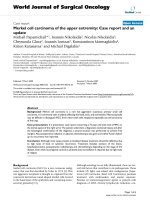
high power H & E stain and S100 stain of biopsyFigure 3
high power H & E stain and S100 stain of biopsy.
FDG PET CTFigure 1
FDG PET CT.
high power H & E stain and S100 stain of biopsyFigure 2
high power H & E stain and S100 stain of biopsy.
Journal of Medical Case Reports 2007, 1:50 />Page 3 of 3
(page number not for citation purposes)
tologically melanoma is composed of epithelioid cells
arranged in nests or spidle cells arranged in fasicles, with
or without melanin deposition of melanin pigment. If a
tumour is amelanotic, it may be difficult to recognise as
malignant melanoma without ancillary immunohisto-
chemical staining [2].
At the time of presentation, metastatic disease is present in
approximately 50% of patients, 31% hepatic, 29% medi-
astinal, 18% pulmonary, and 13% cerebral [3].
Other sites of primary melanoma must be excluded [4].
The Breslow thickness of tumour invasion for cutaneous
melanoma is a good predictor of outcome. However,
given that primary melanoma of the oesophagus is so
rare, it is difficult to apply this staging tool in this setting.
The principles involved in staging any histological type of
oesophageal malignancy are to distinguish between loco-
regional and systemic disease, to assess the extension of
local disease and to determine the possible response to
neo-adjuvant therapy.
Flurodeoxyglucose positron emission tomography (FDG
PET/CT) has proved to be an excellent method for staging
of metastatic melanoma. Due to its high sensitivity for
malignant lesions and the possibility of covering the
whole body in one examination, it can supplement other
staging tools.
Because of the high tumour-to-background ratio, FDG-
PET can highlight metastases at unusual sites that are
missed with conventional imaging modalities.
Furthermore, it provides information on the malignant
potential of the detected lesion. Given the relative scarcity
of primary melanomas of the oesophagus little is known
about its application for these tumours [5,6].
Ott et al recently reported that in oesophageal cancer FDG
PET/CT has been shown to detect metastatic disease in
approximately 20% of patients who are considered as
having only locoregional disease on CT, similar to the
patient reported in this case. The sensitivity of computed
tomography (CT) for detection of distant metastases
ranges between <50% and >90%. They reported a specifi-
city of 80% for locoregional pretherapeutic tumour stag-
ing [7].
Classical histological appearances are seen with tumour
marker staining. In the diagnosis of cutaneous melanoma
many specialized immunohistochemical stains may be
applied. S-100 staining has 95% sensitivity for melanoma.
HMB-45 staining is used for detecting active melanocytes.
Primary treatment is surgical excision with discretionary
lymphadenectomy for operable melanomas, but total or
near-total oesophagectomy offers the best survival out-
come (about 5 years, versus 9 months for local resection)
[4]. Additional immunomodulatory therapy may be used
if there is evidence of metastatic disease.
Adjuavant or neoadjuvant radiotherapy has been used but
its utility is unproven [8].
Interferon alpha is used in a range of neoplastic condi-
tions, including renal cell carcinoma, cutaneous
melanoma and chronic myeloid leukaemia. It stimulates
humoral and cell mediated response and thus has anti-
proliferative effects. However it is not curative. Well
known side effects to this therapy exist include flu-like
symptoms and fatigue but fortunately our patient toler-
ated his treatment well.
Competing interests
The author(s) declare that they have no competing inter-
ests.
Authors' contributions
JK collected all the included data, conceived of the study,
carried out detailed literature review and coordinated the
multi disciplinary approach to the manuscript.
PB oversaw all aspects of the reports' design and helped to
draft the manuscript.
ML carried out and reported on this rare histopathological
diagnosis.
All authors read and approved the final manuscript
Acknowledgements
Written consent was obtained from the patient for publication of this study.
References
1. [].
2. Chang F, Deere H: Esophageal melanocytosis morphologic fea-
tures and review of the literature. Arch Pathol Lab Med 2006,
130:552-557.
3. Chalkiadakis G, Wihlm JJM, Morand G, Weill-Bousson M, Witz JP:
Primary malignant melanoma of the esophagus. Ann Thorac
Surg 1985, 39:472-475.
4. Sabanathan S, Eng J, Pradhan GN: Primary malignant melanoma
of the esophagus. Am J Gastroenterol 1989, 84:1475-1481.
5. Kumar R, Alavi A: Clinical applications of flurodeoxyglucose-
positron emission tomography in the management of malig-
nant melanoma. Curr Opin Oncol 2005, 17(2):154-9.
6. Kumar R, Mavi A, Bural G, Alavi A: Flurodeoxyglucose-PET in the
management of malignant melanoma. Radiol Clin North Am
2005, 43(1):23-33.
7. Ott K, Weber W, Siewert : The importance of PET in the diag-
nosis and response evaluation of esophageal cancer. Dis
Esophagus 2006, 19(6):433-42.
8. Cadwell CB: Unusual malignant neoplasm of the esophagus. J
Thorac Cardiovasc Surg 1991, 101:100-107.