Báo cáo y học: "Giant hepatocellular adenoma as cause of severe abdominal pain: a case report" pot
Bạn đang xem bản rút gọn của tài liệu. Xem và tải ngay bản đầy đủ của tài liệu tại đây (867.58 KB, 4 trang )
BioMed Central
Page 1 of 4
(page number not for citation purposes)
Journal of Medical Case Reports
Open Access
Case report
Giant hepatocellular adenoma as cause of severe abdominal pain: a
case report
Luigi Sandonato
1
, Calogero Cipolla*
1
, Giuseppa Graceffa
1
,
Tommaso V Bartolotta
2
, Sergio Li Petri
1
, Oriana Ciacio
1
, Fabio Cannizzaro
2
and Mario A Latteri
1
Address:
1
Department of Oncology, Division of General and Oncological Surgery, University of Palermo, Palermo, Italy and
2
Department of
Radiology Interdepartmental Unit for Hepatic Neoplasia Group, University of Palermo, Palermo, Italy
Email: Luigi Sandonato - ; Calogero Cipolla* - ; Giuseppa Graceffa - ;
Tommaso V Bartolotta - ; Sergio Li Petri - ; Oriana Ciacio - ;
Fabio Cannizzaro - ; Mario A Latteri -
* Corresponding author
Abstract
The authors describe the case of a large hepatocellular adenoma diagnosed in a 30-year old woman
who came to us complaining of acute pain in the upper abdominal quadrants. The patient had been
taking an oral contraceptive pill for the last ten years. We present the clinical features, the
diagnostic work-up and the treatment prescribed.
Background
During the last 30–40 years, there has been an increase in
the incidence of hepatocellular adenoma (HCA), a rare
primary benign hepatic tumour, in young and middle-
aged women, which has been associated with the use of
oral contraceptives (OCs) [1].
These tumours may be found accidentally, or they may
present with pronounced symptoms, such as acute
abdominal pain or haemorrhage due to the rupture of the
tumour.
We describe the case of a 30 year-old female patient, who
had been taking oral contraceptives for the last ten years
and who came to our observation with severe upper
abdominal pain as the main symptom of a large HCA.
Case presentation
A 30-year-old woman was referred to us for the appear-
ance of frequent attacks during the previous week of acute
pain in the upper abdominal region; she also complained
of nausea and had vomited after eating on several occa-
sions. The patient had no history of abdominal disease
and reported that she had been taking a contraceptive pill
for the last ten years.
Clinical examination of the abdomen revealed a painful,
palpable mass with regular margins of about 6–8 cms. in
diameter located in the upper quadrants, between the epi-
gastric region and the left hypochondrium.
Laboratory tests at hospital admittance showed a slight
increase in serum transaminase (AST 56 U/1, ALT 73 U/
1), whereas haemochrome (RBC 4,70 × 10
6
× µl; Hb 12,9
g/dl), γGT, total and fractionated bilirubin, cholineste-
rase, glycaemia and serum electrolytes were all within nor-
mal limits; markers for hepatitis B and C were negative.
A direct X-ray did not show the presence of any abdomi-
nal dropsy, while an ultrasound examination (US)
Published: 27 July 2007
Journal of Medical Case Reports 2007, 1:57 doi:10.1186/1752-1947-1-57
Received: 19 April 2007
Accepted: 27 July 2007
This article is available from: />© 2007 Sandonato et al; licensee BioMed Central Ltd.
This is an Open Access article distributed under the terms of the Creative Commons Attribution License ( />),
which permits unrestricted use, distribution, and reproduction in any medium, provided the original work is properly cited.
Journal of Medical Case Reports 2007, 1:57 />Page 2 of 4
(page number not for citation purposes)
revealed a neoformation of about 8 cms in the left hepatic
lobe, with clear margins; the mass presented a dyshomo-
geneous echostructure, with hypoechogenic areas alter-
nating with hyperechogenic zones.
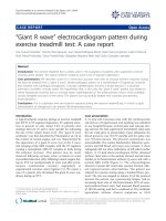
An abdominal CT scan using a contrast medium and
triphasic techniques, performed 48 hours after admit-
tance, confirmed the presence of a nodular lesion of about
10 cms in diameter in the left lobe of the liver compress-
ing the gastric corpus and fundus but with no signs of
infiltration. The lesion showed a dyshomogeneous den-
sity with irregular enhancement in the arterial phase and
wash-out in the late phase. No infiltrations were observed,
either in the parenchyma or within the vascular structure
(Fig. 1a).
The diagnostic work-up, together with the medical history
of the long-term use of OCs, led us to suspect a large HCA.
We therefore performed a liver biopsy for histopathologic
examination which showed a shaft of hepatic tissue con-
taining a few vacuolated hepatocytes within an area of
widespread necrosis; no portal or biliary structures were
present. Morphological examination of the specimen sug-
gested a diagnosis of hepatic adenoma, although this
could not be considered as conclusive.
Four days after admittance, the pain was still present and
there was also a reduction of red blood cells and haemo-
globin (Rbc 3,69 × 10
6
× µl; Hb 9,90 g/dl), together with
a further increase in transaminase (AST 156 U/1, ALT 473
U/1) and an increase of LDH (810 IU/l).
Magnetic resonance (MR) was therefore performed with
the use of a paramagnetic contrast medium, and this con-
firmed the presence of a lesion of 12 cms, with clear mar-
gins, taking up all the II and III segments of the liver (Fig.
1b) and also another nodule of 1 cm with the same fea-
tures in the VI segment. Several interlesional areas show-
ing up as spontaneously hyperintense in the T1-w
sequences indicated probable recent bleeding.
Not only was the tumour extremely large and causing con-
siderable pain, most probably due to the distension of
Glisson's capsule, but laboratory tests and imaging also
revealed endotumoral bleeding, indicating a probable
rupture of the neoplasia, and we thus decided that surgery
was indicated.
At this point it was thus decided to perform an angio-
graphic study in order to check hepatic vascularisation.
Catheterisation of the coeliac tripod, performed during
preoperative angiographic examination of the hepatic vas-
cularisation, showed the presence of a large dyshomoge-
neously hypervascular neoformation within the left
hepatic lobe associated with the presence of an anatomic
variant in which the left hepatic artery originated from the
right gastric artery. Selective catheterisation of the com-
mon hepatic artery did not reveal any opacity within the
neoformation (Fig. 1c).
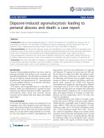
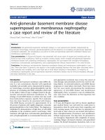
The patient therefore underwent left hepatic lobectomy
(Fig. 2) and focal resection of about 1.5 cms of the VI seg-
ment (Fig. 3). Intraoperative ultrasound did not reveal
any further lesions. There were no post-operative compli-
cations and the patient was sent home on Day VII.
The anatomopathological examination showed a mass of
11.5 cms with a smooth, regular external surface and well-
Intraoperative view of the tumourFigure 2
Intraoperative view of the tumour.
A: Neoformation in the left lobe (CT imaging) B: Neofoma-tion in the left lobe (MR imaging)) C: Angiography: no vision of the left hepatic lobe or neoformationFigure 1
A: Neoformation in the left lobe (CT imaging) B: Neofoma-
tion in the left lobe (MR imaging)) C: Angiography: no vision
of the left hepatic lobe or neoformation.
Journal of Medical Case Reports 2007, 1:57 />Page 3 of 4
(page number not for citation purposes)
defined margins; the walls were of a yellowish colour and
in the centre there was a wide area of necrotic, haemor-
rhagic tissue extending almost as far as Glisson's capsule.
Microscopic examination showed the presence of mature,
vacuolated hepatocytes; no portal or biliary structures
were present, which confirmed the diagnosis of hepatic
adenoma. The smaller nodule in the VI segment presented
exactly the same histopathological features.
Following surgery, the patient stopped taking OCs and
ultrasound follow-up examination at six months did not
reveal the presence of any further focal lesions.
Conclusion
HCA is a primary benign tumour of hepatocellular origin
rarely seen before the introduction of OCs in the 1960s
[1]. In 1973, Baum et al were the first to suspect a link
between HCA and use of OCs [2]. More often than not,
patients with HCA have no symptoms and present a nor-
mal liver function with no rise in alpha-foetoprotein
serum level. Large adenomas, however, may cause anae-
mia because of tumoral bleeding, or pain in the upper
abdominal quadrants with abdominal distress, and may
lead to spontaneous rupture or haemorrhage and, in cer-
tain rare cases, even death.
Several diagnostic procedures, such as US, CT and MR, can
indicate the presence of an HCA, but this diagnosis must
be confirmed by the histopathological examination.
Most HCAs are usually first detected at US. The US HCA
may appear as a hyperechoic lesion or else as a hypo- or
anechoic solid mass, well-circumscribed and rarely capsu-
lated. A mixed appearance is typical of voluminous and
dyshomogeneous masses presenting haemorrhage or
necrosis; calcifications are rarely present.
Colour Doppler US may provide some clues for distin-
guishing HCA from FNH, since the former shows a con-
tinuous venous flow in the central portion and either a
pulsatile or continuous peripheral flow ('basket pattern').
These findings are absent in FNH, in which colour Dop-
pler US may show a typical spoke-wheel arterial pattern of
vessels [3].
On pre-contrast CT scans, HCA has a varied and non-spe-
cific appearance, possibly with hypoattenuating areas due
to the presence of fat, previous haemorrhage or necrosis,
whereas recent haemorrhage or large amounts of glycogen
are observed as hyperdense areas. On contrast CT, HCA
often shows substantial enhancement during the arterial
phase, decreasing during the portal phase and gradually
becoming iso- or hypodense in the liver on delayed scans.
In some cases, there may be a thin, continuous hyper-
dense rim due to the presence of a peripheral capsule [4].
HCAs frequently show heterogeneous hypointensity both
on un-enhanced T1-weighted and T2-weighted images
due to the presence of fat, haemorrhage, or necrosis [5].
Sometimes, a peripheral rim, corresponding histologi-
cally to a pseudocapsule, is seen as a low signal-intensity
rim on both T1-weighted and T2-weighted images [6,7].
After the administration of gadolinium-chelates, most
HCAs show intense enhancement in the arterial phase
and are isointense in liver tissue on portal-venous and
equilibrium images [8]. Hepatocellular-specific contrast
agents may provide useful clues for distinguishing HCA
from FNH. After the injection of such an agent, for exam-
ple gadolinium benzyloxypropionictetraacetate (Gd-
BOPTA), HCA typically appears hypointense on delayed
phase imaging, due to the lack of biliary ducts, whereas
FNH generally appears isointense or slightly hyperintense.
When reticuloendothelial cell-targeted contrast agents are
used, such as ferumoxides (superparamagnetic iron
oxides), some HCAs may show some signal intensity loss,
which might be explained by the presence of Kupffer cells
in the lesions.
The various imaging techniques, which are extremely
important for the diagnosis of HCA, are particularly useful
in all those cases where pain is among the symptoms. The
lesion should be kept under strict observation and any
rapid increase in volume with endotumoral bleeding
linked to a reduction of haemochrome parameters should
suggest an immediate surgical approach in order to avoid
complications which might be brought about by possible
rupture of the tumour.
In the case observed by us, the US and the CT scan using
a contrast medium and triphasic techniques were essential
for reaching a diagnosis. The ultrasound and radiographic
features of the lesion, correlated to a medical history of
the long-term use of Ocs, made it possible right from the
Removal of the nodule from the VI segmentFigure 3
Removal of the nodule from the VI segment.
Journal of Medical Case Reports 2007, 1:57 />Page 4 of 4
(page number not for citation purposes)
beginning of the diagnostic work-up to suspect the pres-
ence of an HCA. Nevertheless, we considered that this
diagnostic hypothesis should be confirmed by means of a
biopsy, since, although the risk of malignant transforma-
tion of an HCA is fairly low, it may occur, and would be
an important indication for a surgical approach. In any
case, this treatment is not particularly invasive, is well-tol-
erated and does not generally involve a high rate of com-
plications.
During hospitalisation, an MR examination performed
because the patient was becoming progressively more and
more anaemic showed a further increase in the size of the
lesion and probable endotumoral bleeding. Since this fact
indicated a surgical approach due to the high risk of
endoperitoneal rupture of the lesion, we immediately per-
formed selective angiography of the liver in order to eval-
uate vascularisation of the organ and of the tumour. This
examination revealed the presence of an anatomic variant
in which the left hepatic artery originated from the right
gastric artery, and showed that the neoplasia was vascular-
ised by the left hepatic artery.
The therapeutic approach to HCA is still not clear. For
asymptomatic patients, conservative treatment requires
stringent follow-up with ultrasonography of the liver and
only in the case of further growth is surgical treatment
indicated. In a recent review regarding the indications for
a surgical approach towards benign hepatic neoplasias[9],
it has been pointed out that no randomised clinical trials
have ever been conducted and that most published
reports involve a small number of cases of various types of
tumours. Vast, long-term randomised clinical trials with
adequate methodology are need for a valid assessment of
the advantages and disadvantages of elective surgery for
benign liver tumours.
The role of elective surgical resection for HCA is still con-
troversial and mainly depends upon the risk of complica-
tions, the uncertain diagnosis and the presence of
symptoms related to tumour size and site, particularly
with regard to the risk of rupture and resulting haemor-
rhage [1,9]. Elective resection of HCA has a mortality rate
of less than 1%, whereas the mortality rate with free rup-
ture is 5 to 10%.
In the case observed by us, the persistent pain caused by
the tumoral growth and the resulting distension of Glis-
son's capsule, together with progressive anaemia brought
about by endotumoral bleeding, led us to suspect the pos-
sible rupture of the tumour with consequent haemoperi-
toneum; immediate surgery was thus considered the
treatment of choice.
In conclusion, HCA is a rare benign tumour of the liver,
generally involving a history of a prolonged use of OCs.
Accurate diagnostic imaging almost always provides a cor-
rect differential diagnosis from other benign tumours of
the liver. Although there is still some doubt regarding the
therapeutic approach to asymptomatic patients, surgery is
probably indicated in large-size HCAs with or without
abdominal symptoms in order to avoid certain complica-
tions such as haemorrhage or rupture of the tumour.
Competing interests
The author(s) declare that they have no competing inter-
ests.
Authors' contributions
LS, CC, and GG performed the operation, assessed the
didactic importance of the clinical case and therefore
acquired the data; TVB and FC were responsible for the
imaging; SLP and OC. TVB and FC were involved in draft-
ing the manuscript and its revision; MAL gave the final
approval of the version to be published.
All authors read and approved the final manuscript.
Acknowledgements
The Authors thank the patient involved for permitting the publication of the
data regarding her case.
The Authors thank Prof. Antonio Craxì for helpful discussion.
References
1. Soe KL, Soe M, Gluud S: Liver pathology associated with the use
of anabolic-androgenic steroids. Liver 1992, 12:73-79.
2. Baum JK, Bookstein JJ, Holtz F, Kein EW: Possible association
between benign hepatomas and oral contraceptives. Lancet
1973, ii:926-29.
3. Bartolozzi C, Lencioni R, Paolicchi A, Moretti M, Armillotta N, Pinto F:
Differentiation of hepatocellular adenoma and focal nodular
hyperplasia of the liver: comparison of power Doppler imaging
and conventional color Doppler sonography. Eur Radiol 1997,
7:1410-5.
4. Ichikawa T, Federle MP, Grazioli L, Marsh W: Hepatocellular ade-
noma: multiphasic CT and histopathologic findings in 25
patients. Radiology 2000, 214:861-8.
5. Paulson EK, McClellan JS, Washington K, Spritzer CE, Meyers WC,
Baker ME: Hepatic adenomas: MR characteristics and correla-
tion with pathologic findings. Am J Roentgenol 1994,
163(1):113-116.
6. Chung KY, Mayo-Smith WW, Saini S, Rahmouni A, Golli M, Mathieu D:
Hepatocellular adenoma: MR imaging features with patho-
logic correlation. Am J Roentgenol 1995, 165(2):303-308.
7. Bartolozzi C, Cioni D, Donati F, Lencioni R: Focal liver lesions: MR
imaging-pathologic correlation. Eur Radiol 2001, 11:1374-88.
8. Grazioli L, Kirchin M, Pirovano G, Spinazzo A: MultiHance in the
dynamic phase of contrast enhancement: a pictorial assess-
ment. J Comput Assist Tomogr 1999, 23(Suppl 1):61-4.
9. Colli A, Flaquelli M, Massironi S, Colucci A, Paggi S, Conte D: Elective
surgery for benign liver tumours. Cochrane Database Syst Rev Jan
24 :CD005164.