Báo cáo khoa hoc:" Perigraft air is not always pathological: a case report" docx
Bạn đang xem bản rút gọn của tài liệu. Xem và tải ngay bản đầy đủ của tài liệu tại đây (223.06 KB, 3 trang )
BioMed Central
Page 1 of 3
(page number not for citation purposes)
Journal of Medical Case Reports
Open Access
Case report
Perigraft air is not always pathological: a case report
Elizabeth Ball, Gareth Morris-Stiff*, Mari Coxon and Michael H Lewis
Address: Department of Surgery, Royal Glamorgan Hospital, Ynysmaerdy, Llantrisant, UK
Email: Elizabeth Ball - ; Gareth Morris-Stiff* - ; Mari Coxon - ;
Michael H Lewis -
* Corresponding author
Abstract
Background: The presence of perigraft air is a common finding in the immediate post-operative
phase following abdominal aortic aneurysm repair whilst the later appearance of air, in association
with elevated inflammatory markers, is regarded as being indicative of the serious complication of
graft infection. What is not known is at what timepoint following surgery does the perigraft air
become a significant finding.
Case Presentation: We report the case of a 71 year old man who underwent a computed
tomography scan 15 days following repair of an abdominal aortic aneurysm because of the presence
of unexplained pyrexia. The scan showed the presence of perigraft air and a small haematoma. The
patient was managed conservatively and after 6 weeks the air and haematoma had resolved
completely.
Conclusion: The presence of perigraft air in the early postoperative phase is probably a normal
finding, is not associated with graft infection and can be managed non-operatively.
Background
Intra-abdominal free gas is a normal finding in the imme-
diate postoperative period following a laparotomy. Stud-
ies evaluating the role of computerised tomography (CT)
scanning in the early postoperative period (within 7 days
of surgery) following aortic aneurysm repair have simi-
larly shown that periprosthetic air is a not uncommon
finding and simply represents air trapped in the tissue
planes between the graft and the aneurysm sac [1].
However, periprosthetic gas later in the postoperative
period following abdominal aortic aneurysm repair is not
such a benign finding and is said to be a reliable indicator
of graft infection. This complication is associated with a
mortality rate of 25–75% [1,2]. Few investigators have
looked at the early postoperative period following resolu-
tion of the 'laparotomy' air.
Case presentation
A 71 year old retired driver presented to the vascular clinic
with a calf claudication distance of three hundred yards.
He also complained of rest pain in his toes. He had been
a non-smoker for thirteen years. His risk factors for
peripheral vascular disease included diet-controlled dia-
betes and hypertension. He had suffered a left-sided
stroke fifteen years prior to admission, from which he had
made a full recovery. He was taking regular aspirin, allop-
urinol and an oral hypoglycaemic.
Examination of his abdomen revealed a tender pulsatile
epigastric mass. All his peripheral pulses were present and
Published: 12 August 2007
Journal of Medical Case Reports 2007, 1:63 doi:10.1186/1752-1947-1-63
Received: 9 March 2007
Accepted: 12 August 2007
This article is available from: />© 2007 Ball et al; licensee BioMed Central Ltd.
This is an Open Access article distributed under the terms of the Creative Commons Attribution License ( />),
which permits unrestricted use, distribution, and reproduction in any medium, provided the original work is properly cited.
Journal of Medical Case Reports 2007, 1:63 />Page 2 of 3
(page number not for citation purposes)
there were no bruits in either his femoral arteries or
adductor canals. An ultrasound scan confirmed the pres-
ence of an abdominal aortic aneurysm measuring 5.2 ×
5.5 cm, with normal calibre iliac vessels. A duplex scan
showed a fifty percent stenosis in the mid-portion of the
left superficial femoral artery. Because of the tenderness,
the patient was admitted and underwent elective repair of
his aneurysm using a woven polyester graft. The proce-
dure was uncomplicated and no haemostatic agents were
used.
On the fifth post-operative day he developed pyrexia of
38°C. His white cell count at that time was 9.2 × 10
9
/l.
Clinical examination was unremarkable. Urine, blood
and wound cultures were sterile and a chest radiograph
was unremarkable.
On the eighth post-operative day the patient complained
of pain and loss of feeling in his left leg. On examination
his foot was cool, and pedal pulses were impalpable. A
duplex scan showed that the left superficial femoral artery
was occluded throughout its length. The patient was taken
to theatre for an emergency left femoral embolectomy.
The procedure was successful, he made a good recovery
and regained both sensation and movement in his leg.
Over the course of the subsequent week, the patient con-
tinued to exhibit a mild pyrexia despite absence of symp-
toms of graft infection such as malaise or back pain, and
he did not experience any gastrointestinal tract bleeding.
Furthermore, inflammatory markers including full blood
count, C-reactive protein and erythrocyte sedimentation
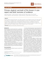
rate were normal and all cultures were sterile. A computed
tomography scan on the fifteenth post-operative day
using both intravenous and oral contrast (Figure 1)
showed a cuff of abnormal soft tissue, consistent with a
small perigraft haematoma, with gas bubbles surrounding
the lower end of the graft.
He was closely observed for a further week. Repeat blood
cultures were negative, and inflammatory markers
remained within normal limits. As the patient was now
apyrexial and continued to be asymptomatic, the decision
was made to discharge the patient. A repeat CT scan per-
formed six weeks following his aortic surgery showed that
the perigraft air had completely resolved and the hae-
matoma had organised. The patient has been followed up
for two year post-operatively and remains asymptomatic.
Discussion
Graft infection is a recognised but catastrophic complica-
tion of aortic bypass surgery, with mortality between 25
and 75% [1,3]. The corrective treatment also carries a high
morbidity and mortality. Graft sepsis can be difficult to
identify in the early post-operative period. The clinical
presentation may be straightforward. However it can also
present with non-specific symptoms such as malaise, back
pain and fever. With such a high mortality it is vital to
diagnose this potentially life-threatening condition as
quickly as possible and CT is the imaging modality of
choice.
There is very little data detailing the natural history of
periprosthetic air in the early post-operative period. Two
prospective studies have been performed with similar
results. Qvardfordt et al. [2] studied 29 patients who
underwent reconstructive aortic surgery, performing a CT
scan at 7, 48 and 102 days post-operatively. Only 4
patients had perigraft air at 7 days, and this air had com-
pletely resolved by day 28. O'Hara et al. looked at 26
patients, scanning them on days 3, 7 and 52. Seventeen
patients had perigraft air on day 3, and seven on day 7. No
patient had residual perigraft air on the final scan.
There is however no data regarding perigraft air in the
period 2–4 weeks following abdominal aortic surgery
such as in our case. O'Hara et al. found that patients with
larger aneurysms (especially over 6 cm) have a higher inci-
dence of perigraft air being detected on an early CT scan.
Our patient had loculated perigraft air on a CT scan per-
formed two weeks after surgery which had resolved by 6
weeks.
CT scan on 15
th
postoperative day demonstrating a rim of air around the graftFigure 1
CT scan on 15
th
postoperative day demonstrating a rim of air
around the graft.
Publish with BioMed Central and every
scientist can read your work free of charge
"BioMed Central will be the most significant development for
disseminating the results of biomedical research in our lifetime."
Sir Paul Nurse, Cancer Research UK
Your research papers will be:
available free of charge to the entire biomedical community
peer reviewed and published immediately upon acceptance
cited in PubMed and archived on PubMed Central
yours — you keep the copyright
Submit your manuscript here:
/>BioMedcentral
Journal of Medical Case Reports 2007, 1:63 />Page 3 of 3
(page number not for citation purposes)
In this case, as the inflammatory markers were normal, it
is likely that the air identified on the CT scan simply rep-
resented air remaining following the initial repair that had
not completely resolved. Another option is that this may
have been indicative of a subclinical infection although
this is less likely as no infection has become evident dur-
ing 2 years of follow-up and there were no other signs of
graft infection such as: persisting perigraft fluid; or pseu-
doaneurysm [1,4,5]. There was a little perigraft soft tissue
attenuation which was believed to be due to a resolving
haematoma in keeping with the recent surgery. In addi-
tion there were no signs associated with the presence of an
aortoenteric fistula such as focal bowel wall thickening or
paraprosthetic extravasation of enteric contrast or of intra-
venous contrast.
Had there been systemic evidence of infection then addi-
tional radiological investigations, in particular isotope
studies such as indium-111 white blood cell, gallium-67
citrate, or Tc-99m hexametazime scanning could have
been performed to try and identify perigraft infection [1].
We suggest that there is a need for further studies to accu-
rately record the natural history of perigraft air in the first
month following surgery, as not all cases may represent
infected grafts. The question arises as to how often you
repeat a CT scan having found post-operative perigraft air,
and whether the finding of perigraft air with non-specific
clinical symptoms indicates early graft infection, or a nor-
mal stage in the healing process.
Competing interests
The author(s) declare that they have no competing inter-
ests.
Authors' contributions
All authors have read and approved the final manuscript.
Acknowledgements
The original idea was that of G Morris-Stiff and MH Lewis (consultant in
charge of the case). The manuscript was written by E Mall and M Coxon
and the manuscript was edited by G Morris-Stiff.
The authors confirm that informed written consent was received for pub-
lication of the manuscript.
References
1. Orton DF, LeVeen RF, Saigh JA, Culp WC, Fidler JL, Lynch TJ,
Gertzen TC, McCowan TC: Aortic prosthetic graft infections:
radiologic manifestations and implications for management.
Radiographics 2000, 20:976-993.
2. Qvarfordt PG, Reilly LM, Mark AS, Goldstone J, Wall SD, Ehrenfeld
WK, Stoney RJ: Computerised tomographic assessment of
graft incorporation after aortic reconstruction. American Jour-
nal of Surgery 1985, 150:227-231.
3. O'Hara PJ, Borkowski GP, Hertzer NR, O'Donovan PB, Brigham SL,
Beven EG: Natural history of periprosthetic air on computer-
ized axial tomographic examination of the abdomen follow-
ing abdominal aortic aneurysm repair. Journal of Vascular
Surgery 1984, 1:429-433.
4. Low RN, Wall SD, Jeffrey RB Jr, Sollitto RA, Reilly LM, Tierney LM:
Aortoenteric fistula and perigraft infection: evaluation with
CT. Radiology 1990, 175:157-162.
5. Peirce RM, Jenkins RH, MacEneaney P: Paraprothetic extravasa-
tion of enteric contrast: a rare and direct sign of secondary
aortoenteric fistula. American Journal of Roentgenology 2005,
184:S73-S74.