Báo cáo khoa hoc:" Occlusal management for a patient with aural symptoms of unknown etiology: a case report" pdf
Bạn đang xem bản rút gọn của tài liệu. Xem và tải ngay bản đầy đủ của tài liệu tại đây (246.77 KB, 4 trang )
BioMed Central
Page 1 of 4
(page number not for citation purposes)
Journal of Medical Case Reports
Open Access
Case report
Occlusal management for a patient with aural symptoms of
unknown etiology: a case report
Kengo Torii*
1
and Ichiro Chiwata
2
Address:
1
Torii dental clinic, 1-23-2 Ando, Aoi-ku, Shizuoka-shi, 420-0882, Japan and
2
Chiwata dental clinic, 2-1-3 Gofuku-cho, Aoi-ku, Shizuoka-
shi, 420-0031, Japan
Email: Kengo Torii* - ; Ichiro Chiwata -
* Corresponding author
Abstract
Background: Although the discrepancy between the habitual occlusal position (HOP) and the flat
bite plate-induced occlusal position (BPOP) (regarded as the muscular physiological reference
position) has been recently reported to be related to symptoms of temporomandibular disorders
(TMDs), it still remains unclear whether the occlusal equilibration in the reference position is
effective to resolve TMD-related discrepancy and symptoms. Aural symptoms (otalgia, tinnitus,
vertigo et cetera) have been included under TMD symptoms.
Methods: To examine the effect of occlusal equilibration for the treatment of TMDs, occlusal
equilibration was performed for a patient with aural symptoms (otalgia, tinnitus and vertigo) of
unknown etiology in the right ear. An occlusal analysis was performed on this patient with dental
models mounted on an articulator after relieving painful symptoms by an appliance therapy and a
discrepancy was identified (p < 0.005). Occlusal equilibration in the BPOP was then performed for
the patient by selective tooth grinding, because it was estimated that the interocclusal space
between upper and lower occlusal surfaces would be rectified by selective grinding.
Results: At completion of treatment, the discrepancy was not significant (p > 0.25), and the
patient's right condyle had shifted 2.8 mm posteromedially in the horizontal plane, and the left
condyle had shifted 1.0 mm laterally in the voluntarily closed position from the previous HOP. The
aural symptoms of the patient were resolved, and there has been no recurrence to date after a
two-year follow-up period.
Conclusion: An occlusal analysis should be performed in patients exhibiting TMD symptoms to
identify the presence or absence of any discrepancy between the HOP and the BPOP. If a
discrepancy exists, occlusal equilibration should be attempted in the reference position.
Background
Aural symptoms have been reported to sometimes occur
in temporomandibular disorders (TMDs) [1,2], and there
have been many reports of improvement of aural symp-
toms after various treatments for TMD [3,4]. A variety of
hypotheses have been proposed to explain the aural
symptoms [5,6]. The relation between TMD and occlu-
sion has been the subject of considerable debate and con-
troversial. However, it has been recently demonstrated
that a discrepancy between the habitual occlusal position
Published: 12 September 2007
Journal of Medical Case Reports 2007, 1:85 doi:10.1186/1752-1947-1-85
Received: 30 June 2007
Accepted: 12 September 2007
This article is available from: />© 2007 Torii and Chiwata; licensee BioMed Central Ltd.
This is an Open Access article distributed under the terms of the Creative Commons Attribution License ( />),
which permits unrestricted use, distribution, and reproduction in any medium, provided the original work is properly cited.
Journal of Medical Case Reports 2007, 1:85 />Page 2 of 4
(page number not for citation purposes)
(HOP) and the flat bite plate-induced occlusal position
(BPOP) is associated with TMD symptoms [7]. The HOP
is obtained by voluntary jaw closing while a patient is sit-
ting in the upright position. The BPOP is obtained during
voluntary jaw closing while the patient is sitting in the
upright position and after wearing an anterior flat bite
plate that covers the 6 upper anterior teeth and both first
premolars for a brief time, and then removing it. The
BPOP has been regarded as the physiologic masticatory
muscular position and a reference position for occlusal
analysis. The effect of occlusal equilibration in BPOP on
TMD symptoms has also been inferred [7,8]. Therefore, it
is worth to examine the occlusal equilibration in the
BPOP for TMD patient. The following case report
describes a patient who presented with aural symptoms of
unknown etiology. Occlusal equilibration in the BPOP
was found to be effective against aural symptoms, and it
might be effective against other TMD symptoms.
Case presentation
A 73-year old woman presented with the chief complaint
of severe pain and tinnitus in the right ear. She reported
that when performing karaoke 2 years previously, she sud-
denly felt a strong impact on her right ear and developed
such severe rotatory vertigo that she was unable to remain
standing and had to crawl on the floor. She was diagnosed
as having Ménière's disease by two independent otorhi-
nolarygologists and was treated with isosorbide, meco-
balamin and ibudilast. However, when the drugs failed to
improve her severe symptoms, she came to our dental
clinic. The patient's medical history was unremarkable.
No impairment of mouth opening, no deviation of the
opening path, and no noises in either of the temporoman-
dibular joints (TMJs) was detected. The patient reported
pain on palpation of the right TMJ and tenderness of the
right lateral pterygoid muscle. Twenty-nine teeth present,
and the upper right third molar was slightly extruded
because there was no opposing tooth. Occlusion was ana-
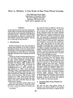
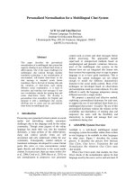
tomically normal. The right TMJ appeared unclear on the
TMJ computed tomography (CT) images in the HOP from
the first consultation (Fig. 1). The cause of the lack of clar-
ity was unknown, but it seemed to be attributable to
inflammation, or to the eccentricity the condyle in the gle-
noid fossa.
For this patient, three HOP records were obtained by vol-
untary jaw closing at the first visit, while the patient was
seated in an upright position with the occlusal plane par-
allel to the floor using a vinyl polysiloxane bite registra-
tion material (GC, Tokyo, Japan). Since an anterior flat
bite plate has been recommended as a provisional appli-
ance to decrease painful symptoms [4], an anterior flat
bite plate was directly fabricated in the patient's mouth
using clear self-curing acrylic resin (GC, Tokyo, Japan),
which the patient wore every night for a week. At the sec-
ond visit, (one week after the first visit), her otalgia had
ceased, and the intensity of the tinnitus was greatly dimin-
ished. The BPOP records were obtained during voluntary
jaw closing, in the upright position and after wearing the
bite plate for five minutes using the same material as in
the HOP. Three BPOP records were obtained. To examine
the difference between the HOP and BPOP, three-dimen-
sional measurements were performed on the modified
articulator using previous records [7]. The difference
between the HOP and BPOP was significant using an
analysis of variance (ANOVA) for a two factor experiment
with repeated measurements for both position factors (p
< 0.005). An occlusal analysis was performed on dental
models mounted on an articulator (Gnatho-Tech, Shi-
zuoka, Japan) with a BPOP wax (Kerr, Romulus, MI,
U.S.A.) record. One of several therapies (selective tooth
grinding, prosthodontic and orthodontic treatments and
orthognathic surgery) may be selected for occlusal equili-
bration in the BPOP, according to the degree of discrep-
ancy between the HOP and BPOP. In the present case,
extraction of the right upper third molar was selected first,
because of premature occlusal contact in the BPOP. Sub-
sequently, tooth grinding was selected, because it was esti-
mated that the interocclusal space between the upper and
lower occlusal surfaces would be rectified by selective
grinding. The instability of the patients' occlusion in
BPOP and the need of occlusal equilibration were
explained to the patient using the articulated dental mod-
els on an articulator. She gave oral informed consent for
performing occlusal equilibration in the BPOP. The upper
right third molar was extracted. After verifying the consist-
Tomographyic image showing an unclear right TMJ in the HOP before treatmentFigure 1
Tomographyic image showing an unclear right TMJ in the
HOP before treatment.
Journal of Medical Case Reports 2007, 1:85 />Page 3 of 4
(page number not for citation purposes)
ency of the BPOP using a split cast method on 3 consecu-
tive visits at 3-day intervals on the articulator, selective
grinding in the BPOP was performed during 5 appoint-
ments over 3 weeks until occlusal contacts of the premolar
and molar teeth were achieved on both sides. For selective
grinding in the patient's mouth, the anterior bite plate was
worn in the moth and the patient was asked to tap and
slide her anterior teeth against the plate for five minutes
while in an upright position. The plate was removed, and
the patient was asked to close her jaw until tooth contact
was made and then hold that position. Premature contact
was located in the mouth by marking and pulling with
occlusal tape (Hanel, Langenau, Germany). The consist-
ency of the BPOP was confirmed at every selective grind-
ing time.
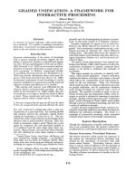
At the completion of the selective tooth grinding, the dif-
ference between the HOP and BPOP was not significant
(p > 0.25), and the right condyle in the voluntarily closed
position had shifted 2.8 mm posteromedially in the hori-
zontal plane, and the left condyle had shifted 1.0 mm lat-
erally from the previously recorded HOP at the first visit
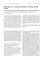
(Fig. 2), and the right TMJ on tomography images
appeared to be clear than that obtained at the first exami-
nation (Fig. 3). The patient's aural symptoms resolved,
and there has been no recurrence in symptoms and no
reappearance of the discrepancy between the HOP and
BPOP to date after a two-year follow-up period.
Discussion
There have been many reports of relief or complete elimi-
nation of otalgia, tinnitus, vertigo, and deafness by vari-
ous treatments for TMDs [3,4]. However, the outcomes of
these various conservative therapies for TMDs in relation
to the aural symptoms have always been an improvement
in symptoms rather than a cure, because they were not
established on the basis of the pathogenic mechanism
underlying the development of the aural symptoms. The
occlusal equilibration in our patient was performed based
on evidence of a significant association between the dif-
ference between the HOP and BPOP and TMJ sounds [7].
The large shift in the position of the right condyle in the
HOP in our patient after treatment means that the right
condyle had deviated anterolaterally before treatment. In
regard to the development of aural symptoms, William-
son [5] hypothesized that vertigo was caused by noxious
pain stimuli to the peridiskal tissues inducing constriction
of the internal auditory and posterior auricular arteries
thus decreasing the blood supply. Myers [6] described the
disk displacement in the TMJ as being accompanied by
stretching of the posterior laminae and stretching or tear-
ing of the lateral collateral ligament. Myers claimed that
this injury caused chronic inflammation with extension of
the inflammatory areas. This occurs especially in the
carotid sheath area, and causes adhesion of arteries, veins,
and nerves of the sheath, such that the indirect tension
along the carotid sheath caused by the adhesion interferes
with the pressure-regulating function of the saccus endol-
ymphaticus. The end result of this process is increased
endolymphatic pressure and development of aural symp-
toms. In our patient, it was considered that the patholog-
ical conditions described by Williamson [5] or Myers [6]
probably occurred with large deviations of the right con-
dyle. Moyers [8] described that the persistence of occlusal
disharmony in centric relation might cause a new reflex
pattern of pathways to be repeatedly used, causing the
The shifts recorded on both condylar areas of the articulatorFigure 2
The shifts recorded on both condylar areas of the articula-
tor. The degree of the shift indicates the actual change in the
patient's condyles because the intercondylar distance of the
articulator is adjusted to the same distance as that of the
patient.
Tomographic image showing a clear right TMJ in the HOP after treatmentFigure 3
Tomographic image showing a clear right TMJ in the HOP
after treatment.
Publish with BioMed Central and every
scientist can read your work free of charge
"BioMed Central will be the most significant development for
disseminating the results of biomedical research in our lifetime."
Sir Paul Nurse, Cancer Research UK
Your research papers will be:
available free of charge to the entire biomedical community
peer reviewed and published immediately upon acceptance
cited in PubMed and archived on PubMed Central
yours — you keep the copyright
Submit your manuscript here:
/>BioMedcentral
Journal of Medical Case Reports 2007, 1:85 />Page 4 of 4
(page number not for citation purposes)
new position of the mandible to represent the intercuspal
position. The discrepancy between the HOP and BPOP in
our patient might have emerged as a result of persistent
premature occlusal contact of the upper right third molar
during its extrusion.
A proper occlusal analysis should be performed with the
BPOP record obtained after relief of the painful symptoms
by appliance therapy. Selective tooth grinding in volun-
tary jaw closing has been considered unreliable; however,
the reproducibility of the jaw closing can be improved by
wearing a bite plate. When performing selective grinding
in the BPOP, it is important to check to see whether the
BPOP is consistent, because the BPOP in TMD patients is
variable [7]. The consistency of BPOP wax records on
three consecutive visits at 3- to 7-day intervals needs to be
confirmed on an articulator. In the present study, the
selective tooth grinding in the BPOP seemed to provide a
"cure" for our patient who had vertigo, tinnitus and otal-
gia.
Conclusion
An occlusal analysis should be carried out in patients with
TMD-related symptoms to identify whether a discrepancy
exists between the HOP and BPOP (the latter being
regarded as a physiological reference position). If a dis-
crepancy does in fact exist, the occlusal equilibration in
the reference position should be attempted.
Competing interests
The author(s) declare that they have no competing inter-
ests.
Authors' contributions
KT conceived of the study, and participated in the design-
ing the study and perfomed the occlusal analysis and
selective grinding. IC carried out the three-dimensional
measurements and performed the statistical analysis.
The authors read and approved the final manuscript.
Acknowledgements
Written consent was obtained from the patient for publication of the study.
References
1. Rubinstein B: Tinnitus and craniomandibular disorders: Is
there a link. Swed Dent J 1993, 96(Suppl):1-46.
2. Chan SWY, Reade PC: Tinnitus and temporomandibular pain-
dysfunction disorder. Clin Otolaryngol Allied Sci 1994, 19:370-380.
3. Erlandsson SI, Rubinstein B, Carlsson SG: Tinnitus: evaluation of
biofeedback and stomatognathic treatment. Br J Audiol 1991,
25:151-161.
4. Wright EF, Bifano SL: Tinnitus improvement through TMD
therapy. J Am Dent Assoc 1997, 128:1424-1432.
5. Williamson EH: The interrelationship of internal derangement
of the derangement of the temporomandibular joint, head-
ache, vertigo, and tinnitus: A survey of 25 patients. J Cranio-
mandib Pract 1990, 8:301-306.
6. Myers LJ: Possible inflammatory pathways relating temporo-
mandibular joint dysfunction to otic symptoms. J Craniomandib
Pract 1988, 6:64-70.
7. Torii K, Chiwata I: Relationship between habitual occlusal posi-
tion and flat bite plane induced occlusal position in volun-
teers with and without temporomandibular joint sounds. J
Cranoimandib Pract 2005, 23:16-21.
8. Moyers RE: Some physiological consideration of centric and
other jaw relations. J Prosthet Dent 1956:183-194.