Báo cáo khoa hoc:" Vitamin C-induced hyperoxaluria causing reversible tubulointerstitial nephritis and chronic renal failure: a case report" pot
Bạn đang xem bản rút gọn của tài liệu. Xem và tải ngay bản đầy đủ của tài liệu tại đây (598.6 KB, 6 trang )
BioMed Central
Page 1 of 6
(page number not for citation purposes)
Journal of Medical Case Reports
Open Access
Case report
Vitamin C-induced hyperoxaluria causing reversible
tubulointerstitial nephritis and chronic renal failure: a case report
Shradha Rathi
1
, William Kern
2
and Kai Lau*
3
Address:
1
Department of Medicine, The University of Oklahoma Health Sciences Center, 1100 N. Lindsay, Oklahoma City, OK 73104, USA,
2
Department of Pathology, The University of Oklahoma Health Sciences Center, 1100 N. Lindsay, Oklahoma City, OK 73104, USA and
3
Department of Medicine, The University of Oklahoma Health Sciences Center, 1100 N. Lindsay, Oklahoma City, OK 73104, USA
Email: Shradha Rathi - ; William Kern - ; Kai Lau* -
* Corresponding author
Abstract
Vitamin C is a precursor of oxalate and promoter of its absorption, potentially causing
hyperoxaluria. Malabsorption causes Calcium (Ca) chelation with fatty acids, producing enteric
hyperoxaluria.
Case
A 73-year-old man with both risk factors was hospitalized with serum creatinine of 8.4 mg/dL
(versus 1.2 mg/dL four months earlier) (normal 0.6–1.3 mg/dL). Given his oxalate-rich diet, chronic
diarrhea, and daily 680 mg vitamin C and furosemide, we postulated Ca oxalate-induced
nephropathy, a diagnosis confirmed by documenting hyperoxaluria, and finding of diffuse
intraluminal crystals and extensive interstitial fibrosis on biopsy. He was hemodialysed 6 times to
remove excess oxalate. Two weeks off vitamin C, his creatinine spontaneously fell to 3.1 mg/dL.
Three months later, on low oxalate diet and 100 mg vitamin B6, urine oxalate to creatinine ratio
decreased from 0.084 to 0.02 (normal < 0.035), while creatinine fell and stayed at 1.8 mg/dL.
Conclusion
1) High-dose vitamin C can induce hyperoxaluric nephropathy and progressive renal failure,
especially if aggravated by diarrhea, oxalate-rich diet, metabolic acidosis, and dehydration. 2) The
diagnosis should be suspected in unexplained renal insufficiency when associated with these risk
factors. 3) Since prompt treatment could avert end-stage renal disease, we recommend monitoring
urinary oxalate in patients on high-dose vitamin C and renal biopsy if necessary.
Introduction
In humans, oxalate is an end product of metabolism.
Absorbed from gut and produced endogenously, it must
be excreted to prevent systemic oxalosis. Due to low solu-
bility of Ca oxalate (CaOx), primary hyperoxaluria,
caused by enzymatic deficiency, produces nephrolithiasis,
Published: 27 November 2007
Journal of Medical Case Reports 2007, 1:155 doi:10.1186/1752-1947-1-155
Received: 30 May 2007
Accepted: 27 November 2007
This article is available from: />© 2007 Rathi et al; licensee BioMed Central Ltd.
This is an Open Access article distributed under the terms of the Creative Commons Attribution License ( />),
which permits unrestricted use, distribution, and reproduction in any medium, provided the original work is properly cited.
Journal of Medical Case Reports 2007, 1:155 />Page 2 of 6
(page number not for citation purposes)
nephrocalcinosis, and progressive renal failure. Less rec-
ognized is subacute insidious nephropathy from second-
ary causes like excessive vitamin C and malabsorption.
Vitamin C is a precursor of oxalate, liable to produce
hyperoxaluria [1-5]. It can also increase oxalate absorp-
tion, further accentuating the hyperoxaluria. In malab-
sorption, Ca chelates with fatty acids, generating enteric
hyperoxaluria. Chronically, these risk factors predispose
to nephrolithiasis and progressive renal failure, similar to
primary hyperoxaluria [6,7]. We here report a patient with
severe renal failure due to these acquired risk factors, plus
dehydration and hypocitraturia from diarrhea-induced
metabolic acidosis. He responded well to stopping vita-
min C and low oxalate diet, regaining enough function to
avoid chronic dialysis.
Case Presentation
A 73-year-old man was hospitalized for chronic diarrhea
and serum creatinine of 8.4 mg/dL (vs. 1.2, 1.8, and 3.1,
respectively, 4 months, 5 weeks, and 8 days earlier) (Fig
1a). He had a past history of chronic alcoholism, atrial
fibrillation, hypertension, heart failure, and hypothy-
roidism (all resolved or controlled).
On examination, he appeared chronically ill, afebrile,
alert, and fully oriented. Blood pressure was 121/75
mmHg. O
2
saturation was normal. He weighed 68 Kg, 2
Kg lighter than 3 months ago. His lungs were clear and
heart rhythm was sinus. His abdomen and extremities
were normal, without edema or asterixis.
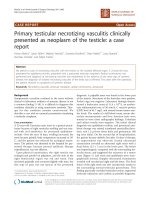
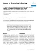
Serum creatinine, urine oxalate:creatinine ratio, and creatinine clearance vs. clinical timelineFigure 1
Serum creatinine, urine oxalate:creatinine ratio, and creatinine clearance vs. clinical timeline. (a) The chart
shows the trend of serum creatinine (gray bars, with the values shown on the left axis), starting from a baseline of 1.2 mg/dL
just over 4 months ago, gradually increasing up to 3.1 mg/dL 8 days ago, and rapidly increasing to 8.4 mg/dL on admission (day
0). The urine oxalate:creatinine ratio (red squares connected by lines, with values shown on the right axis) clearly shows
hyperoxaluria at admission (0.084 at day 1, compared to a normal of 0.035). Vitamin C was stopped on day 4, and creatinine
started improving after 2 days. (b) Renal function in terms of creatinine clearance (% of normal) is also shown.
a) Serum Creatinine and Urine Ox:Cr Ratio b) % Creatinine Clearance vs. Clinical Timeline
0.084
0.052
0.039
0.028
0
2
4
6
8
10
12
14
16
18
20
22
24
-150 -48 -38 -28 -18 -8 2 12 22 32 42 52 62
Days from admission (day 0 = day of admission)
Serum Creatinine (mg/dL)
0
0.012
0.024
0.036
0.048
0.06
0.072
0.084
0.096
0.108
0.12
0.132
0.144
Urine Oxalate:Creatinine Ratio
Serum Creatinine (mg/dL) Creatinine Clearance (% of normal)
Urine Ox:Cr Ratio
Hospital Stay
Sub-acute phase
Acute phase
Normal Urine Ox:Cr Ratio = 0.035
25%
50%
75%
100%
% Cr Clearance
Vit C Stopped,
Low oxalate diet + Vit B6
(a)
(b)
Vit C (680 mg/d)
Dialysis Sessions
Journal of Medical Case Reports 2007, 1:155 />Page 3 of 6
(page number not for citation purposes)
Admission blood tests were remarkable for creatinine of
8.4 mg/dL, which quickly became 11 mg/dL despite fluids
(Fig 1a). HCO
3
was 19 mM (22–29 mM). Anion gap was
14. Ca was 10.4 mg/dL (normal 8.5–10.5 mg/dL). Ionized
Ca was high at 1.24 mM. Phosphorus was 4 mg/dL (nor-
mal 2.5–8.5 mg/dL). CBC was abnormal for hemoglobin
of 9.2 g/dL and persistent megaloblastosis. Urine pH was
5. Specific gravity was 1.020, without casts or eosinophils.
Urine protein:creatinine ratio was 1.2. Kidneys were 10.1
× 7.4 × 5.1 cm (right) and 10.6 × 3.8 × 4 cm (left) on ultra-
sound.
Serial creatinine (Fig 1a) suggested chronic slowly-pro-
gressive failure, until 5 weeks ago, when creatinine rose
abruptly. Since there was no history or evidence for diabe-
tes, accelerated hypertension, nephritis, contrast dye, sep-
sis, allergic reactions, obstruction, thrombo-emboli, or
volume overload or depletion, we postulated nephrotox-
ins exposure at home to explain his slow, insidious relent-
less renal impairment. Diet scrutiny revealed excessive
beans and chocolate, rich in oxalate. Medication review
showed daily 5 mg lisinopril, 75 µg levothyroxine, 800
mg MgO, 650 mg CaCO3, 500 mg niacin, 10,000 IU vita-
min A, 500 mg vitamin C, and a multivitamin with 180
mg of vitamin C.
Since urinalysis was negative and proteinuria was mini-
mal, oxalate-induced tubulointerstitial nephritis was pro-
posed, given history of alcoholism, chronic diarrhea, and
high-dose vitamin C. On day 2, 24-hour urine was sub-
mitted for oxalate. Given his weight loss, renal failure, and
hypercalcemia, multiple myeloma was considered
unlikely given negative serum and urine protein electro-
phoresis and negative bone marrow exam. Other causes
for hypercalcemia were excluded by normal PTH, PTH-
related peptide, 1,25 (OH)
2
vit D, 25 OH vit D, alkaline
phosphatase, and free T4. Neoplasms were excluded by
CT scans of chest, abdomen, and pelvis, and endoscopies.
His creatinine continued to rise despite drinking plenty of
fluids and stopping lasix and lisinopril (Fig 1a). Salt
depletion was excluded by lack of fall in creatinine despite
several liters of saline, arguing against dehydration as the
cause for his acute renal failure. While still waiting for
urine oxalate results, which were delayed due to errone-
ous submission of un-acidified aliquot, we stopped vita-
min C on day 4. Hypercalcemia was attributed to CaCO
3
,
vitamins A and D, as it resolved off these agents. Diarrhea
was attributed to MgO, vitamin A, and niacin, all known
to have cathartic potential. Indeed, diarrhea resolved once
they were stopped. These responses reinforced the suspi-
cion of nephrotoxic "over-the-counter" medications.
He received 6 hemodialysis treatments to correct meta-
bolic acidosis (that otherwise reduces urine citrate and
predisposes to Ca precipitation [8] and to remove excess
oxalate (from presumed enhanced absorption and
impaired excretion). Discontinuation of vitamin C
resulted in slow steady recovery of renal function. Within
days, serum creatinine started falling and continued spon-
taneously (Fig 1a). Nine days after his last dialysis, creati-
nine was 3.1 mg/dL. Six weeks later, while on metolazone
(to reduce urine Ca), a low oxalate diet, and 100 mg daily
vitamin B6, his creatinine was 1.8 mg/dL. It stayed there 3
months after presentation. Urine oxalate to creatinine
ratio steadily decreased, from 0.084 pre-treatment to 0.02
(normal < 0.035) (Fig 1a).
The presumptive diagnosis of oxalate-induced interstitial
nephritis was confirmed on day 14 by a renal biopsy,
showing extensive interstitial fibrosis, tubular atrophy
(Fig 2a), and diffuse intraluminal birefringent crystals
under polarized light (Fig 2b). CAT scan documented
scattered renal calcifications and a 1-cm calculus (Fig 2c).
Urine from day 2 showed increased oxalate:creatinine
ratio excretion of 0.084 (Fig 1a), biochemically corrobo-
rating the clinical diagnosis and biopsy findings.
The hyperoxaluria was attributable to vitamin C, as it
resolved on discontinuation of vitamin C (urine
oxalate:creatinine ratio 0.052, Fig 1a). A low oxalate diet
further reduced this ratio to 0.039 and to 0.028 (Fig 1a).
We did not measure urine citrate during his peak renal
failure since severe metabolic acidosis is expected to non-
specifically suppress excretion. However, 11 and 30 days
post-discharge, when serum HCO
3
approached normal
(22 and 21 mM) and creatinine substantially improved
(4.6 and 2.9 mg/dL), we still documented markedly
reduced urine citrate (respectively, 14 mg and 18 mg per
gram of creatinine; normal being ≥ 250). These results
suggest significant ongoing metabolic acidosis preceding
or paralleling the steady rise in serum creatinine in previ-
ous months.
Discussion
Urinary oxalate is derived from endogenous production
and absorption from exogenous sources. Hyperoxaluria
(urine oxalate above the normal range of 10–35 mg/24
hr) can be primary or secondary. Primary hyperoxaluria
results from genetic defects in glyoxylate metabolism, pro-
ducing nephrolithiasis, nephrocalcinosis, and progressive
renal insufficiency [9]. Secondary hyperoxaluria is
acquired from enteric causes or ethyl glycol intoxication.
Normally, Ca binds most of the intestinal oxalate, with
subsequent stool CaOx elimination. Accordingly, only 4
to 12% of enteric oxalate is normally absorbed. After
small bowel bypass, Ca complexes with poorly absorbed
fatty acids, leaving behind excess unbound oxalate for
absorption, 65 to 80% of which occurs in the colon [6,7].
Journal of Medical Case Reports 2007, 1:155 />Page 4 of 6
(page number not for citation purposes)
This mechanism also explains the enteric hyperoxaluria in
chronic diarrhea and malabsorption.
Another important but under-appreciated etiology of sec-
ondary hyperoxaluria is increased synthesis from vitamin
C [1-5]. We previously reported the quantitative dose-
response relationship in a patient receiving vitamin C
solely from parenteral nutrition [5]. If coexisting, malab-
sorption and high-dose vitamin C could potentiate the
hyperoxaluria induced by each other.
Clinically, hyperoxaluria presents in one of several ways,
depending on the severity, chronicity, etiologies and co-
factors. First, at one extreme, as in ethylene glycol intoxi-
cation, fulminant acute renal failure from intraluminal
obstruction can develop, due to excessive oxalate produc-
tion and profound hyperoxaluria. Second, at the other
end, intermittent hyperoxaluria could cause episodic
painful renal colic from small punctuate Ca Ox calculi.
Despite short-term morbidities, few suffer from serious
long-term renal insufficiency. The third mode of clinical
presentation is illustrated by primary hyperoxaluria.
Our patient demonstrates the fourth mode of manifesta-
tion in that insidious renal failure, easily missed, slowly
evolves over weeks to months, unbeknown to patients
and physicians until renal function is compromised by
>50–70 %. Typically, for diffuse intraluminal crystal
deposits and extensive interstitial fibrosis to develop, the
course is protracted. While the first three modes (ethylene
glycol intoxication, episodic stone attacks, and primary
hyperoxaluria) are clinically symptomatic and promptly
treated, the 4
th
mode is generally quiescent, asympto-
matic, undiagnosed, and untreated for months, like our
patient, unless tests reveal incidental unexpected progres-
sive renal failure.
In our patient, intratubular luminal precipitation of Ca
oxalate was promoted by four pathogenic factors (Fig 3):
(1) high urine specific gravity (due to diarrhea-induced
dehydration), (2) hyperoxaluria from all three potentiat-
ing mechanisms (oxalate-rich diet, 680 mg daily vitamin
C, and possible malabsorption), (3) relative hypercalciu-
ria (due to furosemide, chronic metabolic acidosis, and
hypercalcemia, caused by CaCO3 pills and vitamin A and
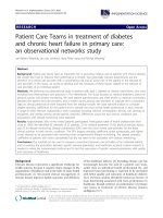
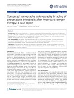
Slides from left kidney biopsy and CT image of abdomenFigure 2
Slides from left kidney biopsy and CT image of abdomen. There is extensive interstitial fibrosis and tubular atrophy,
with marked medial fibrosis in an artery on the lower right (trichrome/10x) (a); bright crystals are seen under partial polariza-
tion within the lumens of two tubules (arrows) (H&E/20x) (b). A 10 mm calculus is seen in the right extra renal pelvis (arrow)
in the CT (c).
(a)
(b)
(c)
Journal of Medical Case Reports 2007, 1:155 />Page 5 of 6
(page number not for citation purposes)
D), and (4) hypocitraturia (due to metabolic acidosis, ini-
tially from chronic diarrhea and later aggravated by pro-
gressive renal failure).
The pathophysiology of hyperoxaluria-induced nephrop-
athy is schematized in Fig 3. Tubular fluid supersaturated
with Ca oxalate crystals causes luminal obstruction, mac-
rophage recruitment, up-regulated reactive oxygen spe-
cies, pro-inflammatory & pro-fibrogenic cytokines,
creating a vicious cycle of interstitial nephritis, relentless
injuries, apoptosis, necrosis, fibrosis, with eventual neph-
ron drop-out and tubular atrophy. Initially, hypocitraturia
from stool alkali losses is modest, but with worsening
renal failure, metabolic acidosis and hypocitraturia
become more severe. Nephron losses further elevate
blood and filtered oxalate, raising tubular fluid oxalate
concentration in surviving nephrons. These changes exac-
erbate the risks for luminal crystals precipitation, tubular
obstruction, inflammation, and interstitial nephritis. As
more tubules die out, the vicious cycle is exaggerated. This
could explain the steep increase in serum creatinine from
1.8 to 8.4 mg/dL (representing 44 % loss in creatinine
clearance in 5 weeks before admission) vs. the gentle rise
from 1.2 to 1.8 mg/dL (representing 28% loss in clearance
over the preceding 4 months) (Fig 1b).
Treatment of hyperoxaluria-induced nephropathy
depends on interrupting the pathophysiology (Fig 3), spe-
cifically, by increased fluids, low oxalate diet, discontinu-
ing vitamins A, C, D, and CaCO
3
, prescribing thiazide (or
metolazone if creatinine is elevated, to reduce urine Ca),
and NaHCO
3
to increase urine citrate. Metabolic acidosis
is known to markedly decrease tubular fluid citrate, the
predominant endogenous inhibitor of Ca oxalate precipi-
tation [8]. In patients with malabsorption, underlying eti-
ologies should be addressed, and Na or K alkali salts
(citrate or bicarbonate) should minimize metabolic aci-
dosis, increase urine citrate, and reduce urine Ca. (Fig 3).
We counseled continued alcohol abstinence in our
patient. All identifiable causes of hypercalcemia and
Pathophysiology and management of hyperoxaluric nephropathyFigure 3
Pathophysiology and management of hyperoxaluric nephropathy.
Pathophysiology & management of secondary hyperoxaluria-induced nephropathy
Intraluminal crystallization of
Ca oxalate & / or Ca phosphate
Tubular obstruction; interstitial inflammation & fibrosis, tubular
cells apoptosis, atrophy & nephron drop-out: renal failure
Hyperoxaluria Hypercalciuria HyperphosphaturiaHypocitraturia
Urinary
concentration
Tubular fluid supersaturation
with Ca, Po4, and oxalate
Dehydration
from diarrhea
& / or fluids
avoidance
Metabolic
acidosis
from
diarrhea
&/or renal
failure
Malabsorption
[chelation of Ca by
fatty acids & enteric
hyperoxaluria]
High-dose
vitamin C (>200
mg/d) even with
normal kidneys
High oxalate diet
(nuts, beans,
coffee, cocoa,
chocolates)
Hypercalcemia
(vit. D & Ca pills)
Fluids
Low
oxalate
diet
d/c
vit. C
d/c
vit. D
& Ca
NaHCO
3
NaCO
3
thiazide
-
Publish with BioMed Central and every
scientist can read your work free of charge
"BioMed Central will be the most significant development for
disseminating the results of biomedical researc h in our lifetime."
Sir Paul Nurse, Cancer Research UK
Your research papers will be:
available free of charge to the entire biomedical community
peer reviewed and published immediately upon acceptance
cited in PubMed and archived on PubMed Central
yours — you keep the copyright
Submit your manuscript here:
/>BioMedcentral
Journal of Medical Case Reports 2007, 1:155 />Page 6 of 6
(page number not for citation purposes)
hypercalciuria were eliminated, and we empirically added
vitamin B6, which promotes conversion of glyoxylate to
glycine instead of oxalate.
Unfortunately, vitamin C-induced hyperoxaluria is often
missed or diagnosed late in the course of renal failure, and
for several reasons. One, increasingly individuals take
daily multi-vitamins or high-dose vitamin C on their own.
Second, current commercial preparations contain vitamin
C typically several-fold over the adult daily requirement of
60 mg. Most juices, soft drinks, and diet supplements con-
tain > 120 mg of vitamin C per 8 oz. These add greatly to
food-derived oxalate. Third, self-prescribed medications
are not routinely checked or discovered by physicians, as
the potential adverse hyperoxaluric consequence is gener-
ally unrecognized. Fourth, the causal relationship
between hyperoxaluria and renal failure is also under-
appreciated. Finally, when advanced renal insufficiency
emerges, the focus is shifted to uremia management. Iden-
tifying and defining the etiology is seldom exhaustively
undertaken or usually unsuccessful.
Conclusion
1) High-dose vitamin C can induce hyperoxaluric neph-
ropathy and progressive renal failure, especially with
diarrhea, oxalate-rich diet, metabolic acidosis, and dehy-
dration. 2) The diagnosis should be suspected in unex-
plained, slowly evolving renal insufficiency, particularly if
additional risk factors coexist. 3) Since prompt treatment
could prevent end-stage renal disease, we recommend a
high index of suspicion, careful review of diets and all
medications, closely monitoring renal function and
oxalate excretion in patients on vitamin C.
Competing interests
The author(s) declare that they have no competing inter-
ests.
Authors' contributions
SR did the initial workup and follow-up. KL made the
diagnosis and treated the patient. Both SR and KL plotted
the graphs and drafted the manuscript. WK was responsi-
ble for interpretation of biopsy findings. All authors read
and approved the final manuscript.
Consent
Written consent for the publication was obtained from
patient's wife as the patient died of a cardiac event 5
months after presentation.
Acknowledgements
We thank all the nurses and physicians involved in the care of our patient
before and during his hospital stay, in particular Drs. Rhett Jackson, Lukas
Haragsim, Henry Reed, and Mohammad Cherry.
References
1. Canos HJ, Hogg GA, Jeffery JR: Oxalate nephropathy due to gas-
trointestinal disorders. Can Med Assoc J 1981, 124(6):729-733.
2. Hassan I, Juncos LA, Milliner DS, Sarmiento JM, Sarr MG: Chronic
renal failure secondary to oxalate nephropathy: a preventa-
ble complication after jejunoileal bypass. Mayo Clin Proc 2001,
76(7):758-760.
3. Lau K, Wolf C, Nussbaum P, Weiner B, DeOreo P, Slatopolsky E,
Agus Z, Goldfarb S: Differing effects of acid versus neutral
phosphate therapy of hypercalciuria. Kidney Int 1979,
16(6):736-742.
4. Leumann E, Hoppe B: The primary hyperoxalurias. J Am Soc
Nephrol 2001, 12(9):1986-1993.
5. Massey LK, Liebman M, Kynast-Gales SA: Ascorbate increases
human oxaluria and kidney stone risk. J Nutr 2005,
135(7):1673-1677.
6. Chai W, Liebman M, Kynast-Gales S, Massey L: Oxalate absorption
and endogenous oxalate synthesis from ascorbate in calcium
oxalate stone formers and non-stone formers. Am J Kidney Dis
2004, 44(6):1060-1069.
7. Mashour S, Turner JF Jr., Merrell R: Acute renal failure, oxalosis,
and vitamin C supplementation: a case report and review of
the literature. Chest 2000, 118(2):561-563.
8. Alkhunaizi AM, Chan L: Secondary oxalosis: a cause of delayed
recovery of renal function in the setting of acute renal fail-
ure. J Am Soc Nephrol 1996, 7(11):2320-2326.
9. Swartz RD, Wesley JR, Somermeyer MG, Lau K: Hyperoxaluria
and renal insufficiency due to ascorbic acid administration
during total parenteral nutrition. Ann Intern Med 1984,
100(4):530-531.