Báo cáo khoa hoc:" Topical latanoprost causes posterior movement of lens in a patient with exfoliation syndrome and subluxated lens: a case report" ppt
Bạn đang xem bản rút gọn của tài liệu. Xem và tải ngay bản đầy đủ của tài liệu tại đây (586.1 KB, 3 trang )
BioMed Central
Page 1 of 3
(page number not for citation purposes)
Journal of Medical Case Reports
Open Access
Case report
Topical latanoprost causes posterior movement of lens in a patient
with exfoliation syndrome and subluxated lens: a case report
Takashi Kanamoto*, Michiya Takamatsu and Yoshiaki Kiuchi
Address: Department of Ophthalmology and Visual Sciences, Graduate School of Biomedical Sciences, Hiroshima University, Japan
Email: Takashi Kanamoto* - ; Michiya Takamatsu - ; Yoshiaki Kiuchi - ykiuchi@hiroshima-
u.ac.jp
* Corresponding author
Abstract
Introduction: To report the effect of topical latanoprost on the position of a subluxated lens.
Case presentation: After 0.005% latanoprost was administered topically to a patient with ocular
hypertension due to a pseudoexfoliation syndrome and a subluxated lens, the position of the lens
was examined by slit-lamp biomicroscopy, and the ciliary body thickness by ultrasound
biomicroscopy. The lens had moved posteriorly, and the thickness of the ciliary body had
decreased after the latanoprost.
Conclusion: We suggest that the decrease in the thickness of the ciliary body resulted in an
increase in the tension of the zonule of Zinn fibers, thus pulling the subluxated lens posteriorly.
Case presentation
An 80-year-old woman complained of visual disturbances
in her right eye that began in July 2002. She did not have
a history of any systemic illness, and there was no family
medical history of any disease. In 2001, she had under-
gone a peripheral iridotomy on the right eye for angle clo-
sure glaucoma, and she developed the pseudoexfoliation
syndrome. Her postoperative intraocular pressure (IOP)
in the right eye was 12 mmHg. The depth of the anterior
chamber of the left eye was normal and the IOP was 11
mmHg.
In May 2002, although the IOP in her left eye was 12
mmHg, the right IOP was 22 mmHg, and we concluded
that she had ocular hypertension secondary to the pseu-
doexfoliation syndrome. We began topical latanoprost in
the right eye, and the IOP decreased to 13 mmHg by June.
The IOP in the left eye remained at 12 mmHg. In July, she
returned reporting visual disturbances and monocular
double vision. The lens was partially dislocated in the
right eye. At this time, the IOP in the right eye was 20
mmHg and the left eye was 10 mmHg. The right lens had
a mild cataract. There were no clear glaucomatous changes
in the optic discs, and no other specific findings. The
Goldman perimetric fields were full.
To examine the effect of latanoprost on the position of the
lens, we stopped the latanoprost for two weeks. The IOP
was measured with a Goldman applanation tonometer
one hour before and after topical latanoprost, and the
position of the lens was assessed by slit-lamp biomicros-
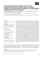
copy. In addition, ultrasound biomicroscopy (UBM) was
performed to measure any changes in the thickness of the
ciliary body [1]. (Figure 1)
One hour after one drop of 0.005% latanoprost, the right
IOP decreased from 20 mmHg to 17 mmHg and the IOP
in the left eye was reduced from 10 mmHg to 7 mmHg.
Published: 5 December 2007
Journal of Medical Case Reports 2007, 1:172 doi:10.1186/1752-1947-1-172
Received: 19 May 2007
Accepted: 5 December 2007
This article is available from: />© 2007 Kanamoto et al; licensee BioMed Central Ltd.
This is an Open Access article distributed under the terms of the Creative Commons Attribution License ( />),
which permits unrestricted use, distribution, and reproduction in any medium, provided the original work is properly cited.
Journal of Medical Case Reports 2007, 1:172 />Page 2 of 3
(page number not for citation purposes)
Slit-lamp biomicroscopy showed a large empty space
between the lens and iris indicating a movement of the
lens posteriorly. The lens in the right eye had not shifted
(Figure 2). In addition, UBM showed that the thickness of
the ciliary body had decreased significantly (Figure 3).
Latanoprost is a prostagrandin F2-alpha receptor antago-
nist [2] that increases the efflux of aqueous humor
through the uveoscleral route [3,4]. The increase results
from a re-organization of the extracellular matrix includ-
ing the matrix metalloproteinases (MMPs) [5]. In the
pseudoexfoliation syndrome, changes in the MMPs are
associated with the loss of the zonules of Zinn fibers.
Latanoprost is widely used to reduce the intraocular pres-
sure (IOP) in eyes with glaucoma, [6] and latanoprost has
been used safely as a first line therapy in eyes with pseu-
doexfoliation glaucoma [7,8]. Our patient with the pseu-
doexfoliation syndrome and subluxated lens offered us an
opportunity to examine the effect of topical latanoprost
on the position of the lens.
The presence of desquamative material on the zonules of
Zinn fibers can lead to abnormalities which may account
for the subluxation. The increased aqueous humor efflux
through the uveoscleral route by latanoprost is probably
aided by the relaxation of the ciliary body muscle [9-11].
In our case, a decrease in the thickness of the ciliary body
was detected by UBM. Although a previous report states
that the mean ciliary body thickness increases two weeks
after latanoprost administration [12], our data showed a
rapid decrease in the thickness of the ciliary body in an eye
with a subluxated lens. Approximately two-third of the
anterior part of the ciliary body moved posteriorly which
would increase the tension of the zonule of Zinn fibers
[13]. Thus, latanoprost relaxes the ciliary body muscle and
increases the tension on the zonule of Zinn as with topical
atropine sulfate.
Conclusion
We suggest that the subluxated lens was due to the loss of
the zonule of Zinn fibers in the superior margin of the
lens, and this loss would make it easier for the lens to
move posteriorly. Although the movement of the lens was
not sizeable, any increase in the distance between the cor-
nea and lens will reduce the overall refractive power of the
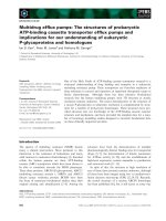
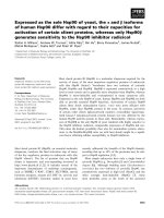
Change in lens position after topical latanoprostFigure 2
Change in lens position after topical latanoprost. Photographs before latanoprost (pre-latanoprost), and the movement of the
subluxated lens posteriorly after latanoprost. Left panel shows a control eye.
Measurement of the thickness of ciliary body by UBM (arrows)Figure 1
Measurement of the thickness of ciliary body by UBM
(arrows).
Publish with BioMed Central and every
scientist can read your work free of charge
"BioMed Central will be the most significant development for
disseminating the results of biomedical research in our lifetime."
Sir Paul Nurse, Cancer Research UK
Your research papers will be:
available free of charge to the entire biomedical community
peer reviewed and published immediately upon acceptance
cited in PubMed and archived on PubMed Central
yours — you keep the copyright
Submit your manuscript here:
/>BioMedcentral
Journal of Medical Case Reports 2007, 1:172 />Page 3 of 3
(page number not for citation purposes)
eye. UBM is useful to determine the mechanism of unex-
pected symptoms such as the monocular diplopia in our
patient, and UBM should be considered for patients with
pseudoexfoliation syndrome following topical medica-
tion. In spite of these changes, latanoprost can be used in
patients with weakened zonules of Zinn, but careful fol-
low-up examinations are recommended especially for
lens subluxation.
Competing interests
The author(s) declare that they have no competing inter-
ests.
Authors' contributions
TK examined the patient and drafted the manuscript. MT
examined the patient. YK performed a literature review.
All authors read and approved the final manuscript.
Consent
Written informed consent was obtained from the patient
for publication.
Acknowledgements
Takashi Noma, Ph.D. M.D. (Department of Ophthalmology, Kure Saiseikai
Hospital, Japan) contributed to examination of the patient.
References
1. Mishima HK, Shoge K, Takamatsu M, Kiuchi Y, Tanaka J: Ultrasound
Biomicroscopic study of ciliary body thickness after topical
application of pharmacogic agents. Am J Ophthalmol 1996,
121:319-321.
2. Mishima HK, Masuda K, Kitazawa Y, Azuma I: A comparison of
latanoprost and timolol in primary open-angle glaucoma and
ocular hypertension. A 12-week study. Arch Ophthalmol 1996,
114:929-932.
3. Toris CB, Camras CB, Yablonski ME: Effect of PhXA41, a new
prostaglandin F2α analog, on aqueous humor dynamics in
human eyes. Ophthalmology 1993, 100:1297-1304.
4. Yousufuzai SY, Zheng P, Abdel-Latif AA: Protaglandin F2α and its
analogs induce release of endogenous prostaglandins in iris
and ciliary muscles isolated from cat and other mammalian
species. Exp Eye Res 1996, 63:305-310.
5. Lindsey JD, Kashiwagi K, Kashiwagi F, Weinreb RN: Prostaglandins
alter extracellular matrix adjacent to human ciliary muscle
cells in vitro. Invest Ophthalomol Vis Sci 1997, 38:2214-2223.
6. Ocklind A: Effect of latanoprost on the extracellular matrix of
ciliary muscle. A study on cultured cells and tissue sections.
Exp Eye Res 1998, 67:179-191.
7. Konstas AG, Kozobolis VP, Tersis I, Leech J, Stewart WC: The effi-
cacy and safety of the timolol/dorzolamide fixed combina-
tion vs latanoprost in exfoliation glaucoma. Eye 2003,
17:41-46.
8. Alm A, Schoenfelder J, McDermott J: A 5-year, multicenter, open-
label, safety study of adjunctive latanoprost therapy for glau-
coma. Arch Ophthalmol 2004, 122:957-965.
9. Goh Y, Hotehama Y, Mishima HK: Characterization of ciliary
muscle relaxation induced by various agents in cats. Invest
Ophthalmol Vis Sci 1995, 36:1188-1192.
10. Poyer JF, Millar C, Kaufman PL: Protaglandin F2α effects on iso-
lated rhesus monkey ciliary muscle. Invest Ophthalmol Vis Sci
1995, 36(12):2461-5.
11. Fujimoto N, Zhao C, Shichi H: The effects of Protaglandin E2
and F2α on porcine ciliary muscle cells in culture. Curr Eye Res
1995, 14(12):1155-63.
12. Marchini G, Ghilotti G, Bonadimani M, Babighian S: Effects of
0.005% Latanoprost on Ocular Anterior Structures and Cili-
ary Body Thickness. J Glaucoma 2003, 12:295-300.
13. Nishida Y: Anatomy of ciliary body. Glaucoma 1993:43-51 [http:/
/www.nakayamashoten.co.jp/cgi-bin/menu.cgi?ISBN=4-521-42043-5].
Tokyo: Nakayama-Shoten [Masuda K (Series Editor): Current encyclo-
pedia of ophthalmology, chapter 3A.]
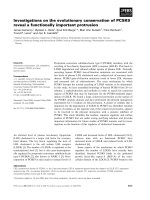
Relaxation of the ciliary body muscles after treatment of latanoprostFigure 3
Relaxation of the ciliary body muscles after treatment of
latanoprost. Before and after latanoprost on right eye, lens
subluxation, thickness of ciliary body was measured in four
directions, vertical and horizontal phase (average ± SD).
Control means left eye, non-lens subluxation eye. (*: P <
0.01, paired t test).