Báo cáo khoa hoc:" Post coital aortic dissection: a case report" pdf
Bạn đang xem bản rút gọn của tài liệu. Xem và tải ngay bản đầy đủ của tài liệu tại đây (236.38 KB, 4 trang )
BioMed Central
Page 1 of 4
(page number not for citation purposes)
Journal of Medical Case Reports
Open Access
Case report
Post coital aortic dissection: a case report
Gareth Morris-Stiff*, Mari Coxon, Elizabeth Ball and Michael H Lewis
Address: Department of Surgery, Royal Glamorgan Hospital, Ynysmaerdy, Llantrisant, Wales, UK
Email: Gareth Morris-Stiff* - ; Mari Coxon - ; Elizabeth Ball - ;
Michael H Lewis -
* Corresponding author
Abstract
Background: Sudden onset peri- or post-coital cardiovascular disease is well documented in the
literature including myocardial infarction, pulmonary embolus and subarachnoid haemorrhage. The
occurrence of aortic dissection in this setting has been reported only once previously.
Case presentation: We report the case of a 47 year old man who developed sudden onset right
leg pain during coitus. This was initially believed to be neurological due to nerve impingement but
an MRI failed to identify a prolapse. On further review after 6 weeks, pulses were noted to be
absent in the patient's right leg and an urgent vascular review with investigation identified a
dissection of the aorta which was subsequently successfully treated.
Conclusion: This case illustrates a rare presentation of aortic dissection and demonstrates the
importance of a thorough vascular assessment in the presence of sudden onset limb pain.
Introduction
Aortic dissection is characterised by separation of the lay-
ers of the aortic wall by extraluminal blood that enters the
aortic wall, almost invariably through a luminal tear [1].
Despite a reduction in its incidence as a result of improved
pharmacological control of hypertension, when it
presents acutely, aortic dissection usually has a cata-
strophic outcome. This case reports an unusual mode of
presentation and illustrates the multidisciplinary aspects
of the pre-, peri- and post-operative care of an unusual
presentation of the condition.
Case presentation
A 47 year old gentleman presented to his general practi-
tioner with acute onset lower back pain. The pain had
commenced during coitus and radiated down the right
leg. The initial diagnosis was of acute disc prolapse and he
was referred for an urgent neurosurgical opinion. The neu-
rosurgeon concurred that the pain may well have been of
neurological origin and arranged an MRI scan. This was
reported as showing no evidence of spinal cord pathol-
ogy. The patient was reassured with the results of the MRI
findings and was advised the pain was probably muscu-
loskeletal in origin and should settle. Over the subsequent
6 weeks, the pain persisted and indeed increased in sever-
ity. The patient noted claudication-type pain in his right
leg after approximately 100 metres. As the pain had not
resolved after 6 weeks he revisited his general practitioner.
During the subsequent examination the pulses in his right
leg were noted to be absent and he was referred for an
urgent vascular surgical opinion.
The patient was seen the following day in the vascular
clinic where a history of severe acute claudication-type
pain was noted in the right leg. There was a past medical
history of marked hypertension and hyperlipidaemia, for
Published: 16 January 2008
Journal of Medical Case Reports 2008, 2:6 doi:10.1186/1752-1947-2-6
Received: 9 March 2007
Accepted: 16 January 2008
This article is available from: />© 2008 Morris-Stiff et al; licensee BioMed Central Ltd.
This is an Open Access article distributed under the terms of the Creative Commons Attribution License ( />),
which permits unrestricted use, distribution, and reproduction in any medium, provided the original work is properly cited.
Journal of Medical Case Reports 2008, 2:6 />Page 2 of 4
(page number not for citation purposes)
which he took relevant medications, but none of angina,
myocardial infarct or valvular heart disease. On clinical
examination the heart rate was 68 beats per minute regu-
lar. The blood pressure in the right arm 130/70 mmHg
was lower than that of the left arm 160/80. Cardiac exam-
ination was normal. There was no clinical evidence of an
abdominal aortic aneurysm. Examination of the limbs
revealed that the right lower limb pulses were all absent
whilst those of the left leg were present and of good vol-
ume. An urgent abdominal ultrasound scan was arranged
which demonstrated dissection of the intra-abdominal
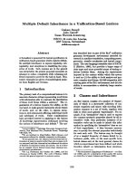
aorta and a subsequent CT scan (Figures 1, 2, 3) con-
firmed that the dissection was a Type A dissection extend-
ing from the aortic valve down to the aortic bifurcation. A
dissection flap was identified in the ascending aorta and
also in the postero-inferior aspect of the descending aorta.
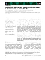
Both lumens were noted to have flow within them with
the true lumen supplying the celiac axis, superior
mesenteric artery and right renal artery and the false
lumen supplying the left renal artery and inferior
mesenteric artery. Immediately below the inferior
mesenteric artery the false lumen obliterated.
An immediate opinion was sought at the regional cardiot-
horacic unit and the patient was transferred urgently
under their care. A transthoracic echocardiogram was per-
formed which confirmed the CT findings demonstrating
turbulent flow in the ascending aorta suggestive of an inti-
mal tear in the region although the lesion itself was not
seen. The arch was mildly dilated but with no visible flap.
Flow in the descending aorta was turbulent in the initial
2–3 cm suggesting intimal disruption.
He underwent operative repair of the thoracic dissection
on the next available theatre list. The aortic valve was
resuspended and the ascending aorta were replaced using
an elephant trunk graft with reimplantation of the brachi-
CT scans demonstrating the dissection at the level of the aortic valveFigure 1
CT scans demonstrating the dissection at the level of the
aortic valve.
CT scans demonstrating the dissection at the level of the coeliac axisFigure 2
CT scans demonstrating the dissection at the level of the
coeliac axis.
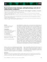
CT scans demonstrating the dissection at the level of the renal arteriesFigure 3
CT scans demonstrating the dissection at the level of the
renal arteries.
Journal of Medical Case Reports 2008, 2:6 />Page 3 of 4
(page number not for citation purposes)
ocephalic artery on 1 patch and the left common carotid
and subclavian arteries on a second patch.
After 48 hours in the intensive care unit the patient was
transferred to the ward where he made an uneventful
recovery. Cardiac and cerebral functions were not
impaired by the procedure as evidenced by return to pre-
operative state and no requirements for chronotropic
medications.
A routine postoperative CT scan demonstrated that the
repair was satisfactory. There was thrombosis within the
false lumen of the descending aorta but persistence of
flow within both lumens of the abdominal aorta.
Discussion
Aortic dissection is characterised by separation of the lay-
ers of the aortic wall due to extraluminal blood that has
entered the aortic wall through an intimal tear. Tears are
seen at areas of high stress, the most common being in the
anterior aortic wall just above the aortic valve (66%), and
the posterior wall of the proximal descending aorta
(33%). When blood enters through an intimal tear it
passes longitudinally along the tunica media separating
the intima from the adventitia.
An acute dissection of the aorta is one which presents
within 14 days of the onset of the disease process. In this
case, presentation with new onset back and right leg pain
occurred on the first day of symptoms and urgent investi-
gations were instituted.
There are several different formats of classification for tho-
racic dissection, the most commonly used being that of
DeBakey [2], which divides the dissections into 3 types: I
– involving the ascending aorta and a variable amount of
descending or thoraco-abdominal aorta; II – dissection
limited to the ascending aorta; and III – dissection of the
descending aorta either without (IIIa) or with (IIIb)
involvement of the abdominal aorta. Our patient clearly
had a type I dissection with 2 lumina identified running
from the aortic valve all the way to the bifurcation of the
aorta into common iliac arteries.
The typical presentation of acute dissection is with sudden
onset, unexpected, intense pain in the interscapular
region radiating to the lower back or abdomen [3].
Patients are typically hypertensive middle-aged or elderly
men and the diagnosis should certainly be entertained in
patients with such symptoms along with other differential
diagnoses which include myocardial ischaemia and
abdominal aortic aneurysm. However, as the dissection
can affect any of the arteries arising from the aorta, other
presentations include stroke, a pulseless limb or abdomi-
nal organ dysfunction such as renal failure or intestinal
ischaema. In this case, whilst the demographics were typ-
ical, the site of the pain was not, being a lot lower than for
a typical dissection. Dissection involving the ascending
aorta (types I and II) is more hazardous than type III
because of the risks of intra-pericardial rupture, acute aor-
tic insufficiency or occlusion of the coronary arteries. For
all dissections there is the risk of aortic rupture and ischae-
mic complications, particularly if the abdominal aorta is
affected by the dissection. The patient reported was there-
fore fortuitous not to have developed complications dur-
ing the 6 week interval from injury to diagnosis of
dissection.
This case has illustrated the importance of taking a
detailed history and performing a thorough clinical exam-
ination in all patients with acute onset limb pain. In this
case, the temporal relationship between the onset of pain
and radiation down the right leg would suggest that the
dissection into the intra-abdominal portion of the aorta
occurred during coitus although it is impossible to prove
this. Had the dissection progressed over a period of weeks
it is likely that the pain would have progressed gradually
rather than arising acutely and persisting at a constant
intensity. However, whilst the main injury may have
occurred with the initial pain, there may have been slow
progression over a period of 6 weeks with loss of the right
limb pulses during this period.
The identification of impaired peripheral pulses is
another important finding in relation to a diagnosis of
aortic dissection. In an extensive examination of the med-
ical literature, Klompas [4] highlighted the importance of
obtaining an accurate history noting that 31% of patients
had evidence of a pulse deficit, and that for patients with
a history highly suggestive of dissection who underwent
advanced imaging studies, the positive likelihood ratio of
a pulse deficit between contralateral limbs was 5.7 and the
negative likelihood ratio was 0.7.
The mode of presentation of our patient's dissection is not
classical; however, this is not the first such case. An unfor-
tunate individual reported by Lovas and Silver [5] also
ruptured a berry aneurysm during his activities and his
outcome was less satisfactory. Furthermore, it would
appear from the literature that peri-coital acute vascular
disease is not uncommon including myocardial infarc-
tion, pulmonary embolism and intracranial haemor-
rhage. Less surprising is the fact that much of the data has
accumulated from the Scandinavian countries.
The pathophysiology of the dissection during coitus is
probably related to the well-recognised increases of blood
pressure seen during vigorous exercise [6]. It has been
demonstrated in a rat model that central aortic pressure
increases by up to 19% during exercise [7]. Furthermore,
Publish with BioMed Central and every
scientist can read your work free of charge
"BioMed Central will be the most significant development for
disseminating the results of biomedical research in our lifetime."
Sir Paul Nurse, Cancer Research UK
Your research papers will be:
available free of charge to the entire biomedical community
peer reviewed and published immediately upon acceptance
cited in PubMed and archived on PubMed Central
yours — you keep the copyright
Submit your manuscript here:
/>BioMedcentral
Journal of Medical Case Reports 2008, 2:6 />Page 4 of 4
(page number not for citation purposes)
it has been shown in an animal aortic aneurysm model
that exercise leads to increased turbulent flow within the
aorta and this in turn increases shear pressures on the aor-
tic wall [8].
Once the diagnosis is suspected, the initial management is
to initiate full monitoring including heart rate, blood
pressure, urine output and central venous pressure. The
systolic blood pressure should be reduced to around 100–
120 mmHg to prevent further dissection and β-blockade
should be instituted. The diagnosis needs to be confirmed
by means of a CT scan or trans-oesophageal echocardiog-
raphy. This provides an assessment of the risk of impend-
ing rupture and allows a decision to be made with regards
the urgency and type of operation necessary. Options
include open replacement of the aorta with reimplanta-
tion of arteries with or without valve replacement depend-
ing upon the location and extent of the dissection. More
recently, the use of endovascular stenting has been docu-
mented for aortic dissection. In a proportion of cases, in
particular those with a more chronic history and minimal
symptoms, conservative management may be adequate.
In our patient there was good flow in both the true and
false lumina and so the cardiothoracic surgeons decided
not to replace the infra-diaphragmatic aorta. We are there-
fore left with the dilemma of what to do this segment of
aorta – to replace or not?
Conclusion
We report an unusual mode of presentation of a rare and
often fatal condition. This case illustrates the importance
of a thorough vascular assessment in the presence of sud-
den onset limb pain. It also emphasises the importance of
multidisciplinary care within surgery vital inputs from car-
diologists, radiologists, anaesthetists and intensivists as
well as cardiothoracic and vascular surgeons.
Competing interests
The author(s) declare that they have no competing inter-
ests.
Authors' contributions
The original idea was that of G Morris-Stiff and MH Lewis
(consultant in charge of the case). The manuscript was
written by M Coxon and E Ball and the manuscript was
edited by G Morris-Stiff. The manuscript was approved by
all 4 authors prior to submission.
Consent
The authors confirm that written informed consent was
obtained from the patient for publication of the manu-
script.
References
1. DeSanctis RW, Doroghazi RM, Austen WG, Buckley MJ: Aortic dis-
section. N Engl J Med 1987, 317:1060-1067.
2. De Bakey ME, Henly WS, Cooley DA, Morris GC Jr, Crawford ES,
Beall AC Jr: Surgical management of dissecting aneurysms of
the aorta. J Thorac Cardiovasc Surg 1965, 19:130-149.
3. Spittell PC, Spittell JA Jr, Joyce JW, Tajik AJ, Edwards WD, Schaff HV,
Stanson AW: Clinical features and differential diagnosis of
acute aortic dissection: experience with 236 cases (1980
through 1990). Mayo Clin Proc 1993, 68:642-651.
4. Klompas M: Does this patient have an acute thoracic aortic
dissection? JAMA 2002, 287:2262-2272.
5. Lovas JG, Silver MD: Coincident rupture of berry aneurysm and
aortic dissection during sexual intercourse. Arch Pathol Lab Med
1984, 108:271-272.
6. Shepherd JT: Circulatory response to exercise in health. Circu-
lation 1987, 76:VI3-10.
7. Niederhoffer N, Kieffer P, Desplanches D, Lartaud-Idjouadiene I, Sor-
nay M-H, Atkinson J: Physical exercise, aortic blood pressure,
and aortic wall elasticity and composition in rats. Hypertension
2000, 35:919-924.
8. Khanafer KM, Bull JL, Upchurch GR Jr, Berguer R: Turbulence sig-
nificantly increases pressure and fluid shear stress in an aor-
tic aneurysm model under resting and exercise flow
conditions. Ann Vasc Surg 2007, 21:67-74.