Báo cáo khoa hoc:" Late presentation of superior mesenteric artery syndrome following scoliosis surgery: a case report" ppt
Bạn đang xem bản rút gọn của tài liệu. Xem và tải ngay bản đầy đủ của tài liệu tại đây (1.15 MB, 5 trang )
BioMed Central
Page 1 of 5
(page number not for citation purposes)
Journal of Medical Case Reports
Open Access
Case report
Late presentation of superior mesenteric artery syndrome
following scoliosis surgery: a case report
Athanasios I Tsirikos*
1,2
, Raymond E Anakwe
1
and Alexander DL Baker
1
Address:
1
Scottish National Spine Deformity Centre, Royal Hospital for Sick Children, Edinburgh, UK and
2
Consultant Orthopaedic and Spinal
Surgeon, Honorary Clinical Senior Lecturer-University of Edinburgh, c/o Scottish National Spine Deformity Centre, Royal Hospital for Sick
Children, Sciennes Road, Edinburgh, EH9 1LF, UK
Email: Athanasios I Tsirikos* - ; Raymond E Anakwe - ;
Alexander DL Baker -
* Corresponding author
Abstract
Introduction: Obstruction of the third part of the duodenum by the superior mesenteric artery
(SMA) can occur following surgical correction of scoliosis. The condition most commonly occurs
in significantly underweight patients with severe deformities during the first few days to a week
following spinal surgery.
Case presentation: We present the atypical case of a patient with normal body habitus and a 50°
adolescent idiopathic thoracolumbar scoliosis who underwent anterior spinal arthrodesis with
instrumentation and developed SMA syndrome due to progressive weight loss several weeks
postoperatively. The condition manifested with recurrent vomiting, abdominal distension, marked
dehydration, and severe electrolyte disorder. Prolonged nasogastric decompression and
nasojejunal feeding resulted in resolution of the symptoms with no recurrence at follow-up. The
spinal instrumentation was retained and a solid spinal fusion was achieved with good spinal balance
in both the coronal and sagittal planes.
Conclusion: SMA syndrome can occur much later than previously reported and with potentially
life-threatening symptoms following scoliosis correction. Early recognition of the condition and
institution of appropriate conservative measures is critical to prevent the development of severe
complications including the risk of death.
Introduction
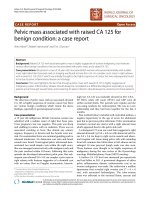
Vascular compression of the third part of the duodenum
by the SMA results in the development of a rare condition
of gastric outlet occlusion known as SMA syndrome. The
etiology of the syndrome is connected to the anatomy of
the third part of the duodenum in relation to the aortome-
senteric angle (Figure 1). Obstruction of the small bowel
by the SMA has been previously associated with spinal
manipulation in the surgical or conservative management
of scoliosis and has also been described in cases of exten-
sive burns, major surgeries such as ileoanal pouch anasto-
mosis, multiple combat injuries, severe trauma,
considerable weight loss in patients with malignancies,
anorexia nervosa or other eating disorders, as well as fol-
lowing the application of spica casts [1-3].
In scoliosis, the syndrome occurs most commonly in thin
and asthenic patients with a low body mass index (BMI)
Published: 19 January 2008
Journal of Medical Case Reports 2008, 2:9 doi:10.1186/1752-1947-2-9
Received: 18 June 2007
Accepted: 19 January 2008
This article is available from: />© 2008 Tsirikos et al; licensee BioMed Central Ltd.
This is an Open Access article distributed under the terms of the Creative Commons Attribution License ( />),
which permits unrestricted use, distribution, and reproduction in any medium, provided the original work is properly cited.
Journal of Medical Case Reports 2008, 2:9 />Page 2 of 5
(page number not for citation purposes)
who undergo spinal manipulation and correction of the
curvature by instrumentation, skeletal traction, casting or
bracing; these corrective techniques all result in significant
lengthening of the vertebral column and an extrinsic com-
pression of the distal duodenum as it passes through the
sharp angle formed by the aorta and the spine posteriorly
and the SMA anteriorly. Following scoliosis surgery, the
condition usually develops during the first postoperative
week [4-6].
We present a patient with an adolescent idiopathic scolio-
sis who underwent anterior spinal arthrodesis and devel-
oped severe SMA syndrome 6.5 weeks following surgery.
To the authors' knowledge this patient constitutes the lat-
est presentation of SMA syndrome following spinal
deformity surgery reported in the literature.
Case presentation
A previously healthy 16.8-year-old caucasian girl pre-
sented to our institution with an adolescent idiopathic
thoracolumbar scoliosis. Her past medical history was
non-contributory in regard to operations, medication, or
allergies. In admission, her body weight was 56.6 kg and
her body height was 164 cm, which were both at the 50
th
percentile for sex- and age-matched normal population.
Her BMI was 21 kg/cm/cm, which was also at the 50
th
per-
centile for her age and gender. There was no family history
of scoliosis or gastrointestinal pathology.
Our patient developed a flexible left thoracolumbar curve
extending from T10 to L2 and measuring 50° (Figure 2).
This was producing a moderate deformity due to listing of
the trunk and thoracic translocation to the left and a sig-
nificant waistline asymmetry. The preoperative blood
screening revealed no pathological findings. Albumin,
white blood cell, and lymphocyte counts were all within
normal limits. She underwent an anterior spinal arthrod-
esis extending from T10 to L2 with the use of third gener-
ation instrumentation [AO-Universal Spine System (USS)
II, Stratec Medical, Oberdorf] applying a spinal derota-
tional effect and autologous rib bone graft performed
through a left thoracoabdominal retroperitoneal
approach to the spine. This achieved a very satisfactory
correction of her scoliosis to 7° and a well-balanced spine
in both the coronal and the sagittal planes. Intraoperative
blood loss was 200 mls.
The patient's immediate postoperative course was uncom-
plicated and she was started on oral feedings at postoper-
ative day 1. The chest drain was removed on postoperative
day 3 and the patient was fitted with a thoracolumbar
plaster jacket to provide additional support to the spine
and mobilise out of her bed as per routine procedure in
our institution. Eight days following surgery she had no
complaints of her spine, was mobilising satisfactorily and
had a body weight of 52 kg (20
th
percentile). She was sub-
sequently discharged and was prescribed dietary supple-
ments high in calories to achieve gradual increase in her
body weight to preoperative values. Follow-up in the out-
patient clinic was arranged at 3 weeks post-discharge.
The patient failed to attend her clinical appointment and
was readmitted acutely at the hospital 45 days after spinal
fusion due to the development of severe nausea and per-
sistent vomiting. At the time of admission, she was mark-
edly dehydrated with associated oliguria and severe
electrolyte disorders including hypokalemia and meta-
bolic alkalosis. Her body weight had dropped further to
45.2 kg, which was at the 3
rd
percentile for gender- and
age-matched normal population. This indicated a total
loss of 11.4 kg post-surgery, which corresponded to 20%
of her preoperative body weight. Her BMI was reduced to
16.8 kg/cm/cm, which was also below the 3
rd
percentile
for her age and gender. The spinal jacket was removed and
on clinical examination her abdomen was found to be
considerably distended but soft and non-tender, with nor-
Graphical representation of the anatomical relations between the duodenum and the aortomesenteric angleFigure 1
Graphical representation of the anatomical relations
between the duodenum and the aortomesenteric angle.
Journal of Medical Case Reports 2008, 2:9 />Page 3 of 5
(page number not for citation purposes)
mal bowel sounds. A barium contrast study was obtained
and confirmed the clinical diagnosis of SMA syndrome
(Figure 3).
A nasogastric (NG) tube was placed for drainage of the bil-
ious gastric contents and a nasojejunal (NJ) tube under
fluoroscopic guidance for feedings. Attention was drawn
to correct electrolyte deficiencies through the administra-
tion of intravenous fluids. The nasogastric aspirates
decreased gradually over the following 10 days. The
patient received enteral feedings for a total period of 2
weeks. She was discharged 62 days post-surgery. Dietary
supplements (calorific drinks) were prescribed for an
additional period of 3 weeks. At the latest follow-up, 3.5
years after scoliosis correction, the patient was free of
symptoms, had increased her body weight and BMI to the
preoperative value of 50
th
percentile of normal and her
spine was fused without evidence of residual or recurrent
deformity.
Discussion
The incidence of SMA syndrome after surgical procedures
to correct spinal deformities has been reported to vary
between 0.5 and 4.7% [1,4,5,7-10]. The condition affects
predominantly adolescent patients who are tall, slimly
built with a thin asthenic body habitus. Children usually
present for surgical correction of an adolescent idiopathic
scoliosis during the phase of their most rapid longitudinal
growth. This accelerated skeletal growth may alter the rela-
tion between the SMA and the spine by decreasing the aor-
tomesenteric angle and, therefore, increase the risk for
duodenal compression. The mechanism is that of an acute
lengthening of the spinal column, which results in a
cephalad displacement of the aorto-SMA junction at the
expense of lateral mobility, due to either rapid height gain
occurring during adolescence, or following correction of
spinal deformities using either conservative (body casts
and braces) or surgical methods.
The duodenum is surrounded by a mesenteric fat pad and
lymphatic tissue as it crosses the aortomesenteric interval,
which both serve as a cushion, allowing for sufficient
space and preventing extrinsic obstruction of the bowel
caused by the SMA. Any factors that obliterate this inter-
vening space or disturb the relation between the adjacent
anatomical structures may result in compression or occlu-
sion of the duodenum. Underweight patients have less
periduodenal fat to cushion and protect the duodenum in
the SMA angle.
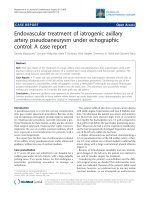
A barium contrast study shows dilatation of the stomach and the proximal duodenum, and occlusion of the third part of the duodenum by the SMA (white arrow)Figure 3
A barium contrast study shows dilatation of the stomach and
the proximal duodenum, and occlusion of the third part of
the duodenum by the SMA (white arrow).
Preoperative anteroposterior radiograph of the spine shows a left thoracolumbar scoliosis extending from T10 to L2 and measuring 50°Figure 2
Preoperative anteroposterior radiograph of the spine shows
a left thoracolumbar scoliosis extending from T10 to L2 and
measuring 50°.
Journal of Medical Case Reports 2008, 2:9 />Page 4 of 5
(page number not for citation purposes)
Surgical correction of the scoliotic spine produces vertical
tension on the SMA and the mesentery and narrows even
further the space available for the duodenum. In addition,
the majority of patients with an adolescent idiopathic tho-
racic scoliosis have associated thoracic hypokyphosis,
which by itself leads to a more extended spine and a
reduced aortomesenteric angle. A developmentally short
suspensory ligament of Treitz will also hold the duode-
num in an elevated position, precipitating further con-
striction between the aorta and the SMA.
In a recent study, Braun et al. [9] reported on a group of
patients who underwent scoliosis surgery and identified a
staged procedure to the spine, a lumbar modifier of B or
C as opposed to A, a low preoperative BMI, and increased
stiffness of a thoracic scoliosis as the most predictive fac-
tors for the development of SMA syndrome.
The classic symptomatology of the condition includes
nausea, bilious vomiting or increased bilious NG aspi-
rates, postprandial abdominal fullness and distension,
epigastric pain, while the bowel sounds are normal or
hyperactive. The abdomen is soft with occasional tender-
ness in the epigastrium to deep palpation. The vomiting
decompresses the stomach and produces asymptomatic
intervals lasting several hours prior to the next episode.
The symptoms most commonly develop within a few days
following scoliosis surgery and are associated with the use
of both Harrington and third generation instrumentation
techniques [4-6,11,12]. However, Kennedy and Cooper
[13] reported a 14-year-old male patient who developed
SMA syndrome and progressed rapidly to death 40 days
after scoliosis correction with Harrington instrumentation
and application of a body cast. Death in SMA syndrome
can result from inhalation of vomitus or can be occasion-
ally the consequence of gastric perforation [13]. In con-
trast, the patient described in the present study had a
favourable outcome after conservative treatment, even
though she developed the condition later than previously
reported following an anterior spinal arthrodesis with
third generation derotational instrumentation. The pre-
operative scoliotic curvature was not particularly severe
and, therefore, our patient would not be regarded at high
risk. The delay in the development of the syndrome can be
attributed to the significant progressive weight loss that
occurred in the postoperative period and resulted in grad-
ual loss of the retroperitoneal fat in the aortomesenteric
interval and subsequent obstruction of the duodenum.
The role of progressive postoperative weight loss has been
increasingly identified as a critical factor predisposing in
the occurrence of the condition [7,8]. This is in accord-
ance with the current report of our patient who developed
severe SMA syndrome resulting in significant medical ill-
ness 6.5 weeks following an anterior spinal arthrodesis.
This patient was at the 50
th
percentile for height, weight
and BMI preoperatively for sex- and age-matched normal
population. Both the weight and BMI progressively
dropped to the 3
rd
percentile following surgery. We, there-
fore, believe that the predominant etiological factor in the
development of the SMA syndrome in this particular
patient was the severe weight loss that occurred during the
postoperative period. It is possible that the application of
the spinal jacket could have caused extrinsic pressure to
the abdomen, resulting in further decrease in the aor-
tomesenteric angle and contributing to the onset of the
symptoms. In addition, Crowther et al. [1] hypothesized
that disruption of the autonomic nerve supply to the
small intestine, which commonly occurs during the retro-
peritoneal dissection to approach anteriorly the thoraco-
lumbar spine, can precipitate the development of the
condition.
Initial treatment of SMA syndrome should involve con-
servative measures. The aim is to reverse the pathological
cascade, which involves a primary partial duodenal
obstruction secondary to anatomical features that
progresses to complete occlusion through duodenal
edema caused by the persistent vomiting and abdominal
distension. Oral intake should be restricted. A NJ feeding
tube should be passed distal to the site of the duodenal
obstruction using radiographic assistance to provide
enteral feedings and achieve gradual weight gain. It is
important to note that gastric dilatation and recurrent
vomiting can ultimately lead to progressive dehydration,
severe hypovolemia, oliguria, electrolyte disorders, such
as hypokalemia and metabolic alkalosis as occurred in
our patient, or even death. Most of the reported deaths by
the condition involve patients in whom the diagnosis was
markedly delayed or was completely missed.
With appropriate conservative treatment, the symptoms
usually regress after 2-3 days and oral intake with soft sol-
ids can be restarted under careful monitoring. If enteral
feedings are not possible and the symptomatology per-
sists, total parenteral nutrition should be introduced in
order to provide adequate nutritional supplementation.
Hospital stay is markedly protracted causing significant
patient and parental anxiety. Surgical management
should be considered only if conservative methods fail.
Operative methods include open or laparoscopic duode-
nojejunostomy, division of the ligament of Treitz, and
open derotation of the duodenum.
Conclusion
We believe that it is essential to identify those patients
who are at greater risk of developing duodenal obstruc-
tion and initiate intensive preoperative dietary supple-
mentation in undernourished patients scheduled to
Publish with BioMed Central and every
scientist can read your work free of charge
"BioMed Central will be the most significant development for
disseminating the results of biomedical research in our lifetime."
Sir Paul Nurse, Cancer Research UK
Your research papers will be:
available free of charge to the entire biomedical community
peer reviewed and published immediately upon acceptance
cited in PubMed and archived on PubMed Central
yours — you keep the copyright
Submit your manuscript here:
/>BioMedcentral
Journal of Medical Case Reports 2008, 2:9 />Page 5 of 5
(page number not for citation purposes)
undergo complex spine deformity surgery as a preventa-
tive measure. In addition, the significance of a close mon-
itoring of any marked postoperative weight loss and the
need for an early intervention cannot be overemphasised.
We have described a patient who demonstrates that SMA
syndrome can develop late following scoliosis surgery, a
finding that is consistent with etiology of gradual onset. A
high index of suspicion will lead to an early diagnosis of
the condition at a stage when conservative measures are
more likely to produce a good outcome. If the diagnosis is
delayed or missed, SMA syndrome can cause considerable
morbidity and may result in potentially life-threatening
complications.
Abbreviations
SMA: superior mesenteric artery
BMI: body mass index
NG: nasogastric
NJ: nasojejunal
Competing interests
The author(s) declare that they have no competing inter-
ests.
Authors' contributions
1). A.I. Tsirikos: conception and design, analysis of data,
preparation of the manuscript, final approval of the ver-
sion to be published.
2). R.E. Anakwe: acquisition and analysis of data, drafting
the manuscript.
3). A.D.L. Baker: acquisition and analysis of data.
Consent
Written informed consent was obtained from the patient
for publication of the study. A copy of the written consent
is available for review by the Editor-in-Chief of this jour-
nal.
References
1. Crowther MA, Webb PJ, Eyre-Brook IA: Superior mesenteric
artery syndrome following surgery for scoliosis. Spine 2002,
27:528-533.
2. Wilson-Storey D, MacKinlay GA: The superior mesenteric
artery syndrome. J R Coll Surg Edinb 1986, 31:175-178.
3. Welsch T, Buchler MW, Kienle P: Recalling superior mesenteric
artery syndrome. Dig Surg 2007, 24:149-156.
4. Altiok H, Lubicky JP, DeWald CJ, Herman J: The superior
mesenteric artery syndrome in patients with spinal deform-
ity. Spine 2005, 30:2164-2170.
5. Tsirikos AI, Jeans LA: Superior mesenteric artery syndrome in
children and adolescents with spine deformities undergoing
corrective surgery. J Spinal Disord Tech 2005, 18:263-271.
6. Zhu ZZ, Qiu Y: Superior mesenteric artery syndrome follow-
ing scoliosis surgery: its risk indicators and treatment strat-
egy. World J Gastroenterol 2005, 11:3307-3310.
7. Hutchinson DT, Bassett GS: Superior mesenteric artery syn-
drome in pediatric orthopedic patients. Clin Orthop 1990,
250:250-257.
8. Munns SW, Morrissy RT, Golladay ES, McKenzie CN: Hyperalimen-
tation for superior mesenteric-artery (cast) syndrome fol-
lowing correction of spinal deformity. J Bone Joint Surg (Am)
1984, 66:1175-1177.
9. Braun SV, Hedden DM, Howard AW: Superior mesenteric artery
syndrome following spinal deformity correction. J Bone Joint
Surg (Am) 2006, 88:2252-2257.
10. Schwartz A: Scoliosis, superior mesenteric artery syndrome,
and adolescents. Orthop Nurs 2007, 26:19-24. quiz 25-6.
11. Vitale MG, Higgs GB, Liebling MS, Roth N, Roye DP Jr: Superior
mesenteric artery syndrome after segmental instrumenta-
tion: a biomechanical analysis. Am J Orthop 1999, 28:461-467.
12. Zadegan F, Lenoir T, Drain O, Dauzac C, Leroux R, Morel F, Guigui
P: Superior mesenteric artery syndrome following correc-
tion of spinal deformity: case report and review of the liter-
ature. Rev Chir Orthop Reparatrice Appar Mot 2007, 93:181-185.
13. Kennedy RH, Cooper MJ: An unusually severe case of the cast
syndrome. Postgrad Med J 1983, 59:539-540.