Báo cáo khoa hoc:" Giant hepatic hydatid cyst with sub-fascial extension treated by open minimally invasive surgery: a case report" potx
Bạn đang xem bản rút gọn của tài liệu. Xem và tải ngay bản đầy đủ của tài liệu tại đây (543.7 KB, 5 trang )
BioMed Central
Page 1 of 5
(page number not for citation purposes)
Journal of Medical Case Reports
Open Access
Case report
Giant hepatic hydatid cyst with sub-fascial extension treated by
open minimally invasive surgery: a case report
Dipesh D Duttaroy*
1
, Samir Kacheriwala
1
, Bithika Duttaroy
2
,
Jitendra Jagtap
1
, Gunjan Patel
1
and Nikhil Modi
1
Address:
1
Department of Surgery, Government Medical College & Sir Sayajirao General Hospital, Baroda, Gujarat, 390001, India and
2
Department
of Microbiology, Government Medical College & Sir Sayajirao General Hospital, Baroda, Gujarat, 390001, India
Email: Dipesh D Duttaroy* - ; Samir Kacheriwala - ; Bithika Duttaroy - ;
Jitendra Jagtap - ; Gunjan Patel - ; Nikhil Modi -
* Corresponding author
Abstract
Introduction: Hepatic hydatid disease can be successfully treated by a variety of modalities.
Case Presentation: We report a case of a 60 year old male with giant hepatic hydatid disease
who presented with a huge cystic mass in the upper abdomen. Diagnosis was confirmed by
serology, ultrasonography and CT scan. The patient was treated successfully by open minimally
invasive surgery with minimum breaching of the peritoneal cavity using a laparoscopic trocar to
evacuate the cyst.
Conclusion: The use of a laparoscopic trocar through a small abdominal incision in selected
patients with hepatic hydatid disease with subfascial extension can be a safe, minimally-invasive
option of treatment
Introduction
Cystic hydatid disease (echinococcosis) is an important
zoonotic disease caused in humans by Echinococcus gran-
ulosus, a cestode that usually inhabits the intestine of
dogs and other canines as a definitive host. Humans are
accidental intermediate hosts due to ingestion of the par-
asitic eggs. The liver is the most common site for the
occurrence of the larval form of cystic hydatid disease, the
others being lung, brain and other viscera [1]. Though a
variety of treatment modalities have been successfully
employed, there is a lack consensus as to the most appro-
priate method. Medical therapy in the form of benzoimi-
dazole carbamates alone or in combination with
praziquantel has been advocated for the treatment of
hydatid disease [2-4]. Interventional radiologists and gas-
troenterologists have used minimal invasive procedures
such as PAIR (puncture, aspiration, injection, re-aspira-
tion) [5-7] and PEVAC (percutaneous evacuation of cyst
content) [8] for treating hepatic echinococcosis. An array
of surgical procedures has been recommended. In recent
times, laparoscopic surgery and the use of laparoscopic
instruments (trocar and suction) have been found to be
safe and effective in the management of hepatic hydatid
disease [9-11]. We report a patient with giant hepatic
hydatid disease with subfascial extension into the abdom-
inal wall who was treated successfully by open minimal
invasive surgery with minimum violation of the perito-
neal cavity.
Published: 28 January 2008
Journal of Medical Case Reports 2008, 2:26 doi:10.1186/1752-1947-2-26
Received: 29 August 2007
Accepted: 28 January 2008
This article is available from: />© 2008 Duttaroy et al; licensee BioMed Central Ltd.
This is an Open Access article distributed under the terms of the Creative Commons Attribution License ( />),
which permits unrestricted use, distribution, and reproduction in any medium, provided the original work is properly cited.
Journal of Medical Case Reports 2008, 2:26 />Page 2 of 5
(page number not for citation purposes)
Case presentation
A 60-year-old male presented with continuous dull aching
upper abdominal pain of four months duration and a
gradually increasing visible upper abdominal lump over
the past two months. Clinical examination revealed an
afebrile non-icteric man with mild pallor and pedal
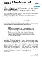
edema. The patient had a huge lobulated liver (span 22
cm) occupying both hypochondria and the right lumbar,
epigastrium and umbilical regions with a localized cystic
subfascial projection in the epigastrium of 8 × 8 cm. (Fig-
ure-1A &1B) Laboratory investigations revealed haemo-
globin 10 gm%, white blood cell count 7500/µl and
eosinophils 900/µl. Serological test with an enzyme-
linked immunosorbent assay (ELISA) for echinococcus
was positive. Liver function tests were within normal
range. Radiography showed elevation of the right dia-
phragm with a soft tissue shadow in the upper abdomen.
Ultrasonography (USG) of the abdomen revealed a 19 ×
12 × 13 cm cystic lesion in the right lobe of the liver with
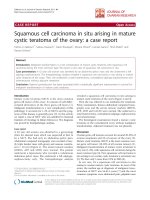
multiple anechoic cysts within it. Spiral CT scan of the
liver (Figure-2A &2B) confirmed a hydatid cyst in the right
lobe (segments – V, VI, VII, VIII), with multiple daughter
cysts within, compressing the portal vein, inferior vena
cava, hepatic veins, gallbladder, intra and extra hepatic
biliary tree and the right kidney. The anterior aspect of the
cyst demonstrated a cystic projection in the midline
stretching the fascial aponeurotic layer. (Black arrow –
Figure-2A)
The patient received Albendazole 10 mg/kg/day for 28
days with the aim of sterilizing the cyst contents. His
abdomen was then explored under general anaesthesia
using a 6 cm midline incision over the epigastric cystic
swelling. After dividing the linea alba a close continuous
suture of 2-0 silk was taken all around between the
divided fascial aponeurosis and the projecting cyst wall
A) Anterior view of abdomen showing the globular cystic lump in the epigastriumFigure 1
A) Anterior view of abdomen showing the globular cystic lump in the epigastrium. B) Left lateral view abdomen showing the
lateral profile of the lump. C) Inset: Sutured incision with Foleys catheter in situ, draining the cyst cavity.
Journal of Medical Case Reports 2008, 2:26 />Page 3 of 5
(page number not for citation purposes)
(Figure-3A) to prevent the spillage and entry of cyst con-
tents into the peritoneal cavity during the process of evac-
uation. Using a 16-gauge needle, a three-way stopcock,
and a 50 ml syringe, we attempted aspiration of the cyst
prior to instillation of a scolicidal agent through the
exposed cyst wall. Due to the thick contents of the cyst, the
attempt failed and was abandoned. We introduced a 10
mm laparoscopic trocar into the cyst cavity after stabiliz-
ing the exposed cyst wall with tissue forceps and isolating
the area with gauze packs soaked in 0.5% Cetrimide solu-
tion. A suction cannula was applied to the mouth of the
laparoscopic sleeve keeping the valve open. Alternate use
of the suction tube applied to the mouth of the sleeve
(Figure-3B) and a high pressure laparoscopic suction irri-
gation apparatus introduced into the cyst cavity resulted
in a drainage of three liters of thick, viscid, cream-colored
cyst contents containing abundant daughter cysts (Figure-
3B inset). After near total evacuation of the cyst, a 30° tel-
escope was introduced through the trocar sleeve to visual-
ize the cavity for adherent membranes and biliary leak.
Adherent daughter cysts and membranes were then evac-
uated manually by a long thin spoon introduced through
the trocar site after its removal.
We could not visualize any gross biliary leak. The cyst cav-
ity was irrigated with 2.5 liters of 1% povidone iodine
solution twice. A 24 F self retaining Foleys catheter was
then introduced into the cyst cavity as a drain, the course
of which was routed with the help of a long Robert forceps
passed though the opening into the cyst cavity and then
rail-roaded into position. (Figure-1C inset) The trocar site
A) Intraoperative view showing the exposed cystic projec-tion of the hydatid cyst with a purse string all aroundFigure 3
A) Intraoperative view showing the exposed cystic projec-
tion of the hydatid cyst with a purse string all around. B)
Daughter cysts being sucked through the laparoscopic trocar
sheath. (Inset: suction bottle containing cyst contents.)
A) & B) Axial spiral CT scan of abdomen through two differ-ent levels showing a hydatid cyst in the left lobe (Segments V, VI, VII, VIII) with a cystic projection anteriorlyFigure 2
A) & B) Axial spiral CT scan of abdomen through two differ-
ent levels showing a hydatid cyst in the left lobe (Segments V,
VI, VII, VIII) with a cystic projection anteriorly.
Journal of Medical Case Reports 2008, 2:26 />Page 4 of 5
(page number not for citation purposes)
on the cyst was closed by continuous 2-0 Polyglactin
sutures. After local irrigation of the site with 0.5% Cetrim-
ide solution, the silk suture anchoring the cyst wall to the
fascial layer was then detached and the linea alba was
closed with continuous 1-0 Polypropylene sutures.
Patient received liquids orally in the evening and solid
food next morning onwards. The patient had received an
injection of cefotaxime perioperatively. He was dis-
charged on the third postoperative day with the drain in
situ.
The cyst contents, which were sterile on culture, showed
protoscolices, fragments of laminated membrane and
hooklets. Skin sutures were removed on the eighth post-
operative day. Other than postoperative biliary drainage
of 500 ml/day that gradually decreased over one month,
the patient's recovery was uneventful. We removed the
drain after USG confirmed a collapsed cyst cavity with
minimal collection. Follow-up USG after a period of four
months revealed a collapsed residual cavity with no evi-
dence of recurrent disease. Though a CT scan is essential
to compare the pre and postoperative characteristics in
hepatic hydatid disease, it was not feasible due to finan-
cial constraints.
Discussion
Despite a variety of open and minimal invasive tech-
niques being available for the treatment of hydatid dis-
ease of the liver, one of the main concerns of the treating
physician is spillage of cyst contents that can lead to recur-
rence in various forms and anaphylactic reactions. We
cannot apply a single procedure uniformly because
hepatic hydatid disease presents in diverse forms, which
necessitates appropriate measures for each case. PAIR with
benzoimidazole carbamates is recommended as a primary
line of therapy for uncomplicated hepatic echinococcosis
[5-7,12]. Though, there have been reports of successful
percutaneous drainage of giant hepatic hydatid cyst, huge
complicated cysts with impending rupture or fistulization
are poor candidates for such intervention. In such
instances, appropriate surgical management becomes
vital.
Open surgical techniques employed include evacuation
and simple closure, evacuation with drainage, marsupial-
ization, closed total cystectomy, partial pericystectomy,
partial pericystectomy with capitonnage, partial pericys-
tectomy with cavity management (omentoplasty and
internal drainage) and partial hepatectomy [11,12]. The
principle of any surgical procedure for liver hydatid dis-
ease is complete evacuation of the cyst, prevention of
intra-abdominal spillage, detection of major cysto-biliary
communications, and sterilization and early obliteration
of the residual cavity [11-13]. However, surgical proce-
dures are not without complications and are associated
with both morbidity (anaphylaxis, cyst infection, liver or
intra-abdominal sepsis, haemorrhage and biliary fistula)
and rarely mortality [7]. Over the last decade, laparo-
scopic management of liver hydatid disease has been car-
ried out the world over with excellent results [9,11,12,14].
While, laparoscopic surgery follows all the principles of
open surgery it is beneficial to the patient in providing
reduced postoperative discomfort, shorter recovery time
and reduced hospital stay.
In this case we approached the cyst directly since the ante-
rior portion of the huge cyst was herniating into the mid-
line as a diverticulum (Figure-2A Black arrow) and
stretching the linea alba. Apprehensions about the spill-
age of the cyst contents into the peritoneal cavity pre-
vented us from penetrating the cyst directly through the
abdominal wall with a laparoscopic trocar. Our experi-
ence with advanced laparoscopic surgery is limited. Spe-
cialized instruments such as the Palanivelu Hydatid
System [11] or the locking umbrella trocar, [14] which
have been designed to prevent the spillage of hydatid fluid
during laparoscopic surgery, were not available; hence, we
avoided the conventional laparoscopic route. The open
surgical technique adopted by us in this case offered most
of the advantages of laparoscopic surgery. We could evac-
uate a giant hepatic hydatid cyst without intraperitoneal
spillage, visualize the cavity and drain it through a small
abdominal incision. Postoperative recovery time and hos-
pital stay was reduced. The percutaneous laparoscopic
approach has been adopted by Kayalp et al to deal with a
liver abscess pointing onto the anterior abdominal wall in
which the trocar was directly introduced into the abscess
cavity [15]. The same author has used a laparoscopy trocar
for evacuation of a hydatid cyst after conventional
abdominal exploration through an extended subcostal
incision with the aim of preventing spillage [10]. Seven et
al have used the laparoscopic approach to enter the cyst
cavity with a 10 mm trocar having an umbrella locking
mechanism, that was utilized to suspend and fix the cyst
against the abdominal wall [14]. This was subsequently
followed by aspirating the cyst contents through the tro-
car, direct visualization of the cyst by introducing a tele-
scope and drainage of the cyst. The advantage of their
approach was that a biliary communication could be dealt
with by laying open the cyst wall, which was not possible
with our technique.
The advantage of our technique is that gross intra-perito-
neal contamination is eliminated since the cyst is not
exposed to the peritoneal cavity during surgery. The cyst
contents, including daughter cysts, can be evacuated by
high-pressure suction. If adherent membranes are visual-
ized on the wall, they can be manually debrided through
the same opening. The cyst is accessed through a small
abdominal incision and there is no handling of abdomi-
Publish with BioMed Central and every
scientist can read your work free of charge
"BioMed Central will be the most significant development for
disseminating the results of biomedical research in our lifetime."
Sir Paul Nurse, Cancer Research UK
Your research papers will be:
available free of charge to the entire biomedical community
peer reviewed and published immediately upon acceptance
cited in PubMed and archived on PubMed Central
yours — you keep the copyright
Submit your manuscript here:
/>BioMedcentral
Journal of Medical Case Reports 2008, 2:26 />Page 5 of 5
(page number not for citation purposes)
nal viscera other than the liver. Postoperative pain and
ileus is minimal leading to an early recovery. The patient
can be started on oral fluids by the evening of surgery.
One of the drawbacks is the potential risk of puncture of
the cyst wall while taking the circumferential anchoring
sutures between the fascia and the cyst wall leading to leak
of hydatid fluid. Another limitation of the technique is
that if cysto-biliary communications are visualized they
cannot be dealt with intraoperatively without modifying
the procedure. Postoperative biliary leakage has to be
dealt with conservatively on expectant lines as in our
patient, or by further interventional procedures. The intro-
duction of the drain, though guided, is a blind procedure
and can lead to potential injuries to the adjacent organs;
hence utmost care has to be taken during its introduction.
Ultrasound guided drain insertion may be a sound option
if available. Though this method has been tried success-
fully in only a single patient we would like to emphasize
that the same can be replicated in a selected subset of
patients with large superficial palpable hydatid cysts
either stretching or herniating through the abdominal
wall musculature.
Conclusion
The use of a laparoscopic trocar through a small abdomi-
nal incision in selected patients with hepatic hydatid dis-
ease can be a safe, minimally-invasive surgical option of
treatment, which would reduce post operative discomfort
and result in early recovery.
Competing interests
The author(s) declare that they have no competing inter-
ests.
Authors' contributions
DDD is the consultant surgeon responsible for the
patient's care. He conceived this report, drafted the article
and performed the surgery. SK assisted in performing the
surgery, and helped in drafting and revision of the article.
BD performed the investigations, helped in the literature
search and supervised the drafting and overall structure of
the article. JJ did the photography, helped in acquisition
of data and technical support and revision of the article.
GP acquired the radiological images and helped in draft-
ing. NM performed the literature search and helped in
revision. All authors read, appraised and approved the
final manuscript.
Consent
Written informed consent was obtained from the patient
prior to publication of this case report.
References
1. Menezes da Silva A: Hydatid cyst of the liver-criteria for the
selection of appropriate treatment. Acta Trop 2003,
85:237-242.
2. Teggi A, Lastilla MG, De Rosa F: Therapy of human hydatid dis-
ease with mebendazole and albendazole. Antimicrob Agents
Chemother 1993, 37:1679-1684.
3. Silva MA, Mirza DF, Bramhall SR, Mayer AD, McMaster P, Buckels JA:
Treatment of hydatid disease of the liver. Evaluation of a UK
experience. Dig Surg 2004, 21:227-233.
4. Koulas SG, Sakellariou A, Betzios J, Nikas K, Zikos N, Pappas-Gogos
G, Tsimoyiannis EC: A fifteen years experience (1988–2003) on
the management of liver hydatidosis in northwestern
Greece. Int Surg 2006, 91:112-116.
5. Akhan O, Ozmen MN, Dincer A, Sayek I, Gocmen A: Liver hydatid
disease: long-term results of percutaneous treatment. Radi-
ology 1996, 198:259-264.
6. Brunetti E, Filice C, Macpherson C, Meslin F, Vuitton D, et al.: PAIR:
Puncture, Aspiration, Injection, Re-aspiration. An option for
the treatment of Cystic Echinococcosis. WHO/EMC web site
[ />WHO_CDS_CSR_APH_2001.6.pdf]. Accessed on January 10, 2007
7. Smego RA Jr, Bhatti S, Khaliq AA, Beg MA: Percutaneous aspira-
tion injection-reaspiration drainage plus albendazole or
mebendazole for hepatic cystic echinococcosis: a meta-anal-
ysis. Clin Infect Dis 2003, 37:1073-1083.
8. Schipper HG, Lameris JS, van Delden OM, Rauws EA, 281 Kager PA:
Percutaneous evacuation (PEVAC) of multivesicular echino-
coccal cysts with or without cystobiliary fistulas which con-
tain non-drainable material: first results of a modified PAIR
method. Gut 2002, 50:718-723.
9. Bickel A, Eitan A: The use of a large, transparent cannula, with
a beveled tip, for safe laparoscopic management of hydatid
cysts of liver. Surg Endosc 1995, 9:1304-1305.
10. Kayaalp C: Evacuation of hydatid liver cysts using laparoscopic
trocar. World J Surg 2002, 26:1324-1327.
11. Palanivelu C, Jani K, Malladi V, Senthilkumar R, Rajan PS, Sendhilkumar
K, Parthasarthi R, Kavalakat A: Laparoscopic management of
hepatic hydatid Disease. JSLS 2006, 10:56-62.
12. Dervenis C, Delis S, Avgerinos C, Madariaga J, Milicevic M: Changing
concepts in the management of liver hydatid disease. J Gas-
trointest Surg 2005, 9:869-877.
13. Losanoff JE, Richman BW, Jones JW: Organ-sparing surgical
treatment of giant hepatic hydatid cysts. Am J Surg 2004,
187:288-290.
14. Seven R, Berber E, Mercan S, Eminoglu L, Budak D: Laparoscopic
treatment of hepatic hydatid cysts. Surgery 2000, 128:36-40.
15. Kayaalp C, Yol S, Nessar G: Drainage of liver abscess via lapar-
oscopic trocar with local anesthesia. Surg Laparosc Endosc Percu-
tan Tech 2003, 13:121-124.