Báo cáo khoa hoc:" Hepaticocystic duct and a rare extra-hepatic "cruciate" arterial anastomosis: a case report" docx
Bạn đang xem bản rút gọn của tài liệu. Xem và tải ngay bản đầy đủ của tài liệu tại đây (422.29 KB, 3 trang )
BioMed Central
Page 1 of 3
(page number not for citation purposes)
Journal of Medical Case Reports
Open Access
Case report
Hepaticocystic duct and a rare extra-hepatic "cruciate" arterial
anastomosis: a case report
Vasitha Abeysuriya*
1
, Sujatha Salgado
1
, Kemal I Deen
2
and
Sumudu K Kumarage
2
Address:
1
Department of Clinical Anatomy, Faculty of Medicine, University of Kelaniya, Ragama, Sri Lanka and
2
Department of Surgery, Faculty
of Medicine, University of Kelaniya, Ragama, Sri Lanka
Email: Vasitha Abeysuriya* - ; Sujatha Salgado - ; Kemal I Deen - ;
Sumudu K Kumarage -
* Corresponding author
Abstract
Introduction: The variations in the morphological characteristics of the extra-hepatic biliary
system are interesting.
Case presentation: During the dissection of cadavers to study the morphological characteristics
of the extra-hepatic biliary system, a 46-year-old male cadaver was found to have drainage of the
common hepatic duct drains directly into the gall bladder neck. The right and left hepatic ducts
were not seen extra-hepatically. Further drainage of the bile away from the gallbladder and into the
duodenum was provided by the cystic duct. Formation of the common bile duct by the union of
the common hepatic duct and cystic duct was absent. Further more the right hepatic artery was
found to be communicating with the left hepatic artery by a "bridging artery" after giving rise to the
cystic artery. An accessory hepatic artery originated from the "bridging artery" forming a "cruciate"
hepatic arterial anastomosis.
Conclusion: Combination of a Hepaticocystic duct and an aberrant variation in the extra-hepatic
arterial system is extremely rare.
Introduction
The variations in the morphological characteristics of the
extra-hepatic biliary system are numerous. It has been
stated that the extra-hepatic biliary system has more
anomalies in one cubic centimeter of the space around the
region of the cystic duct than any other part of the body
[1,2]. These anomalies add to operative difficulties during
cholecystectomy.
The incidence of congenital anomalies of the extra-hepatic
biliary system varies between 0.58% and 47.2% [3]. Due
to the scarcity of studies of this regional anatomy, the
exact incidence of all the anomalies of the biliary system
is not known but as it has been observed that, vascular
anomalies are more frequent than those of the ductal sys-
tem [2,4]. Anomalies of the extra-hepatic biliary system
can arise from the gallbladder, cystic duct, hepatic ducts or
Published: 6 February 2008
Journal of Medical Case Reports 2008, 2:37 doi:10.1186/1752-1947-2-37
Received: 20 November 2007
Accepted: 6 February 2008
This article is available from: />© 2008 Abeysuriya et al; licensee BioMed Central Ltd.
This is an Open Access article distributed under the terms of the Creative Commons Attribution License ( />),
which permits unrestricted use, distribution, and reproduction in any medium, provided the original work is properly cited.
Journal of Medical Case Reports 2008, 2:37 />Page 2 of 3
(page number not for citation purposes)
the common bile duct as a result of aberrations of normal
embryological development. Therefore, it is essential to
appreciate the extent of anomalies of the extra-hepatic bil-
iary system.
One such rare anomaly is where the right and left hepatic
ducts are not seen in their usual extra hepatic location and
the common hepatic duct drains directly into the gall
bladder neck with absence of the common bile duct. Fur-
ther drainage of the bile away from the gallbladder and
into the duodenum is provided by the cystic duct. It has
also been referred to differently as the cholecystohepatic
duct. Possible anomalies include congenital absence of
the common bile duct, transverse lie of the gallbladder, or
gallbladder interposition [1].
Case presentation
A 46-year-old male cadaver was found to have an extra-
hepatic biliary system with rare morphological anomalies.
This was identified during a descriptive-prospective cross
sectional study of the morphological characteristics of the
extra-hepatic biliary system in humans.
The anterior abdominal wall was opened longitudinally
along the midline. The abdominal wall was separated on
to the right side along the central margin up to the mid
axillary line. Then the abdominal wall was divided from
the right side of the pubic bone up to the anterior superior
iliac spine. The anterior abdominal wall flap was reflected
laterally. The stomach was retracted to the left side and the
second part of the duodenum, free margin of the lesser
omentum, epiploic foramen and gall bladder identified.
Dissection was done to demonstrate the extra-hepatic bil-
iary system and its vascular pattern.
The right and left hepatic ducts were not seen extra-hepat-
ically and the common hepatic duct drained directly into
the gall bladder neck, with absence of the common bile
duct. Further drainage of the bile away from the gallblad-
der and into the duodenum was provided by the cystic
duct. The gall bladder was lying in the gall bladder fossa
in the right lobe of the liver in a transverse plane. The
width and the length of the gall bladder were 2.5 cm and
4.5 cm respectively. The length of the common hepatic
duct was 2.6 cm and the cystic duct that drained the gall
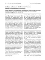
bladder to the duodenum was 6.9 cm (Figure 1).
Apart from the anomalous extra-hepatic biliary morphol-
ogy, a rare abnormal arterial pattern was observed. The
cystic artery was anterior to the common hepatic duct and
it was originating from the right hepatic artery. The divi-
sion of anterior and posterior branches of the cystic artery
was not noted. The right hepatic artery was found to be
communicating with the left hepatic artery by a "bridging
artery" after giving rise to the cystic artery. An accessory
hepatic artery originated from the "bridging artery" form-
ing a "cruciate" hepatic arterial anastomosis (Figure 1).
Discussion
The liver, gallbladder and the extra-hepatic biliary tree
arise from the hepatic diverticulum of the foregut in the
beginning of the fourth week of embryological develop-
ment. This diverticulum rapidly proliferates into the sep-
tum transversum and divides into two parts, the distal
pars hepatica, and the proximal pars cystica [1]. At the
time of appearance of the pars cystic artery, there occurs
proliferation of the cells at the junction of the cystic and
hepatic ducts to form the common bile duct, which is ini-
tially a cylindrical mass that undergoes vacuolation to
canalize and form a single, continuous, epithelium lined
lumen [1,5-7].
Liver (LIV) with partially dissected right lobe is seen craniallyFigure 1
Liver (LIV) with partially dissected right lobe is seen cranially.
The common hepatic duct (CHD) directly drains to the
upper segment of the gall bladder (GB). From the GB the
cystic duct (CD) originates and drains out as the common
bile duct without joining to the CHD. The common hepatic
artery is divided into the proper hepatic artery (PHA) and
the gastro-duodenal artery (GDA). The portal vein (PV) is
seen between the CD and the PHA. The PHA divides into
left and right hepatic arteries (LHA & RHA). The cystic
artery (CA) is originating from the RHA. A "bridging artery"
(BA) connects the right and left hepatic arteries. An acces-
sory hepatic artery (AH) is originating from the bridging
artery, forming a "cruciate" hepatic arterial anastomosis.
Publish with BioMed Central and every
scientist can read your work free of charge
"BioMed Central will be the most significant development for
disseminating the results of biomedical research in our lifetime."
Sir Paul Nurse, Cancer Research UK
Your research papers will be:
available free of charge to the entire biomedical community
peer reviewed and published immediately upon acceptance
cited in PubMed and archived on PubMed Central
yours — you keep the copyright
Submit your manuscript here:
/>BioMedcentral
Journal of Medical Case Reports 2008, 2:37 />Page 3 of 3
(page number not for citation purposes)
Failure of this normal development results in various
anomalies, the rarest amongst which, is the Hepaticocystic
duct. There are various patterns of Hepaticocystic ducts
that have been recognized. Type I refers to the absence of
the common hepatic duct where the right and left ducts
drain separately into the gallbladder; Type II is when the
right and left hepatic ducts unite upon entering the gall-
bladder; Type III refers to a common hepatic duct that
enters the gallbladder, and in Type IV multiple small bile
ducts connect the intrahepatic biliary system with the gall-
bladder. Type III is further subdivided into the common
hepatic duct entering the superior wall of the gallbladder
(III A), neck (III B), posterior gallbladder wall (III C), and,
the fundus (III D) [8].
In our dissections the human cadaver was found to have a
Type III B Hepaticocystic duct. Additionally it was found
that there was no division of the common hepatic duct
into right and left hepatic ducts extra-hepatically. There-
fore we would like to sub-categorise our case as a variant
of Type III B. There have been no previous reported cases
of this nature of combined anomaly.
Although the exact etiology of this rare anomaly is
unknown, it is thought to result either from failure of reca-
nalization, with persistence of fetal communications
between the gallbladder and liver [7] or from delayed divi-
sion of the hepatic antrum into the cystic and hepatic
diverticuli [1,2].
Conclusion
Occurrence of a Hepaticocystic duct and an aberrant vari-
ation in the extra hepatic arterial system is extremely rare.
The knowledge of such variation is important in surgical
procedures related to the extra-hepatic biliary system.
Comprehensive knowledge and clear visualization during
surgery is mandatory for safe surgical procedures related
to this important anatomical region.
Competing interests
The author(s) declare that they have no competing inter-
ests.
Authors' contributions
VA was responsible for the study conception and design,
writing the manuscript, dissections and literature review.
SS was responsible for literature review, dissection and
proofreading the manuscript. KD was responsible for lit-
erature review and proofreading the manuscript. SK was
responsible for literature review and dissection. All
authors read and approved the final manuscript.
Consent
Written informed consent was obtained from the patients'
daughter for publication of this case report and accompa-
nying images. A copy of the written consent is available
for review by the Editor-in-Chief of this journal.
References
1. Robin kaushik MS, Attri AKMS: Hepaticocystic Duct-A Case
Report. The International Journal of Surgery 2005, 21:1528-8242.
2. Walia HS, Abraham TK, Baraka A: Gallbladder Interposition: A
Rare Anomaly of the Extrahepatic Ducts. Int Surg 1986,
71:117-21.
3. Lamah M, Dickson GH: Congenital anatomical abnormalities of
the extrahepatic biliary duct: a personal audit. Surg Radiol Anat
1999, 21:325-7.
4. Lamah M, Karanjia ND, Dickson GH: Anatomical variations of
the Extrahepatic Biliary Tree; Review of the World Litera-
ture. Clin Anat 2001, 14:167-72.
5. Losanoff JE, Kjossev KT, Katrov E: Hepaticocystic duct – A Case
Report. Surg Radiol Anat 1996, 18:339-41.
6. Adkins RB Jr, Chapman WC, Reddy VS: Embryology, Anatomy,
and Surgical Applications of the Extrahepatic Biliary System.
Surg Clin N Am 2000, 80:363-79.
7. Olsha O, Steiner A, Rivkin LA, Sheinfeld A: Congenital absence of
the anatomic bile duct. Acta Chir Scand 1987, 153:387-90.
8. Losanoff JE, Jones JW, Richman BW, Rangnekar NJ: Hepaticocystic
duct: A Rare Anomaly of the Extrahepatic Biliary System.
Clin Anat 2002, 15:314-5.