Báo cáo y học: "Antigenized antibodies expressing Vβ8.2 TCR peptides immunize against rat experimental allergic encephalomyelitis" pps
Bạn đang xem bản rút gọn của tài liệu. Xem và tải ngay bản đầy đủ của tài liệu tại đây (614.32 KB, 12 trang )
BioMed Central
Page 1 of 12
(page number not for citation purposes)
Journal of Immune Based Therapies
and Vaccines
Open Access
Original research
Antigenized antibodies expressing Vβ8.2 TCR peptides immunize
against rat experimental allergic encephalomyelitis
Cristina Musselli, Svetlana Daverio-Zanetti and Maurizio Zanetti*
Address: The Department of Medicine and Cancer Center, University of California, San Diego, La Jolla CA USA
Email: Cristina Musselli - ; Svetlana Daverio-Zanetti - ; Maurizio Zanetti* -
* Corresponding author
EAETCRIdiotypeRegulation
Abstract
Background: Immunity against the T cell receptor (TCR) is considered to play a central role in
the regulation of experimental allergic encephalomyelitis (EAE), a model system of autoimmune
disease characterized by a restricted usage of TCR genes. Methods of specific vaccination against
the TCR of pathogenetic T cells have included attenuated T cells and synthetic peptides from the
sequence of the TCR. These approaches have led to the concept that anti-idiotypic immunity
against antigenic sites of the TCR, which are a key regulatory element in this disease.
Methods: The present study in the Lewis rat used a conventional idiotypic immunization based on
antigenized antibodies expressing selected peptide sequences of the Vβ8.2 TCR (
93
ASSDSSNTE
101
and
39
DMGHGLRLIHYSYDVNSTEKG
59
).
Results: The study demonstrates that vaccination with antigenized antibodies markedly
attenuates, and in some instances, prevents clinical EAE induced with the encephalitogenic peptide
68
GSLPQKSQRSQDENPVVHF
88
in complete Freunds' adjuvant (CFA). Antigenized antibodies
induced an anti-idiotypic response against the Vβ8.2 TCR, which was detected by ELISA and
flowcytometry. No evidence was obtained of a T cell response against the corresponding Vβ8.2
TCR peptides.
Conclusions: The results indicate that antigenized antibodies expressing conformationally-
constrained TCR peptides are a simple means to induce humoral anti-idiotypic immunity against
the TCR and to vaccinate against EAE. The study also suggests the possibility to target idiotypic
determinants of TCR borne on pathogenetic T cells to vaccinate against disease.
Introduction
Experimental allergic encephalomyelitis (EAE) is an
experimentally induced autoimmune disease mediated by
T cells. It can be induced in susceptible animals either by
immunization with myelin basic protein (MBP) or prote-
olipid protein PLP, or by immunization with synthetic
peptides from the MBP sequence [1]. EAE can also be ini-
tiated by the passive transfer of encephalitogenic, MBP-
specific T cell lines or clones [2,3]. In the Lewis rat, EAE is
characterized by a self limiting, ascending, hind limb
paralysis. Histologically, EAE is hallmarked by perivascu-
lar and submeningeal infiltration of inflammatory cells
Published: 12 November 2004
Journal of Immune Based Therapies and Vaccines 2004, 2:9 doi:10.1186/1476-8518-2-9
Received: 24 June 2004
Accepted: 12 November 2004
This article is available from: />© 2004 Musselli et al; licensee BioMed Central Ltd.
This is an Open Access article distributed under the terms of the Creative Commons Attribution License ( />),
which permits unrestricted use, distribution, and reproduction in any medium, provided the original work is properly cited.
Journal of Immune Based Therapies and Vaccines 2004, 2:9 />Page 2 of 12
(page number not for citation purposes)
within the brain and spinal cord [4]. After recovery, ani-
mals become refractory to further induction of paralysis
by immunization with MBP. Owing to similarities in clin-
ical expression and histopathology, EAE has long been
recognized as an animal model for multiple sclerosis, a
demyelinating chronic inflammatory disease in humans
of unknown origin. For this reason, studies on EAE are
thought to elucidate aspects of the pathogenesis and indi-
cate possible ways of immune intervention.
EAE is mediated by MHC class II -restricted, MBP-specific
CD4
+
T lymphocytes bearing an antigen receptor (TCR)
variable (V) regions belonging to a limited set of TCR V
region gene families [5,6] and restricted Vα-Vβ gene com-
binations [7]. Several rational approaches have been used
to prevent EAE, including passive transfer of monoclonal
antibodies that interfere with the recognition of the MHC,
TCR and MBP peptide complex [8,9], antibodies against
CD4 [10] and T regulatory cells [11-14]. Active immunity
against attenuated encephalitogenic T cells was shown to
prevent the induction of disease [15,16] and vaccination
with synthetic peptides of the complementarity-determin-
ing regions (CDR) of the TCR of ecephalitogenic T cells,
confer resistance to EAE in the rat [17-20]. Together these
facts indicated that T cells are crucial to the pathogenesis
of EAE and, in converse, immunity to idiotypic determi-
nants of the TCR of encephalitogenic T cells may be
protective.
Approaches to directly target the TCR of pathogenetic T
cells are an attractive direction for therapy and immu-
nointervention as well as an opportunity to further under-
stand the immunological events involved in protection in
vivo. However, limitations exist to methods available for
TCR vaccination. Vaccination using attenuated encepha-
litogenic T cells requires that these are specifically
expanded in vitro and can only be used in an autologous
system. Synthetic peptides, albeit successful in several
instances [17-20], offer no tri-dimensional conformation
and may even yield to opposite effect, e.g., worsening of
disease [21,22]. Similarly, vaccination with single chain
TCR was shown to either prevent or exacerbate EAE in
mice [23].
In previous work from this laboratory we demonstrated
the induction of anti-receptor immunity using immu-
noglobulins (Ig) expressing discrete peptide portions of
human CD4 [24]. We refer to such Ig as antigenized anti-
bodies, i.e., Ig molecules in which foreign peptide
sequences are conformationally-constrained and
expressed in the complementority-determining region
(CDR) loops [25]. Immunization with antigenized anti-
bodies is an efficient method to focus the immune
response against defined epitopes of foreign antigens. If
CDR sequences of TCRs are functionally comparable to Ig
idiotypes, antigenized antibodies provide a tool to induce
anti-idiotypic responses against TCR. Here, we used anti-
bodies antigenized with TCR sequences as vaccines to
control disease. We engineered two antibodies encom-
passing in the CDR3 of the heavy (H) chain two synthetic
peptides from the sequence of rat Vβ8.2 gene product,
39
DMGHGLRLIHYSYDVNSTEKG
59
(CDR2) and
93
ASSDSSNTE
101
(CDR3, VDJ junction), both reported to
confer protection against EAE in the Lewis rat [17-20]
when used as vaccines. The results show that vaccination
with antigenized antibodies expressing sequences of
encephalitogenic T cells induces anti-idiotypic immunity
against the TCR and high level resistance against EAE.
Material and Methods
Animals
Eight week old, weight-matched female Lewis rats were
purchased from Charles River Laboratories (Wilmington,
MA). Animals were housed (three rats per cage) in the ani-
mal facility of the Universitiy of California, San Diego.
Food and water were provided at libitum.
Antigenized antibodies
The peptide sequences
93
ASSDSSNTE
101
and
39
DMGHGLRLIHYSYDVNSTEKG
59
were engineered into
the CDR3 loop of the murine V
H
62
gene [26] according to
our published methods [27]. The antigenized V
H
was then
ligated in plasmid vector containing a human γ1 constant
(C) region gene. Transfection of the plasmid DNA was
performed on murine J558L cells, a H-chain defective var-
iant of myeloma J558, carrying the rearrangement for a λ1
light (L) chain [28]. The resulting antigenized antibodies
were termed γ1TCR-I and γ1TCR-II, respectively (Figure
1). Wild-type transfectoma antibodies γ1WT and γ2bWT
[26] engineered to have the same C and V regions, but
lacking the TCR peptides in the CDR3 of the H chain,
served as controls. Transfected cells were incubated with-
out selection for 24 hours and then selected in the pres-
ence of 1.2 mg/ml G418 (GIBCO). G418-resistant clones
secreting high level of Ig were identified by enzyme-linked
immunosorbent assay (ELISA) using horseradish peroxi-
dase (HRP)-conjugated goat antibody to human Ig
(Sigma) [29]. Cultures secreting 10–20 µg/ml were
selected, expanded, and their supernatants precipitated by
(NH
4
)
2
SO
4
. Antibodies were purified by affinity chroma-
tography on a Protein A-Sepharose column (Pharmacia-
LKB, Alameda, CA) equilibrated with 3 M NaCl/1M gly-
cine, pH 8.9. Elution was performed using glycine 0.1 M-
HCl/0.5 M NaCl pH 2.8. The eluted fractions were neu-
tralized using 1 M Tris-HCl, pH 8.0, and dialyzed against
0.15 M phosphate-buffered saline (PBS) pH 7.3. The
purity of the antibodies was assessed by electrophoresis
on a 10% Sodium Dodecyl Sulfate (SDS)-Polyacrylamide
Gel (PAGE).
Journal of Immune Based Therapies and Vaccines 2004, 2:9 />Page 3 of 12
(page number not for citation purposes)
Synthetic peptides
Synthetic peptide GSLPQKSQRSQDENPVVHF corre-
sponding to amino acid residues 68–88 of guinea-pig
MBP [30], DMGHGLRLIHYSYDVNSTEKG corresponding
to amino acid residues 39–59 of rat Vβ8.2 (CDR2 pep-
tide), ASSDSSNTE corresponding to amino acid residues
93–101 of rat Vβ8.2 (CDR3 peptide) rat [17,18], and the
(NANP)
3
peptide of Plasmodium falciparum parasite [31]
were all synthesized in the Peptide Synthesis Facility of the
Universitiy of California, San Diego. After synthesis pep-
tides were analyzed by HPLC for purity. Peptide KKSIQF-
HWKNSNQIKILGNQGSFLTKGPS corresponding to
residues 21–49 of the extracellular domain of human
CD4 was described previously [32].
Enzyme-linked immunosorbent assay (ELISA)
Serum antibodies against antigenized antibodies and
their control were determined by ELISA on 96-well poly-
styrene microtiter plates (Costar, Cambridge, MA) coated
(5 µg/ml – 50 µl/well) with γ1TCR-I, γ1TCR-II, γ2bTCR-I
proteins in 0.9% NaCl by drying at 37°C. The wells were
blocked with a 1% bovine serum albumin (BSA) in phos-
phate-buffered saline (PBS), and then incubated over-
night at +4°C with individual rat sera diluted in PBS
containing 1% BSA and 0.05% Tween 20 (PBSA). After
washing, the bound antibodies were detected by adding
peroxidase-conjugated goat antibodies to rat IgG (γ spe-
cific) (Biomeda, CA) at 1:500 dilution in PBSA for 1 hour
at room temperature. After washing, the bound peroxi-
dase was measured by adding o-phenylenendiamine (100
µl/well) and H
2
O
2
. After 30 minutes, the plates were read
in a micro-plate reader (Vmax, Molecular Devices) at 492
nm. Tests were done in duplicate. Antibodies to TCR pep-
tides were detected in ELISA on 96-well polystyrene
microtiter plates coated (10 µg/ml) with the Vβ8.2 syn-
thetic peptides
39
DMGHGLRLIHYSYDVNSTEKG
59
and
93
ASSDSSNTE
101
in 0.1M carbonate buffer, pH 9.6, by
overnight incubation at +4 C. After blocking unreactive
sites, sera (1:25 dilution in PBSA) were added to plates
and incubated overnight at +4°C. The bound antibodies
and reactive peroxidase were detected as detailed above. Ig
reactive with synthetic peptide
21
KKSIQFHWKNSNQIKILGNQGSFLTKGPS
49
of human
CD4 were determined on 96-well polystyrene microtiter
plates coated (2.5 µg/ml) with peptide 21–49 in 0.9%
NaCl by drying at 37 C as previously established [32].
Briefly, sera (1:400 dilution in PBSA) were incubated
overnight at +4°C. After washing, the test was continued
as specified above. Plates were read in a micro-plate reader
(Vmax, Molecular Devices) at 492 nm.
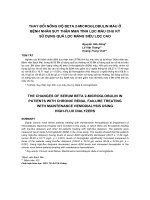
Schematic representation of the two V regions antigenized with TCR sequencesFigure 1
Schematic representation of the two V regions antigenized with TCR sequences. In each case the H chain of the antigenized
antibody is formed of a murine V
H
62
region in which the CDR3 has been engineered to express either
93
ASSDSSNTE
101
or
39
DMGHGLRLIHYSYDVNSTEKG
59
sequence between two Val-Pro (VP) doublets of the unique cloning site in the CDR3 loop
of V
H
62
. The complete H chain is the product of the fusion of the antigenized V
H
region with a human γ1C region. The light (L)
chain (not shown) is the murine λ1 which is provided by the J558L host cell. (H chain not to scale).
Journal of Immune Based Therapies and Vaccines 2004, 2:9 />Page 4 of 12
(page number not for citation purposes)
FACS analysis
Autoantibodies reactive with the Vβ8.2
+
TCR were sought
by flowcytometry on the S23B1E11 T cell hybridoma [33],
derived from the fusion of Vβ8.2
+
CD4 T lymphoblasts
specific for MBP with the murine TCR α/β
-
BW1100.129.237 thymoma cell line [33]. For FACS anal-
ysis the following procedure was utilized. 10
6
hybridoma
T cells in 100 µl of RPMI-1640 containing 1% egg albu-
min, 0.01% NaN
3
and 10 mM Hepes, were incubated with
rat sera (1:10 dilution) for 90 minutes at +4°C. Cells were
washed three times with cold RPMI-1640 and subse-
quently incubated with a fluorescein-isotyocianate
(FITC)-conjugated goat antibody (0.5 µg/10
6
cells) to rat
Ig (H+L) (Caltag, So. San Francisco, CA) for 20 minutes at
+4°C. After incubation, the cells were washed twice, resus-
pended in 1% paraformaldehyde, and analyzed in a FACS
Scan (BD Biosciences). To stain for dead cells, 20 µl of
propidium iodide in PBS were added to unfixed cells
before FACS analysis. R-phycoerythrin conjugated mouse
monoclonal antibody R78 (IgG1, k) specific for the rat
Vβ8.2, the kind gift of Pharmingen (San Diego, CA), was
used to control for the expression of the Vβ8.2 TCR on
S23B1E11 hybridoma cells.
In vitro proliferative response
Poplyteal, inguinal and paraortic lymph nodes were
removed from immunized animals at different times, dis-
sociated and washed in RPMI-1640. Lymph node cells
were plated in round-bottom 96-well plates at 2.5 × 10
5
cells/well in the presence of various (10–100 956;g/ml)
amounts of antigen in 200 µl of RPMI containing 10%
FCS, 100 U/ml penicillin, 100 µg/ml streptomycin, 4 mM
glutamine, 0.1 mM non-essential aminoacids, 1 mM
sodium pyruvate and 0.5 µM 2-β mercaptoethanol. Cul-
tures were incubated for 72 hours in a 10% CO
2
atmos-
phere. The evening before harvest 1 µCi/well of [
3
H]-
thymidine was added to each well. Cells were harvested
onto glass fiber filters and counted on an automatic Beck-
man LS 6000IC β-counter.
Vaccinations and immunizations schedule
Animals were vaccinated with antigenized antibodies
(100 µg/rat) in complete Freunds' adjuvant (CFA) divided
equally between the posterior paws (25 µl each) and two
points in the back subcutaneously. A booster injection (50
µg/rat) in incomplete Freunds' adjuvant (IFA) was given
subcutaneously on day 21. EAE was induced on day 28 by
immunization with MBP peptide
68
GSLPQKSQRSQDENPVVHF
88
(30 µg/rat) in the ante-
rior paws (25 µl each) in CFA (H37RA 10 mg/ml). Con-
trol rats were similarly injected with transfectoma
antibody γ1WT or γ2bWT. Rats inoculated with Freunds'
adjuvant only served as additional control. Serum sam-
ples were collected from the retro-orbital sinus on day 0
before vaccination, day 21 before booster injection, day
28 before EAE induction, and day 50 after recovery from
disease. Sera were stored at -20 C until use.
Clinical evaluation of EAE
EAE was monitored daily by two operators for clinical
signs using the following scale: grade 0 = no appreciable
symptoms; grade 1 = tail atony; grade 2 = paraparesis;
grade 3 = paraplegia; grade 4 = paraplegia with forelimb
weakness, moribund state. Typically symptoms of disease
began to appear on day 11–13 from the injection of the
encephalitogenic peptide. The Disease Index was calcu-
lated according to the formula: [(Maximum Score) ×
(Duration of Disease) × (Incidence)].
Statistical Methods
Statistical analyses was performed using the Fisher's test.
Table 1: Vaccination against antigenized antibodies expressing TCR peptides protects from EAE
Severity of Disease*
Group No. Rats Immunogen Incidence Max Score (mean ± SD) Duration (mean ± SD) Disease Index
I6γ1TCR-I 4/6 1.1 ± 0.9
a
2.5 ± 2.2
b
1.8
II 10 γ1TCR-II 8/10 1.6 ± 1.0
c
3.8 ± 2.3
d
4.9
III 10 γ1WT 10/10 2.2 ± 0.9 5.1 ± 1.0 11.3
IV 5 CFA 5/5 3.4 ± 0.9 6.6 ± 1.3 22.4
V 6 - 6/6 3.5 ± 0.5 7.2 ± 1.3 25.2
* EAE was scored according to incidence, severity and duration. Disease index was calculated as follows: Mean Maximum Score × Mean Duration
Disease × Incidence.
Significance: (
a
) Group I vs Group III p = 0.04 and Group I vs Group V p = 0.0002; (
b
) Group I vs Group III p = 0.009 and Group I vs Group V p =
0.0001; (
c
) Group II vs Group III p = 0.16 and Group II vs Group V p = 0.0005; (
d
) Group II vs Group III p = 0.12 and Group II vs Group V p = 0.001.
Journal of Immune Based Therapies and Vaccines 2004, 2:9 />Page 5 of 12
(page number not for citation purposes)
Results
Vaccination with antigenized antibodies and effect on EAE
Two antigenized antibodies were engineered to express
the CDR3
93
ASSDSSNTE
101
and CDR2
39
DMGHGLRLIHYSYDVNSTEKG
59
sequences, and were
termed γ1TCR-I and γ1TCR-II, respectively (Figure 1). Rats
were vaccinated with an individual antigenized antibody
and received a booster injection 21 days later. EAE was
induced on day 28 by immunization with the encepha-
litogenic MBP peptide
68
GSLPQKSQRSQDENPVVHF
88
.
As shown in Table 1, vaccination with both γ1TCR-I and
γ1TCR-II reduced disease severity. Rats immunized with
γ1TCR-I (group I) had a disease index of 1.8. Within this
group, two out of six rats (33%) did not develop disease,
one had grade 1 and three had grade 2. None proceeded
through grade 3 or 4. Rats immunized with γ1TCR-II
(group II) had a disease index of 4.9. Within this group
two out of ten rats (20%) did not develop the disease, two
had grade 1, four had grade 2 and two had grade 3. In con-
trast, all fifteen control rats vaccinated with γ1WT or given
CFA only (groups III and IV) developed EAE with a disease
index ranging between 11.3 and 22.4. Unmanipulated
rats immunized with the MBP peptide (group V) devel-
oped EAE with a disease index of 25.2. There was a direct
correlation between the severity of the disease and its
duration. In rats immunized with γ1TCR-I, the disease
lasted on average for 2.5 days and in rats immunized with
γ1TCR-II 3.8 days. In contrast, in all the other groups
(groups III-V) the duration of the disease was significantly
longer (6–7 days). Of note, although group III rats had an
overall lower score than unmanipulated rats, they differed
from rats in group I or group II by the above mentioned
parameters and these difference were statistically signifi-
cant (Table 1). CFA did not confer protection. Taken
together, these data indicate that active immunity elicited
with antigenized antibodies expressing rat Vβ8.2 TCR
peptides was effective in markedly reducing the severity of
EAE in the Lewis rat.
Antibody responses after vaccination
Antibodies in response to the immunogen were assessed
by solid-phase ELISA at various times after immunization.
As shown in Table 2, antibody titers against the immuno-
gen developed in each group (group I-III) irrespective of
which antibody was used to detect the antibody response
in sera. This suggests that the human constant region of
the antigenized antibodies is immunogenic in the rat.
Antibody titers increased after the booster immunization
and after challenge with the encephalitogenic MBP pep-
tide. Control rats (group IV-V) did not mount any anti-
body response. No reactivity was found on the 19
mer
MBP
peptide (GSLPQKSQRSQDENPVVHF) used as a control.
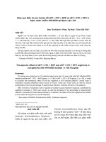
Anti-TCR (anti-idiotypic) antibodies were tested using
two approaches. In the first case, sera of immunized ani-
mals were tested on Vβ8.2 synthetic peptides by ELISA. A
weak but distinct response was detected in both instances
starting on day 21 or 28 (Figure 2). Sera from control ani-
mals did not react with TCR peptides. Together with the
fact that these were tested at a 1:25 dilution it appears that
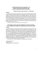
the anti-idiotypic response is weak. In the second case, we
tested anti-idiotypic antibodies for their reactivity with the
TCR in its native configuration. This was done by flowcy-
tometry using the Vβ8.2
+
T cell hybridoma S23B1E11 as
the cell substrate. Two out of six rats in group I had a
bright cellular staining (Figure 3). Reactive antibodies
were detectable on day 21, 28 and day 50. Rats immu-
nized with γ1TCR-II (group II) as well control rats (group
III-V) were negative. Interestingly, the two rats whose sera
reacted with TCR by flowcytometry did not develop symp-
Table 2: Detection of antibodies against γ1TCR-I and γ1TCR-II in vaccinated Lewis rats
Days After Vaccination
a Immunogen Rats (No.) Responders (No.) 0 21 28 50
γ1TCR-I 6 6/6 ≤2.3* 3.9 ± .2 4.2 ± .2 4.5 ± 0.2
γ1TCR-II 10 10/10 ≤2.3 3.7 ± 0.2 4.1 ± 0.1 4.5 ± 0.2
γ1WT 10 10/10 ≤2.3 3.2 ± 0.4 3.9 ± 0.2 4 ± 0.2
CFA 5 0/5 ≤2.3 ≤2.3 2.6 ≤2.3
-6 0/6 ≤2.3 ≤2.3
b
γ1TCR-I 6 6/6 ≤2.3 4 ± 0.2 4 ± 0.2 4.6 ± 0.3
γ1TCR-II 10 10/10 ≤2.3 4.1 ± 0.3 4.4 ± 0.3 4.7 ± 0.5
γ1WT 10 10/10 ≤2.3 3.3 ± 0.3 4 ± 0.2 4.2 ± 0.2
CFA 5 0/5 ≤2.3 ≤2.3 ≤2.3 ≤2.3
-6 0/6 ≤2.3 ≤2.3
* Antibody titers are expressed in log10. Sera were tested on microtiter plates coated with each of the TCR antigenized antibody γ1TCR-I (panel a)
or γ1TCR-II (pane b). End point dilutions were determined as the last serum dilution binding with an OD ≥ 0.200.
Journal of Immune Based Therapies and Vaccines 2004, 2:9 />Page 6 of 12
(page number not for citation purposes)
toms of EAE.
Vaccination with a murine antigenized antibody
To explore the importance of foreigness of the constant
region on the immunogenicity of the Vβ8.2 peptides we
engineered an antigenized antibody with a murine γ2b
constant region. Homology search using the BLAST pro-
gram of the NCBI gene bank indicated that the murine
γ2b C region is 56.7% identical to the rat γ2b C region,
with a homology of 71% between residues 106 and 333.
Because significant protection was found in rats vacci-
nated with the antibody expressing the
93
ASSDSSNTE
101
peptide (γ1TCR-I), we engineered an antibody with the
same V region (γ2bTCR-I). Rats vaccinated with γ2bTCR-I
and subsequently immunized with MBP peptide, were
protected only partially compared to rats vaccinated with
γ1TCR-I (10.2 vs. 1.9) (Table 3). Notably, within the six
rats immunized with γ2bTCR-I, two were grade ≤ 2 and
four developed a grade 3 for an average of two days. On
the other hand, three out of six rats immunized with con-
trol antibody γ2bWT proceeded through a grade 4 disease.
Similarly, all five control rats (group III and IV) developed
a grade 4 disease. Of note, although the severity of the dis-
ease in group I rats was less than in control group II, the
difference was not statistically significant (Table 3). All
rats developed antibodies to the respective immunogen.
However, when compared with the total antibody titer of
rats immunized with γ1TCR-I and γ1TCR-II the titers were
on average lower at single time points (Table 4). All sera
reacted with the synthetic peptide
93
ASSDSSNTE
101
start-
ing from day 21 with a progressive increase over time (Fig-
ure 3).
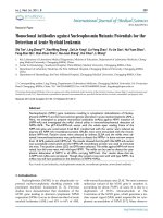
Serum antibodies of vaccinated rats bind a synthetic
peptide of human CD4
In the attempt to correlate the antibody response after vac-
cination with protection, the sera of vaccinated rats and
their controls were tested on a synthetic peptide corre-
sponding to amino acid residues 21–49 of the first extra-
cellular domain of human CD4. This peptide binds Ig irre-
spective of antigen specificity and heavy chain isotype
with an affinity of 10
-5
M (26). It also binds antigen:anti-
body complexes formed at molar equivalence with an
affinity about 100 fold higher [31]. When the sera of
vaccinated rats were assayed on plates coated with the syn-
thetic peptide of human CD4, strong binding was
observed by sera from all rats immunized with γ1TCR-I
whereas sera from rats immunized with γ1TCR-II or γ1WT
bound much less (Figure 5a). Control sera of groups IV
and V did not bind. Binding could be attributed either to
a differential property of the two antigenized V regions or
to differences in the immune response triggered by the V
regions themselves. To distinguish between the two possi-
bilities two experiments were performed. First, we
assessed binding of γ1TCR-I and γ1TCR-II on the CD4
peptide. Both bound equally at saturating and non-satu-
rating concentrations (data not shown). Second, we tested
sera of rats immunized with γ2bTCR-I considering that, if
Antibody response to TCR peptides following vaccination with antigenized antibody γ1TCR-I or γ1TCR-II
39
DMGHGLRLIHYSYDVNSTEKG
59
tested on the ASSDSSNTE (panel a) or (panel b)Figure 2
Antibody response to TCR peptides following vaccination with antigenized antibody γ1TCR-I or γ1TCR-II
39
DMGHGLRLIHYSYDVNSTEKG
59
tested on the ASSDSSNTE (panel a) or (panel b). The number of rats in each group is that
indicated in Table 1. Results are expressed as Log2 ± SD.
Journal of Immune Based Therapies and Vaccines 2004, 2:9 />Page 7 of 12
(page number not for citation purposes)
Sera from rats vaccinated with γ1TCR-I bind Vβ8.2
+
T cells by flowcytometryFigure 3
Sera from rats vaccinated with γ1TCR-I bind Vβ8.2
+
T cells by flowcytometry. Vβ8.2
+
S23B1E11 T cell hybridoma were used as
substrate. Sera were tested at 1:25 dilution. Bound antibodies were revealed using a FITC-conjugated goat antibody to rat Ig.
Table 3: Protection against EAE by vaccination with antigenized antibodies with a murine γ2b constant region
Severity of Disease*
Group No. Rats Immunogen Incidence Max Score (mean ± SD) Duration (mean ± SD) Disease Index
I6γ2bTCR-I 6/6 2.5 ± 0.8
a
4.2 ± 0.4
b
10.5
II 6 γ2bWT 6/6 3.0 ± 1.3 5.7 ± 1.7 17.1
III 2 CFA 2/2 4 6 24
IV 3 - 3/3 4 7.3 ± 0.6 29.2
* EAE was scored according to incidence, severity and duration. Disease index was calculated as follows: Mean Maximum Score × Mean Duration of
Disease × Incidence.
Significance: (
a
) Group I vs Group II p = 0.394 and Group I vs Group IV p = 0.051; (
b
) Group I vs Group III p = 0.05 and Group I vs Group IV p = 0.026.
Journal of Immune Based Therapies and Vaccines 2004, 2:9 />Page 8 of 12
(page number not for citation purposes)
the effect was due to the immune response against
93
ASSDSSNTE
101
, we would have found similar results. As
shown (Figure 5b), the sera of γ2bTCR-I vaccinated rats all
bound to the CD4 peptide comparably to rats vaccinated
with γ1TCR-I. This suggests that binding to the CD4 pep-
tide may reflect differences in the type of V regions utilized
by the antibodies generated in vivo in response to immu-
nization with the TCR peptide
93
ASSDSSNTE
101
as com-
pared with the TCR peptide
39
DMGHGLRLIHYSYDVNSTEKG
59
or the wild type V
region. Further studies will be needed to clarify this issue.
Proliferative response
Spleen cells and draining lymph nodes of rats tested 15 or
30 days after the initial immunization were tested in a
proliferative assay against the Vβ8.2 peptides. No prolifer-
ative response was detected (data not shown).
Discussion
In this report we demonstrate that the severity of EAE in
the Lewis rat can be greatly attenuated, and in some
instances completely prevented, by active immunization
with antigenized antibodies expressing amino acid
sequences of the rat Vβ8.2 gene product. Immunity
against synthetic peptides of the TCR has been shown to
be effective in preventing or reducing the severity of EAE
in the rat [17-20], suggesting that autoimmunity against
the TCR reacting with encephalitogenic sequences of MBP
is key to immunoregulatory events. The control of patho-
genetic T cells may involve both T cells and antibodies.
Autoregulation via T cells in EAE is well established. Thus,
spontaneous recovery from EAE is impaired by splenec-
tomy or thymectomy [34] and EAE can be prevented by
vaccination with "attenuated" pathogenic T cells [15].
Autoregulation in EAE may involve both CD4
+
and CD8
+
T cells [35-38] as well as suppression by cytolytic T-T inter-
actions [39]. A prevailing idea has been that in the rat [40]
and in the mouse [41] idiotypic determinants of the TCR
may be autoimmunogenic and contribute to mechanisms
of immune regulation leading to protection. On the other
hand, at least in a few instances, monoclonal antibodies
against these TcR Vβ region [9,42,43] or against TCR idio-
type [44] have been shown to block or attenuate disease.
Here we show that immunity against idiotopes of anti-
bodies engineered to express TCR peptides is effective in
generating anti-idiotypic immunity directed against rat
Vβ8.2 TCR gene product. Importantly, this type of immu-
nity protected from EAE. The new approach used herein to
Table 4: Detection of antibodies against γ2bTCR-I in vaccinated Lewis rats
Days After Vaccination
Immunogen Rats (No.) Responders (No.) 0 21 28 50
γ2bTCR-I 6 6/6 ≤2.3 3.2 ± 0,2 3.7 ± 0.1 4 ± 0.3
γ2bWT 6 6/6 ≤2.3 3 ± 0.3 3.4 ± 0.3 3.6 ± 0.3
CFA 2 0/2 ≤2.3 ≤2.3 ≤2.3 ≤2.3
- 3 0/3 - - ≤2.3 ≤2.3
* Antibody titers are expressed in log10. Sera were tested on microtiter plates coated with γ2bTCR-I. End point dilutions were determined as the
last serum dilution binding with an OD ≥ 0.200.
Sera from rats vaccinated with γ1TCR-I or γ2bTCR-I bind synthetic peptide 21–49 of human CD4Figure 5
Sera from rats vaccinated with γ1TCR-I or γ2bTCR-I bind
synthetic peptide 21–49 of human CD4.
Journal of Immune Based Therapies and Vaccines 2004, 2:9 />Page 9 of 12
(page number not for citation purposes)
induce anti-TCR immunity is based on conventional idio-
typic immunization in which antigenized antibodies
mimic the immunogenic properties of soluble TCR func-
tioning as a surrogate internal image [45] in much the
same way as previously demonstrated for a non-self anti-
gen [31]. The present approach is reminiscent of
experiments in which induction of anti-idiotypic immu-
nity against TCR with specificity for MHC was obtained by
immunization with soluble alloantibodies of relevant
specificity [46,47], or by immunization with autologous
idiotype positive molecules that are shed from the cell sur-
face in the serum [48]. Thus, antibodies purposely modi-
fied to express selected loops of the TCR obviate the
necessity to purify the receptor, isolate idiotypic TCR mol-
ecules from the serum, or use antigen-specific T cell blasts.
Antibodies reacting with TCR peptides were detected in
every vaccinated rat indicating that immunization with
antigenized antibodies is an efficient method to induce an
anti-idiotypic response specific for a somatic receptor. The
fact that only two out of sixteen vaccinated rats developed
antibodies against the native receptor detectable by flow-
cytometry on Vβ8.2
+
T cells suggests that cross-reactive
anti-idiotypic antibodies may be very low titer. Alterna-
tively, they may be adsorbed on T cells in vivo precluding
their detection in the serum. The first possibility is consist-
ent with the self nature of TCR peptides and a predicted
paucity of self reactive clonotypes within the natural B cell
repertoire. Interestingly, we noted that the anti-idiotypic
response against a non-self peptide expressed in an anti-
genized antibody [31] is much greater than the one
observed here against a self peptide. That only rats vacci-
nated with the antigenized antibody expressing the
93
ASSDSSNTE
101
sequence developed flowcytometry-reac-
tive autoantibodies could reflect difference in conforma-
tion once the two peptides are embedded in the CDR3
loop of an antigenized antibody. For instance,
93
ASSDSSNTE
101
could be better surface exposed and
more stably expressed
39
DMGHGLRLIHYSYDVNSTEKG
59
. A computer-assisted
comparison of hydrophilicity profiles [49] of the
93
ASSDSSNTE
101
peptide in the parental. TCR Vβ8.2 gene
product and in the antibody V region shows that in both
instances the peptide is highly hydrophilic (Figure 5). On
the other hand, the Vβ8.2 CDR2 region shows a highly
hydrophilic profile alternating with large hydrophobic
regions of poorly exposed amino acid residues, both in
the parental TCR and in the antibody CDR (data not
shown).
Our data show that although the process of antibody anti-
genization allows one to conformationally-constrain and
express discrete peptide sequences of somatic receptors,
the induction of anti-receptor antibodies is not directly
predictable. Previously, we demonstrated flowcytometry-
reactive antibodies to human CD4 in a high proportion
(75 %) of cases [24]. We conclude that the physical char-
acteristics of a given receptor peptide (e.g., length,
hydrophilicity, etc.) likely determine its ability to induce
antibodies cross-reactive with the native receptor.
Interestingly, rats immunized with the antigenized anti-
body expressing
93
ASSDSSNTE
101
but not
39
DMGHGLRLIHYSYDVNSTEKG
59
reacted immunologi-
cally with a synthetic peptide of human CD4 previously
described to bind Ig [32]. Because the two antigenized
antibodies reacted equally with the CD4 peptide and only
differ by the composition of their CDR3, we suggest that
binding to CD4 by anti-
93
ASSDSSNTE
101
serum antibod-
ies underscores qualitative differences of the immune
response between rats immunized with γ1TCR-I and
1TCR-II, respectively. Thus, it appears as if
93
ASSDSSNTE
101
induced a different immune response
than
39
DMGHGLRLIHYSYDVNSTEKG
59
. Furthermore,
since vaccination with γ1TCR-I also promoted greater pro-
tection from EAE, it is tempting to speculate that a
component of the anti-idiotypic response against γ1TCR-
I is associated with protection.
In conclusion, three points have emerged from this study.
First, antigenized antibodies expressing conformation-
ally-constrained loops of the Vβ8.2 TCR can be used as
vaccines in the prevention of EAE in the Lewis rat. Our
new approach to generate anti-TCR immunity, confirms
the relevance of anti-idiotypic regulation in controlling rat
Antibody response to TCR peptide
93
ASSDSSNTE
101
follow-ing vaccination with antigenized antibody γ2bTCR-IFigure 4
Antibody response to TCR peptide
93
ASSDSSNTE
101
follow-
ing vaccination with antigenized antibody γ2bTCR-I. The
number of rats in each group are not indicated in Table 3.
Results are expressed as means of Log2 ± SD.
Journal of Immune Based Therapies and Vaccines 2004, 2:9 />Page 10 of 12
(page number not for citation purposes)
EAE [17,18,20]. Second, since a weak antibody anti-idio-
typic response in the apparent lack of a cell proliferative
response was associated with protection, it appears as if a
humoral anti-TCR response is relevant to protection from
disease. Although this contrasts the relevance of T cell
immunity in the regulation of EAE in the rat, reports exist
to support the idea that humoral immunity is also impor-
tant [20,50,51]. EAE was shown to be prevented or atten-
uated by passive transfer of serum from rats recovering
from EAE [52], or by passive transfer of monoclonal anti-
bodies against these TCR Vβ region and its idiotypes
[9,42-44]. However, whether anti-idiotypic antibodies
against the TCR predispose to anergy, apoptosis or killing
of pathogenetic T cells remains to be determined. Finally,
our study indicates that antigenized antibodies can be
used as vaccines in conditions where immunopathology
and disease involve receptors on somatic cells, and anti-
receptor immunity alone could prevent or mitigate a path-
ological condition.
Hydrophilicity profiles of TCR peptides-containing V regionsFigure 6
Hydrophilicity profiles of TCR peptides-containing V regions. Hydrophilic profile of the rat Vβ8.2 TCR, amino acid residues
80–130, inclusive of the CDR3 sequence
93
ASSDSSNTE
101
.
Hydrophilic profile of the mouse VH62, amino acid residues 80–125, engineered with the
93
ASSDSSNTE
101
peptide of the rat Vβ8.2 TCR-CDR3Figure 7
Hydrophilic profile of the mouse VH62, amino acid residues 80–125, engineered with the
93
ASSDSSNTE
101
peptide of the rat
Vβ8.2 TCR-CDR3.
Journal of Immune Based Therapies and Vaccines 2004, 2:9 />Page 11 of 12
(page number not for citation purposes)
Competing interests
The authors declare that they have no competing interests
Acknowledgments
This work was supported by NIH grant PO1AI33204.
References
1. Swanborg RH: Experimental autoimmune encephalomyelitis
in the rat: lessons in T-cell immunology and autoreactivity.
Immunol Rev 2001, 184:129-135.
2. Hoffman PM, Gaston DD, Spitler LE: Comparison of experimen-
tal allergic encephalomyelitis induced with spinal cord, basic
protein, and synthetic encephalitogenic peptide. Clin Immunol
Immunopathol 1973, 1:364-371.
3. Richert JR, Kies MW, Alvord EJ: Enhanced transfer of experi-
mental allergic encephalomyelitis with Lewis rat lymph node
cells. J Neuroimmunol 1981, 1:195-203.
4. Polman CH, Dijkstra CD, Sminia T, Koetsier JC: Immunohistolog-
ical analysis of macrophages in the central nervous system of
Lewis rats with acute experimental allergic
encephalomyelitis. J Neuroimmunol 1986, 11:215-222.
5. Acha-Orbea H, Mitchell DJ, Timmerman L, Wraith DC, Taich GS,
Waldor MK, Zamvil S, McDevitt H, Steinman L: Limited heteroge-
neity of TcRs from lymphocytes mediating autoimmune
encephalomyelitis aloows specific immune intervention. Cell
1988, 54:263-273.
6. Urban J, Kumar V, Kono DH, Gomez C, Horvath SJ, Clayton J, Ando
DG, Sercarz EE, Hood L: Restricted use of T cell receptor V
genes in murine autoimmune encephalomyelitis raises pos-
sibilities for antibody therapy. Cell 1988, 54:577-592.
7. Herber-Katz E: A new hierarchy of TCR specificity: Autoim-
mune diseases are defined by particular VaVb combinations
and not by antigen specificity. Cold Spring Harbor Laboratory Press
1989, 54:875-878.
8. Steinman L, Solomon D, Lim M, Zamvil S, Sriram S: Prevention of
experimental allergic encephalitis with in vivo administra-
tion of anti I-A antibody. Decreased accumulation of radiola-
belled lymph node cells in the central nervous system. J
Neuroimmunol 1983, 5:91-97.
9. Owhashi M, Herber-Katz E: Protection from experimental aller-
gic encephalomyelitis conferred by a monoclonal antibody
directed against a shared idiotype on rat T cell receptors
specific for myelin basic protein. J Exp Med 1988, 168:2153-2164.
10. Brostoff SW, Mason DW: Experimental allergic encephalomy-
elitis: successful treatment in vivo with a monoclonal anti-
body that recognizes T helper cells. J Immunol 1984,
133:1938-1942.
11. Welch AM, Swanborg RH: Characterization of suppressor cells
involved in regulation of experimental allergic
encephalomyelitis. Eur J Immunol 1976, 6:910-912.
12. Adda DH, Beraud E, Depieds R: Evidence for suppressor cells in
Lewis rats experimental allergic encefalomyelitis. Eur J
Immunol 1977, 7:620-625.
13. Beraud E, Varriale S, farnarier C, bernard D: Suppressor cells in
Lewis rats with experimental allergic encephalomyelitis:
prevention of the disease and inhibition of lymphocytes pro-
liferation by suppressor cells or their products. Eur J Immunol
1982, 12:926-930.
14. Miller A, Lider O, Roberts AB, Sporn MB, Weiner HL: Suppressor
T cells generated by oral tolerization to myelin basic protein
suppress both in vitro and in vivo immune responses by the
release of transforming growth factor beta after antigen-
specific triggering. Proc Natl Acad Sci U S A 1992, 89:421-425.
15. Ben-Nun A, Wekerle H, Cohen IR: Vaccination against autoim-
mune encephalomyelitis with T-lymphocyte line cells reac-
tive against myelin basic protein. Nature 1981, 292:60-61?.
16. Rennie J: The body against itself. Scientific American 1990,
263:106-115.
17. Howell MD, Winters ST, Olee T, Powell HC, Carlo DJ, Brostoff SW:
Vaccination against experimental allergic encephalomyelitis
with T cell receptor peptides. Science 1989, 246:668-670.
18. Vandenbark AA, Hashim G, Offern H: Immunization with a syn-
thetic T-cell receptor V-region peptide protects against
experimental autoimmune encephalomyelitis. Nature 1989,
341:541-544.
19. Offner H, Hashim GA, Vandenbark AA: T cell receptor peptide
therapy triggers autoregulation of experimental
encephalomyelitis. Science 1991, 251:430-432.
20. Stevens DB, Karpus WJ, Gould KE, Swanborg RH: Studies of V beta
8 T cell receptor peptide treatment in experimental autoim-
mune encephalomyelitis. J Neuroimmunol 1992, 37:123-129.
21. Desquenne-Clark L, Esch TR, Otvos L Jr, Heber-Katz E: T-cell
receptor peptide immunisation leads to enhanced and
chronic experimental allergic encephalomyelitis. Proc Natl
Acad Sci USA 1991, 88:7219-7223.
22. Matsumoto Y, Tsuchida M, Hanawa H, Abo T: T cell receptor pep-
tide therapy for autoimmune encephalomyelitis: stronger
immunization is necessary for effective vaccination. Cell
Immunol 1994, 153:468-478.
23. Kumar V, Coulsell E, Ober B, Hubbard G, Sercarz E, Ward ES:
Recombinant T cell receptor molecules can prevent and
reverse experimental autoimmune encephalomyelitis: dose
effects and involvement of both CD4 and CD8 T cells. J
Immunol 1997, 159:5150-5156.
24. Lanza P, Billetta R, Antonenko S, Zanetti M: Active immunity
against the CD4 receptor using an antibody antigenized with
residues 41-55 of the first extracellular domain. Proc Natl Acad
Sci USA 1993, 90:11683-11687.
25. Zanetti M: Antigenized antibodies. Nature 1992, 355:466-477.
26. Sollazzo M, Hasemann CA, Meek KD, Glotz D, Capra JD, Zanetti M:
Molecular characterization of the VH region of murine
autoantibodies from neonatal and adult BALB/c mice. Eur J
Immunol 1989, 19:453-457.
27. Sollazzo M, Billetta R, Zanetti M: Expression of an exogenous
peptide epitope genetically engineereed in the variable
domain of an immunoglobulin: implications for antibody and
peptide folding. Protein Eng 1990, 4:215-220.
28. Morrison S: Transfectomas provide novel chimeric
antibodies. Science 1985, 229:1202-1207.
29. Billetta R, Zanetti M: Ligand expression using antigenization of
antibody: Principle and methods. ImmunoMethods 1992,
1:41-51.
30. Hashim GA, Carvalho EF, Sharpe RD: Definition and synthesis of
the essential amino acid sequence for experimental allergic
encephalomyelitis in Lewis rats. J Immunol 1978, 121:665-670.
31. Billetta R, Hollingdale RM, Zanetti M: Immunogenicity of an engi-
neered internal image antibody. Proc Natl Acad Sci USA 1991,
88:4713-4717.
32. Lenert P, Kroon D, Spiegelberg H, Golub ES, Zanetti M: Human
CD4 binds immunoglobulins. Science 1990, 248:1639-1643.
33. Gold DP, Offner H, Sun D, Wiley S, Vandenbark AA, Wilson DB:
Analysis of T cell receptor beta chains in Lewis rats with
experimental allergic encephalomyelitis: conserved comple-
mentarity determining region 3. J Exp Med 1991,
174:1467-1476.
34. Ben-Nun A, Ron Y, Cohen IR: Spontaneous remission of autoim-
mune encephalomyelitis is inhibited by splenectomy,
thymectomy or ageing. Nature 1980, 288:389-390.
35. Karpus WJ, Gould KE, Swanborg RH: CD4+ suppressor cells of
autoimmune encephalomyelitis respond to T cell receptor-
associated determinants on effector cells by interleukin-4
secretion. Eur J Immunol 1992, 22:1757-1763.
36. Koh DR, Ho A, Rahemtulla A, Penninger J, Mak TW: Experimental
allergic encephalomyelitis (EAE) in mice lacking CD4+ T
cells. Eur J Immunol 1994, 24:2250-2253.
37. Jiang H, Zhang SI, Pernis B: Role of CD8+ T cells in murine
experimental allergic encephalomyelitis. Science 1992,
256:1213-1215.
38. Koh DR, Fung LW, Ho A, Gray D, Acha OH, Mak TW: Less mor-
tality but more relapses in experimental allergic encephalo-
myelitis in CD8-/- mice. Science 1992, 256:1210-1213.
39. Sun D, Qin Y, Chluba J, Epplen JT, Wekerle H: Suppression of
experimentally induced autoimmune encephalomyelitis by
cytolytic T-T cell interactions. Nature 1988, 332:843-845.
40. Heber-Katz E, Acha-Orbea H: The V-region disease hypothesis:
evidence from autoimmune encephalomyelitis. Immunol Today
1989, 10:164-169.
Publish with BioMed Central and every
scientist can read your work free of charge
"BioMed Central will be the most significant development for
disseminating the results of biomedical research in our lifetime."
Sir Paul Nurse, Cancer Research UK
Your research papers will be:
available free of charge to the entire biomedical community
peer reviewed and published immediately upon acceptance
cited in PubMed and archived on PubMed Central
yours — you keep the copyright
Submit your manuscript here:
/>BioMedcentral
Journal of Immune Based Therapies and Vaccines 2004, 2:9 />Page 12 of 12
(page number not for citation purposes)
41. Kumar V, Sercarz E: An integrative model of regulation cen-
tered on recognition of TCR peptide/MHC complexes. Immu-
nol Rev 2001, 182:113-121.
42. Hashim GA, Vandenbark AA, Galang AB, Diamanduros T, Carvalho E,
Srinivasa J, Jones R, Vainiene M, Morrison WJ, Offner H: Antibodies
specific for VB8 receptor peptide suppress experimental
autoimmune encephalomyelitis. J Immunol 1990,
144:4621-4627.
43. Matsumoto Y, Tsuchida M, Hanawa H, Abo T: Successful preven-
tion and treatment of autoimmune encephalomyelitis by
short-term administration of anti-T-cell receptor alpha beta
antibody. Immunology 1994, 81:1-7.
44. Zhou SR, Whitaker JN: Specific modulation of T cells and
murine experimental allergic encephalomyelitis by mono-
clonal anti-idiotypic antibodies. J Immunol 1993, 150:1629-1642.
45. Jerne NK, Roland J, Cazenave PA: Recurrent idiotypes and inter-
nal images. EMBO J 1982, 1:243-247.
46. McKearn TJ, Hamada Y, Stuart FP, Fitch FW: Anti-receptor anti-
body and resistance to graft-versus-host disease. Nature 1974,
251:648-650.
47. Binz H, Wigzell H: Successful induction of specific tolerance to
transplantation antigens using autoimmunisation against the
recipient's own natural antibodies. Nature 1976, 262:294-295.
48. Binz H, Wigzell H: Specific transplantation tolerance induced
by autoimmunization against the individual's own, naturally
occurring idiotypic, antigen-binding receptors. J Exp Med 1976,
144:1438-1457.
49. Kyte J, Doolittle RF: A simple method for displaying the hydro-
pathic character of a protein. J Mol Biol 1982, 157:105-132.
50. Sun D: Synthetic peptides representing sequence 39 to 59 of
rat V beta 8 TCR fail to elicit regulatory T cells reactive with
V beta 8 TCR on rat encephalitogenic T cells. Cell Immunol
1992, 141:200-210.
51. Kawano Y, Sasamoto Y, Vacchio MS, Hodes RJ, Gery I: Immune
responses against self-TCR peptides. Cell Immunol 1994,
159:235-245.
52. Paterson PY, Harwin SM: Suppression of allergic encephalomy-
elitis in rats by means of antibody serum. J Exp Med 1963,
117:755-780.