Báo cáo y học: "Age-related waning of in vitro Interferon-γ levels against r32kDaBCG in BCG vaccinated children" ppt
Bạn đang xem bản rút gọn của tài liệu. Xem và tải ngay bản đầy đủ của tài liệu tại đây (349.33 KB, 7 trang )
BioMed Central
Page 1 of 7
(page number not for citation purposes)
Journal of Immune Based Therapies
and Vaccines
Open Access
Original research
Age-related waning of in vitro Interferon-γ levels against
r32kDaBCG in BCG vaccinated children
B Anuradha
1,3
, CM Santosh
2
, V Hari Sai Priya
3
, G Suman Latha
3
, KJR Murthy
3
and Valluri Vijaya Lakshmi*
1,3
Address:
1
LEPRA Society – Blue Peter Research Center, Hyderabad, AP, India,
2
Center for DNA Finger printing and Diagnosis, Hyderabad, AP, India
and
3
Bhagwan Mahavir Medical Research Centre, Hyderabad, AP, India
Email: B Anuradha - ; CM Santosh - ; V Hari Sai Priya - ;
G Suman Latha - ; KJR Murthy - ; Valluri Vijaya Lakshmi* -
* Corresponding author
Abstract
Background: Mycobacterium bovis BCG vaccine has displayed inconsistent efficacy in different
trials conducted in various geographical regions. Nevertheless, it significantly reduces the risk of
severe childhood tuberculosis and continues to be used to prevent tuberculosis in many countries.
Many studies revealed that efficacy of vaccine wanes with age. Most of the studies were based on
in vivo and in vitro responses to tuberculin. With the advent of newer tests such as in vitro interferon-
γ assays and identification of potent immunogenic mycobacterial proteins there is a need to
corroborate the observations. This study aims at ascertaining the need for a booster at a later age
as indicated by in vitro release of IFN-γ while evaluating Ag85A as an antigen.
Methods: Ninety healthy children who were without any clinical evidence of the disease, 45 with
a BCG-scar and the remaining 45 without scar and 25 with tuberculosis were included in the study.
The incidence of TB was analyzed in 216 children attending a DOTS clinic during 1996–2005.
CD3+, CD4+ and CD8+ cell counts were measured by Flow cytometry. r32kDaBCG (Ag85A-
BCG) protein was used to stimulate T cells in in vitro T cell responses and interferon-γ (IFN-γ)
cytokine levels in the supernatants were measured by ELISA.
Results: High incidence of TB was observed in age group 13–14 years followed by children in the
age group 10–12 years (Chi-square 242.22; p < 0.000). T cell subsets were within the normal range
in all subjects. 79% of vaccinated children showed positive proliferative responses with a mean SI
value of 4.98 ± 1.99 while only 39% of the unvaccinated and 58% of the tuberculosis children
showed positive responses with mean values of 2.9 ± 1.6 (p < 0.001) and 2.9 ± 1.7(p < 0.057),
respectively. The stimulation indices in vaccinated children decreased in the older children
concurring with an increase in the incidence of TB.
Conclusion: Significantly high levels of in vitro IFN-γ demonstrated in BCG vaccinated children in
our study substantiate the observation that BCG is effective in children, but the effect may wane
with age. The immunity could be boosted using modified r32kDa (Ag85A) of BCG.
Published: 7 June 2007
Journal of Immune Based Therapies and Vaccines 2007, 5:8 doi:10.1186/1476-8518-5-8
Received: 24 April 2007
Accepted: 7 June 2007
This article is available from: />© 2007 Anuradha et al; licensee BioMed Central Ltd.
This is an Open Access article distributed under the terms of the Creative Commons Attribution License ( />),
which permits unrestricted use, distribution, and reproduction in any medium, provided the original work is properly cited.
Journal of Immune Based Therapies and Vaccines 2007, 5:8 />Page 2 of 7
(page number not for citation purposes)
Background
Mycobacterium bovis BCG (Bacillus Calmette Guerine) vac-
cine has displayed inconsistent efficacy in different trials
conducted in various geographical regions. Nevertheless,
it significantly reduces the risk of tuberculosis by 50% [1]
and the risk of severe childhood tuberculosis by 70% [2,3]
and continues to be used to prevent tuberculosis in many
countries. Moreover, many studies confirm the protective
capacity of neonatal BCG against childhood tuberculosis
[4-6]. Therefore BCG vaccination at birth must remain a
public health priority especially in countries like India
with a high incidence of the disease. However, the results
of our earlier study [7,8], based on in vitro T cell assays
using tuberculin as a stimulant, revealed that the effect of
BCG wanes with age. Waning of the effect of BCG was also
reported by other investigators [9,10], but the results were
based mostly on tuberculin skin tests. Although the test
has proven to be useful in clinical practice, it has several
major limitations. Skin test with tuberculin was not an
ideal indicator of BCG vaccination status [7,11-13]. With
the advent of newer tests such as in vitro interferon-γ (IFN-
γ) assays and identification of potent immunogenic
mycobacterial proteins there is a need to corroborate the
earlier observations.
Though a variety of live vaccines have been developed as
vaccines, until now no booster vaccine has been shown
capable of significantly enhancing the level of protective
immunity. The efficient recruitment of antigen-specific T-
cells principally CD4+ in the lungs, as well the cytokines
that are released particularly IFN-γ against M. tuberculosis
is the sign of improved protective immunity for the devel-
opment of a new vaccine [14]. IFN-γ is the most important
cytokine for inducing the macrophage killing activation
mechanism [15].
Preliminary studies conducted at our center demonstrated
that 30–34 cluster of culture filtrate protein of M. bovis
BCG as the most immunogenic [16]. A triad of related
gene products 30/32-kDa, referred to as Ag85 complex are
the major secretory proteins of M. tuberculosis and have
been shown to be protective in the guinea pig model of
pulmonary tuberculosis. The amino acid sequences of
Ag85A and Ag85C are 100% identical for M. tuberculosis
and M. bovis BCG [17]. Their abundant production either
extracellulary in broth culture or intracellulary in human
monocytes, suggests a vital role in the physiology of the
mycobacteria [1]. Studies report that most effective pro-
tection was observed when mice were immunized with
Ag85A from M. bovis BCG [18]. A single immunization
with Ag85 could induce antigen-specific IFN-γ synthesis
and more effective protection than live BCG vaccine [19-
22].
Reports on 32KDa protein indicating its use as a booster
vaccine, were conducted in animals, leading to clinical tri-
als in adult humans. However, there are no reports in chil-
dren. In India children are vaccinated at birth. This study
aims at ascertaining the need for a booster at a later age as
indicated by in vitro release of IFN-γ while evaluating
r32kDa as an antigen.
Methods
Subjects
The study, cleared by the Institutional Ethical Committee,
included 115 children <12 years of age after obtaining a
written consent from the parents. Ninety children who
were healthy without any clinical evidence of the disease
were included, of these 45 had a scar at the site of BCG
administration, and 45 did not have a scar (henceforth
referred to as scar-positive and scar-negative groups,
respectively). On interrogating the parents, it was
recorded that about 10 children (22%) in the latter group
had a definite history of vaccination. In addition 25 chil-
dren with tuberculosis – confirmed by radiological evi-
dence and sputum smear microscopy for pulmonary
tuberculosis (n = 11) and biopsy for extra-pulmonary
tuberculosis (lymph node; n= 14), visiting the clinics of
'Mahavir Hospital and Research Center' and 'State Tuber-
culosis Center, AP' were included in the study. Retrospec-
tive analysis of data was done to determine the incidence
of tuberculosis in 267 children who attended Mahavir
Hospital-DOTS clinic, Hyderabad, India during 1996–
2005. The population coverage by the clinic was 100000
initially in 1996 and was scaled up to 500000 by 1998.
Informed consents and ethical issues were the major con-
straints in including a larger number of children and per-
forming the tuberculin skin test, which is an invasive
procedure.
Antigen preparation
Genomic DNA of M. bovis was purified from the cultured
cells of M. bovis BCG using standard procedures. Primers
VLV85AFOR: 5'- AATCCGCATATGCAGCTTGTTG ACAG-
GGTTCGTGGC -3' and VLV85AREV: 5'-
AACTGTGGATCCCTAGTGGTGGTGGTGGTGGT-
GGGCTCCCTGGGGCGCGG -3', designed for the ORF
sequence for the Mycobacterium bovis gene for 32kDa pro-
tein (antigen 85 A) (GenBank: D26486) incorporating a
C- terminal His tag. The gene was amplified in GeneAmp
PCR System 2700 (ABI) by Acutaq (Sigma) using
Genomic DNA as template at different MgCl
2
(2 mM–8
mM) concentrations. The conditions used for the amplifi-
cation were: Initial denaturation at 95°C for 10 minutes
followed by 40 cycles of 95°C for 1 minute, 65°C for 1
minute and 72°C for 1 minute 30 seconds. This was fol-
lowed by final extension of 15 minutes at 72°C. The PCR
product was extracted and digested with NdeI and BamHI
(NEB). This was cloned into pET23a (Novagen). The
Journal of Immune Based Therapies and Vaccines 2007, 5:8 />Page 3 of 7
(page number not for citation purposes)
clones were confirmed by restriction digestion and
sequencing with T7 promoter primer on an Applied Bio-
systems Prism 377 DNA sequencer. The clone was
expressed in E. coli BL21 (DE3) plysS (Novagen). The
transformants were cultured in Terrific broth supple-
mented with ampicillin (Sigma, Aldrich, St.Louis, MO,
USA) & chloramphenicol (Sigma, Aldrich, St.Louis, MO,
USA) and was induced at late log phase with 0.7 mM IPTG
for 6 hours. The protein was purified under native condi-
tions using Ni-NTA(Qiagen, Valencia, CA, USA) Chroma-
tography, designed for the purification recombinant
6XHis-tagged proteins and was concentrated using 3 KDa
Molecular Weight cut off Centriplus concentrators (Milli-
pore, Billerica, MA, USA). Integrity of the protein was
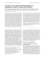
checked on 10% SDS PAGE (Fig. 1). The protein concen-
tration was estimated by Bradford's method.
CD3+, CD4+ and CD8+ cell counts
CD3+, CD4+ and CD8+ cell counts were performed using
four-color BD FACSCalibur system flow cytometer (BD
Biosciences, San Jose, CA, USA). For staining, 50 μl of
whole blood was aliquoted into 12 × 75 mm polystyrene
BD TruCOUNT™ Tubes (BD MultiTEST™, San Jose, CA,
USA) containing 20 μl CD3 FITC/CD8 PE/CD45 Per CP/
CD4 APC antibody (BD Pharmingen™, Franklin Lakes, NJ,
USA). The samples were mixed well and incubated for 20
minutes at room temperature in the dark and then lysed
using 450μl of lysing solution and incubated for 10 min-
utes. The tubes were centrifuged at 90 g (1000 rpm) for 5
minutes and the supernatant discarded. Two ml of PBS
buffer (PH 7.2–7.4, 0.05 M) was added in the tube and it
was centrifuged again at 90 g (1000 rpm) for 5 minutes.
After the supernatant was discarded and the cells resus-
pended in 500ul of PBS, the samples were acquired within
2 hrs after staining and anlysed by using CellQuest Pro
software (BD Biosciences, San Jose, CA, USA).
PBMC assay
For assessing T cell proliferation, blood was drawn in
Heparin (5000 I.U/5 ml; Biological E limited, Hyderabad,
AP, India), diluted with equal volume of RPMI-1640
medium (Invitrogen corporation, Grand Island, N.Y.
USA) without AB serum, layered on Histopaque (Sigma,
St Louis, MO, USA) gradient in 1:3 proportion and centri-
fuged at 200–350 g (1500–2000 rpm) for 30 minutes.
After the peripheral blood mononuclear cells (PBMC)
were isolated, washed twice to remove the cell debris and
platelets each at 90 g (1000 rpm) for 10 minutes, the cell
concentration was adjusted to 1 million cells/ml. Viability
of the cells was checked using Trypan blue (Sigma Aldrich,
St.Louis, MO, USA). To 0.1 ml of cell suspension 0.1 ml
of media for the control wells, 8 μl (3 mg/ml) of recom-
binant protein (r32kda-BCG, also referred to as Ag85A-
BCG) in 0.1 ml of media and 30 μl of 1 mg/ml Concana-
valin A (Con-A, Sigma Aldrich, St.Louis, MO, USA) for the
experimental wells were added in duplicates. Concentra-
tion of the recombinant protein was standardized based
on the optimal proliferation of the cells using children's
blood samples. The plate was incubated at 37°C with 5%
CO
2
and humidified air. At the end of the 3
rd
day for Con-
A and 5
th
day for the antigen, MTT (3-(4-5-dimethyl thia-
zol-2-yl) 2,5, diphenyl tetrazoleum bromide) (Sigma
Aldrich, St.Louis, MO, USA) was added and optical den-
sity (OD) measured at 570 nm and 620 nm reference filter
[3,23] in ELISA reader (BIO-RAD, Hercules, CA, USA).
Stimulation index (SI), a ratio between the OD values of
the test and control, was considered as positive if ≥ 2 [23].
In vitro interferon gamma assay
Sandwich Enzyme Linked Immunosorbent Assay (ELISA)
was performed to measure the IFN-γ levels in the superna-
SDS PAGE representing the purified r32KDa proteinFigure 1
SDS PAGE representing the purified r32KDa protein.
Lane 1 & 2 represents purified r32KDa protein and M repre-
sents molecular weight marker.
Journal of Immune Based Therapies and Vaccines 2007, 5:8 />Page 4 of 7
(page number not for citation purposes)
tants collected from the above cultures. To each well, 50
μl of the capture antibody (BD pharmingen™, Franklin
Lakes, NJ, USA). diluted in coating buffer was added and
incubated over night at 4°C. After 5 washes, the wells
were blocked with 50 μl of 2% BSA for 3 hours at 37°C. A
further 5 washes were followed by the addition of 50 μl of
supernatants and standards and then incubated for 4
hours at room temperature. After another 5 washes, 50 μl
secondary antibody was added, incubated for 3 hours at
37°C and washed 5 times. After addition of 50 μl of work-
ing detector (HRP antigen, BD pharmingen™, Franklin
Lakes, NJ, USA) and incubation for one and half hour and
5 washes, the wells were incubated with the substrate (50
μl) Ortho Phenyl Diamine (OPD, Sigma, St. Louis, MO,
USA) + H
2
O
2
(Qualigen, Mumbai, MH, India) for 20 min-
utes at room temperature in the dark. 50 μl of stop solu-
tion was added and absorbance was measured at 490 nm.
Statistical analysis
Student's-t test was used for comparing the mean values
between the groups. Chi-square test to compare number
of children between the groups was done by cross tabula-
tion method. A p value of ≤ 0.05 was considered as signif-
icant.
Results
Expression and purification of M. bovis BCG r32KDa
protein
The protein was purified under native conditions using
Ni-NTA Chromatography. The purified r32KDa protein
was separated on an 10% gel and visualized for expected
protein band using Coomassie Brilliant Blue. The purity
of the protein was about 95%. To remove the endotoxin
contamination, purified recombinant 32KDa protein was
incubated with 10% v/v polymyxin B-agarose (Sigma-
Aldrich; binding capacity, 200 to 500 μg of LPS from
Escherichia coli serotype O128:B12/ml) for 1 hour at
4°C. For further immunological studies this endotoxin
free r32KDa protein was used.
Cell counts and proliferation
The mean (±SD) numbers of CD3+, CD4+, CD8+ cells
and the SIs in T cell proliferation assay against Con-A,
were comparable in all the three groups of children (val-
ues not shown in the tables).
Age groups
When the 267 children with TB (tuberculosis)were classi-
fied into different age-groups, the highest number (n =
116) was observed in age group 13–14 years followed by
children in the age group 9–12 years (n = 96). The num-
bers declined further with the last group (age < 6 years)
having the least number (n = 20). Chi-Square between the
number of children in different age groups was highly sig-
nificant (242.22; p < 0.000).
Stimulation Indices and their IFN-γ levels in the superna-
tants in PBMC assay according to their age distribution in
vaccinated children were shown in Figure 2. Significant
differences were observed between the SI in <6 years (5.60
± 2.8) and that in 9–12 years groups (2.70 ± 1.13) (p <
0.0002); between 6–8 years (4.36 ± 2.1) and 9–12 years
(2.70 ± 1.13) groups (p < 0.013). Significant differences
were observed between the IFN-γ levels in <6 years (3316
± 718 pg/ml) and that in 9–12 years groups (1360 ± 344
pg/ml) (p < 0.003); between 6–8 years and 9–12 years
groups (2880 ± 733 and 1360 ± 344; p < 0.01).
PBMC assay
Percent number and mean ± SD of negative (<2) and pos-
itive (≥2) stimulation indices (SI) in PBMC assays with
BCGr32Kda protein observed in children (a) with BCG
scar (b) without scar and (c) with TB are shown in Table
1. The proliferation in healthy BCG scar positive children
were significantly high when compared to that in scar neg-
ative- and tuberculosis- children with a p < 0.0004 and
0.0014 respectively. Seventy nine percent of vaccinated
children showed positive proliferative responses with a
mean SI value of 4.98 ± 1.99 while only 39% of the unvac-
cinated and 58% of the tuberculosis children showed pos-
itive responses with mean values of 2.9 ± 1.6 and 2.9+1.7,
respectively. Chi-square test by cross tabulation method
to compare number of children between scar positive and
scar negative children was: 10.465; p < 0.001 and between
scar positive versus tuberculosis, 3.630; p < 0.057.
In vitro IFN-
γ
assay
The overall mean IFN-γ levels (2744 ± 1004pg/ml) in vac-
cinated children was significantly high (p < 0.0008) when
compared to 1556 ± 490 pg/ml in scar-negative and to
2112 ± 988 pg/ml in TB children (Figure 3). 2046 pg/ml
of IFN-γ was considered as the cut-off value (based on the
least arithmetic mean + standard deviation in scar nega-
tive children). 66% of the vaccinated children, 40% of TB
children and only 10% of scar negative children showed
IFN-γ levels greater than 2046 pg/ml.
Discussion
An understanding of protective immune responses in
humans is essential for the rational development and clin-
ical testing of new, effective vaccines [24] which are char-
acterized by bacterial antigen specific lymphoproliferative
responses, IFN-γ production, high Th1 type-Th2 type
ratios and induction of CTL [25]. It is important to under-
stand the quantitative relationship between host
responses and disease and to identify proliferation of T
cells specific to antigen, which contribute to protective
immunity. In vitro cytokine production by peripheral
blood mononuclear cells can be an important and reliable
measure of immunocompetence. Also, spontaneous
release of cytokines by PBMCs may serve as a measure of
Journal of Immune Based Therapies and Vaccines 2007, 5:8 />Page 5 of 7
(page number not for citation purposes)
their activation invivo [26]. In the present study prolifera-
tive responses and in vitro IFN-γ production were thus
demonstrated in different categories of children from
Hyderabad, India. The exposure to environmental myco-
bacteria is presumed to be more or less similar in all the
children since they are from same geographical location.
Comparison has been made between scar positive and
scar negative children to evaluate the influence of BCG
vaccination on the assay.
It was observed in this study that BCG induced immunity
wanes with age, concurring with an increase in the
number of children with tuberculosis. Waning of immu-
nity is of particular public health interest because it may
result in increased susceptibility later in life. The mecha-
nism underlying the gradual loss of effectiveness of BCG
as the individual reaches 10 to 15 years of age is poorly
understood [27,28]. In the study on tuberculosis epidemi-
ology in south India (1961–68), the prevalence of infec-
tion over the study period varied between 1 to 2.1%, 6.4
to 7.9% and 15.4 to 16.9% in the 0–4, 5–9 and 10–14
year age group, respectively [29]. Similar observations
were made in other parts of India [30,31]. Studies
reported that memory immunity slowly declines but can
be recovered by boosting if a candidate antigen that can be
specifically recognized by this immunity is reintroduced
[27]. Therefore, pre-exposure priming with a highly effica-
cious attenuated vaccine strain should be followed by
post exposure boosting with a potent subunit vaccine [9].
The results observed in earlier and present studies proba-
bly necessitate the need for a booster vaccine perhaps at
the age of about four years, much before the waning
begins.
The low responses observed in children without a BCG-
scar in this study, further reiterate that Ag85A may be
effectual not only as a booster-vaccine but also as an anti-
gen in in vitro assays as a correlate of protection. A possible
explanation for children with tuberculosis exhibiting
moderate in vitro immune responses as observed in this
study may be the presence of M. tuberculosis specific T
cells. Thus, Ag85A protein may also be a potential diag-
nostic agent for assessing the immune status of healthy
individuals and predict their clinical outcome.
Furthermore, the nature of the cells responding to M.
tuberculosis infection and how their relative contribution
changes over time is a crucial aspect in vaccine develop-
ment [32]. Animal models suggest that CD4+ T cells are
the most important aspect of the protective response in
primary infection [33,34]. Though the subset of T cells
that released IFN-γ was not identified in this study, there
are reports that BCG stimulates both CD4
+
and CD8
+
cells
in vitro, while PPD stimulates only CD4
+
lymphocytes
[35]. CD8
+
T cells which also respond by releasing IFN-
γ[36] exhibit strong recognition of Ag85A from healthy
Table 1: Percent-number and mean ± SD of Stimulation-Indices in PBMC assays with BCGr32Kda protein in different categories of
children.
Scar +ve
n = 45
Scar -ve
n = 45
TB
n = 25
P value
Mean (± SD) of positive SIs 4.9 ± 1.9
a
2.9 ± 1.17
b
2.9 ± 0.73
c
p < 0.0004
ab
p < 0.0014
ac
Mean (± SD) of negative SIs 1.6 ± 0.29 1.6 ± 0.31 1.7 ± 0.20
%n with positive SI* 79 39 58
%n with negative SI* 21 61 42
Stimulation Indices (SI) < 2: negative & ≥ 2: positive in T cell assays.
Statistical analysis of the mean values of SIs was done by Student's t-test.
Chi-square test to compare number of children was done by cross tabulation method:
*Scar positive Vs. Scar negative, χ
2
= 10.465; p < 0.001
* Scar positive Vs. Tuberculosis, χ
2
= 3.630; p < 0.057
T-cell assays with BCG r32kDa: Stimulation Indices and Interferon-γ levels in different age-groups of BCG-vaccinated children (n = 45)Figure 2
T-cell assays with BCG r32kDa: Stimulation Indices
and Interferon-γ levels in different age-groups of
BCG-vaccinated children (n = 45). Definition of abbrevi-
ation: IFN-γ = Interferon – gamma, SI = Stimulation Index.
Statistical Significance between mean values of IFN-γ
levels (pg/ml): (a) < 6 years & 9–12 years (3316 ± 718 &
1360 ± 344; p < 0.003). (b) 6–8 years and 9–12 years (2880 ±
733 & 1360 ± 344; p < 0.01). Statistical Significance
between mean values of SI: (a)<6 years & 9–12 years
(5.69 ± 2.21 & 2.70 ± 1.13; p < 0.0002) (b) 6–8 years & 9–12
years (4.36 ± 2.10 & 2.70 ± 1.13; p < 0.013)
Journal of Immune Based Therapies and Vaccines 2007, 5:8 />Page 6 of 7
(page number not for citation purposes)
controls [37]. Intranasal vaccination with Mycobacterium
tuberculosis antigen85A against airway Mycobacterium
tuberculosis challenge in mice induced a significantly
higher level of interferon-γ [38]. Further studies should
involve the specific memory cells and their kinetics.
Though it has been demonstrated earlier that Ag85A of
Mycobacterium tuberculosis is immunogenic, the studies
were conducted in animals or adults. A recombinant
modified vaccinia virus Ankara expressing this antigen
(MVA85A) has already entered into clinical trials in 2002
[39]. To conclude, the present study involved antigen 85A
of BCG and was based on observations in children vacci-
nated with BCG at birth. Given their age, it is unlikely that
the children had a prior exposure to environmental myco-
bacteria, significantly high in vitro IFN-γ levels observed in
BCG vaccinated children in our study substantiate that
BCG is effective in children, but that the effect may wane
with age. The immunity could be boosted using modified
r32kDa (Ag85A) of BCG.
Authors' contributions
AB carried out the study, collection of samples, performed
all the experiments, statistical analysis of data and pre-
pared the manuscript. SCM synthesized r32kDa protein.
HSP was associated in standardizing techniques. SLG
supervised sample collection and oversaw clinical docu-
mentation. KJRM and VVL were involved in the concep-
tion of the study. KJRM contributed towards the clinical
aspects. VVL designed the study and edited the manu-
script. All authors read and approved the final manu-
script.
Acknowledgements
We thank Dr. Akbar Yazdani, Prof. Indira Nath, Dr. C.E.Prasad, Dr.
Shekar.C.Mande and Dr. S.E. Hasnain for their support. We are grateful to
Bhagawan Mahvir Trust and LEPRA Society, UK for the funds.
References
1. Castañón-Arreola Mauricio, López-Vidal Yolanda: A second-gener-
ation anti TB vaccine is long overdue. Ann Clin Microbiol Antimi-
crob 2004, 3:10. doi:10.1186/1476-0711-3-10
2. Lanckriet C, Levy-Bruhl D, Bingono E, Siopathis RM, Guerin N: Effi-
cacy of BCG vaccination of the newborn: evaluation by a fol-
low-up study of contacts in Bangui. Int J Epidemiol 1995,
24(5):1042-9.
3. Vijaya Lakshmi V, Kumar Sunil, Surekha Rani H, Suman Latha G,
Murthy KJR: Tuberculin Specific T Cell Responses in BCG Vac-
cinated Children. Indian Pediatr 2005, 42:36-40.
4. Srinivasan Ramesh, Menon Leena, Stevens Pat, Campbell Ian, Alfaham
Mazin: Ethnic differences in selective neonatal BCG immuni-
sation: white British children miss out. Thorax 2006,
61:247-249. doi:10.1136/thx.2004.036269.
5. Kösecik Mustafa, Emiroglu Haldun, Mansur Tatli M, Koc Ahmet, Atas
Ali: Prevalence of Tuberculous Infection and the Impact of
BCG Vaccination on Tuberculin Testing Among Primary
Schoolchildren in Turkey. Indian Pediatrics 2002, 39:362-365.
6. Vijaya Lakshmi V, Murthy KJR: The efficacy of BCG in conferring
protection against tuberculosis. Lung India 1992:38-40.
7. Vijayalakshmi V, Devi PS, Murthy KJR, Rao DV, Jain SN: Cell medi-
ated immune responses in BCG vaccinated children. Indian
Pediatr 1993, 30:899-903.
8. Vijaya Lakshmi V, Murthy KJR: Revaccination with BCG. Lung
India 1998, 16:75-77.
9. Kaufmann SHE: New issues in tuberculosis. Ann Rheum Dis Review
2004, 63:ii50-ii56. doi: 10.1136/ard.2004.028258.
10. Britton WJ, Palendira U: Improving vaccines against tuberculo-
sis. Immunol Cell Biol 2003, 81:34-45.
11. Vijayalakshmi V, Rao DV, Murthy KJR, Jain SN: A study of the
tuberculin test and its correlation with the in vitro
responses. Lung India 1989, 7(2):63-6.
12. Lyon AJ: Sensitivity of neonates to tuberculin after BCG vac-
cination. Br Med J (Clin Res) 1986, 292:1526-1527.
13. Karalliedde S, Katugaha LP, Uragoda CG: Tuberculin response of
Srilankan children after BCG vaccination at birth. Tubercle
1987, 63:33-38.
14. Schluger NW, Rom WN: The host immune response to tuber-
culosis. Am J Respir Crit Care Med 1998, 157:679-91.
15. Ellner JJ: Review: The Immune Response in Human Tubercu-
losis-Implications for Tuberculosis Control. J Infect Dis 1997,
176:1351-1359.
16. Surekha Rani H, Vijaya Lakshmi V, Sumanlatha G, Murthy KJR: Cell-
mediated immune responses in children towards secreted
proteins of Mycobacterium bovis BCG. Tuberculosis 2005,
85:89-93.
17. D'Souza S, Rosseels V, Romano M, Tanghe A, Denis O, Jurion F, Cas-
tiglione N, Vanonckelen A, Palfliet K, Huygen Kris: Mapping of
Murine Th1 Helper T-Cell Epitopes of Mycolyl Transferases
Ag85A, Ag85B, and Ag85C from Mycobacterium tuberculosis.
Infection and Immunity 2003, 71:483-493. No. 1. 0019-9567/03/
$08.00+0 DOI: 10.1128/IAI.71.1.483-493.2003.
18. Huygen K, Abramowicz D, Vandenbussche P, Jacobs F, De Bruyn J,
Kentos A, Drowart A, Van Vooren JP, Goldman M: Spleen cell
cytokine secretion in Mycobacterium bovis BCG-infected
mice. Infect Immun 1992, 60:2880-86.
19. Launois P, DeLeys R, N'Diaye Niang M, Drowart A, Andrien M, Dier-
ckx P, Cartel J-L, Sarthou J-L, Van Vooren J-P, Huygen K: T-cell-
epitope mapping of the major secreted Mycobacterial anti-
gen Ag85A in tuberculosis and leprosy. Infect Immun 1994,
62:3679-87.
20. Munk ME, De Bruyn J, Gras H, Kaufmann SHE: The Mycobacterium
bovis BCG 32-kilodalton protein antigen induces human
cytotoxic T-cell responses. Infect Immun 1994, 62:726-28.
21. Roche PW, Peake PW, Billman-Jacobe H, Doran T, Britton WJ: T-
cell determinants and antibody binding sites on the major
mycobacterial secretory protein MPB59 of
Mycobacterium
bovis. Infect Immun 1994, 62:5319-26.
IFN-γ levels in the supernatants of T cell assays stimulated with BCG r32KDa protein in childrenFigure 3
IFN-γ levels in the supernatants of T cell assays stim-
ulated with BCG r32KDa protein in children. Defini-
tion of abbreviation: IFN-γ = Interferon – γ. Horizontal bars
represent mean values. Statistical Significance between
mean values of IFN-γ levels (pg/ml): Scar positive Vs.
Scar negative: p < 0.0008
Publish with BioMed Central and every
scientist can read your work free of charge
"BioMed Central will be the most significant development for
disseminating the results of biomedical research in our lifetime."
Sir Paul Nurse, Cancer Research UK
Your research papers will be:
available free of charge to the entire biomedical community
peer reviewed and published immediately upon acceptance
cited in PubMed and archived on PubMed Central
yours — you keep the copyright
Submit your manuscript here:
/>BioMedcentral
Journal of Immune Based Therapies and Vaccines 2007, 5:8 />Page 7 of 7
(page number not for citation purposes)
22. Silver RF, Wallis RS, Ellner JJ: Mapping of T cell epitopes of the
30-kDa α antigen of Mycobacterium bovis strain bacillus Cal-
mette-Guérin in purified protein derivative (PPD)-positive
individuals. J Immunol 1995, 154:4665-74.
23. Mosmann TR: Rapid colorimetric assay for cellular growth and
survival: application to proliferation and cytotoxicity assay. J
Immunol Methods 1983, 65:55-63.
24. Pathan Ansar A, Wilkinson Katalin A, Klenerman Paul, McShane
Helen, Davidson Robert N, Pasvol Geoffrey, Hill Adrian VS, Lalvani
Ajit: Direct Ex Vivo Analysis of Antigen-Specific IFN-γ Secret-
ing CD4 T Cells in Mycobacterium tuberculosis -Infected Indi-
viduals: Associations with Clinical Disease State and Effect of
Treatment. The Journal of Immunology 2001, 167:5217-5225.
25. Hussey Gregory D, Watkins Marcia LV, Elizabeth A, Goddard ,
Gottschalk Sean, Hughes Elizabeth J, Iloni Karen, Kibel Maurice A,
Ress Stansley R: Neonatal mycobacterial specific cytotoxic T-
lymphocyte and cytokine profiles in response to distinct
BCG vaccination strategies. Immunology 2002, 105:314-324.
26. Friberg D, Bryant J, Shannon W, Whiteside TL: In vitro cytokine
production by normal human peripheral blood mononuclear
cells as a measure of immunocompetence or the state of
activation. Clin Diagn Lab Immunol 1994, 1(3):261-268.
27. Brooks Jason V, Frank Anthony A, Keen Marc A, Bellisle John T,
Orme Ian M: Boosting Vaccine for Tuberculosis. Infect Immun
2001, 69:2714-2717. doi: 10.1128/IAI.69.4.2714-2717.2001.
28. Olsen W, Brandt Ly, Agger EM, van Pinxteren LAz H, Andersen P:
The Influence of Remaining Live BCG Organisms in Vacci-
nated Mice on the maintenance of Immunity to Tuberculo-
sis. Scand J Immunol 2004, 60:273-277.
29. National Tuberculosis Institute: Tuberculosis in a rural popula-
tion of south India: A five year epidemiological study. Bull
WHO 1974, 51:473-488.
30. Chakraborthy AK, Chaudhuri K, Sreenivasan TR, Krishnamurthy MS,
Shashidhara AN, Channabasaviah R: Tuberculosis infection in a
rural population of South India: 23 years trend. Tubercle Lung
Disease 1992, 73:213-218.
31. Pattanaik D, Singh KB, Guresh K, Anand K, Kant S, Kapoor SK: Prev-
alence of Tuberculosis infection in children below fourteen
years in rural Haryana. Indian Pediatrics 2002, 39:70-74.
32. Mark Doherty T, Andersen Peter: Vaccines for Tuberculosis:
Novel Concepts and Recent Progress. Clin Microbiol Rev 2005,
18:687-702.
33. Rahemtulla A, Fung-Leung WP, Schilham MW, Kundig TM, Sambhara
SR, Narendran A, Arabian A, Wakeham A, Paige CJ, Zinkernagel RM,
et al.: Normal development and function of CD8+ cells but
markedly decreased helper cell activity in mice lacking CD4.
Nature 1991, 353:180-184.
34. xing Zhou, Wang Jun, Croitoru Kenneth, Wakeham Julia: Protec-
tion by CD4 or CD8 T cells against pulmonary Mycobacte-
rium bovis Bacillus Calmette-Gue'rin infection. Infection and
Immunity 1998, 66:5537-5542.
35. Turner J, Dockrell HM: Stimulation of human peripheral blood
mononuclear cells with live Mycobacterium bovis BCG acti-
vates cytolytic CD8
+
T cells in vitro. Immunology 1996,
87:339-342.
36. Szabo SJ, Sullivan BM, Stemmann C, Satoskar AR, Sleckman BP, Glim-
cher LH: Distinct effects of T-bet in TH1 lineage commitment
and IFN-gamma production in CD4 and CD8 T cells. Science
2002, 295:338-42.
37. Smith Steven M, Klein Michèl R, Malin Adam S, Sillah Jackson, Huygen
Kris, Andersen Peter, McAdam Keith PWJ, Dockrell Hazel M:
Human CD8
+
T Cells Specific for Mycobacterium tuberculosis
Secreted Antigens in Tuberculosis Patients and Healthy
BCG-Vaccinated Controls in the Gambia. Infect Immun 2000,
68:7144-7148.
38. Haile M, Hamasur B, Jaxmar T, Gavier-Widen D, Chambers MA,
Sanchez B, Schroder U, Kallenius G, Svenson SB, Pawlowski A: Nasal
boost with adjuvanted heat-killed BCG or arabinomannan-
protein conjugate improves primary BCG-induced protec-
tion in C57BL/6 mice. Tuberculosis 2005, 85:107-114.
39. Ibanga Hannah B, Brookes Roger H, Hill Philip C, Owiafe Patrick K,
Fletcher Helen A, Lienhardt Christian, Hill Adrian VS, Adegbola Rich-
ard A, McShane Helen: Early clinical trials with a new tubercu-
losis vaccine, MVA85A, in tuberculosis-endemic countries:
issues in study design. Lancet Infect Dis 2006, 6:522-28.