báo cáo khoa học: " A rice calcium-dependent protein kinase is expressed in cortical root cells during the presymbiotic phase of the arbuscular mycorrhizal symbiosis" pps
Bạn đang xem bản rút gọn của tài liệu. Xem và tải ngay bản đầy đủ của tài liệu tại đây (2.6 MB, 14 trang )
RESEARC H ARTIC L E Open Access
A rice calcium-dependent protein kinase is
expressed in cortical root cells during the
presymbiotic phase of the arbuscular mycorrhizal
symbiosis
Lidia Campos-Soriano
1
, Jorge Gómez-Ariza
2
, Paola Bonfante
2
and Blanca San Segundo
1*
Abstract
Background: The arbuscular mycorrhizal (AM) symbiosis consists of a mutualistic relationship between soil fungi
and roots of most plant species. This association provides the arbuscular mycorrhizal fungus with sugars while the
fungus improves the uptake of water and mineral nutrients in the host plant. Then, the establishment of the
arbuscular mycorrhizal (AM) symbiosis requires the fine tuning of host gene expression for recognition and
accommodation of the fungal symbiont. In plants, calcium plays a key role as second messenger during
developmental processes and responses to environmental stimuli. Even though calcium transients are known to
occur in host cells during the AM symbiosis, the decoding of the calcium signal and the molecular events
downstream are only poorly understood.
Results: The expression of seventeen Calcium-dependent Protein Kinase (CPK) genes representative of the four
distinct phylogenetic groups of rice CPKs was monitored during the presymbiotic phase of the AM symbiosis.
Among them, OsCPK18 and OsCPK4, were found to be transcriptionally activated in response to inoculation with
the AM fungus Glomus intraradices. OsCPK18 and OsCPK4 gene expression was also up-regulated by fungal-
produced diffusible molecules. Laser microdissection revealed expression of OsCPK18 in cortical cells, and not in
epidermal cells of G. intraradices-inoculated rice roots, suggesting a preferential role of this gene in the root cortex.
Moreover, a plasma membrane localization of OsCPK18 was observed by transient expression assays of green
fluorescent protein-tagged OsCPK18 in onion epidermal cells. We also show that the myristoylation site of the
OsCPK18 N-terminus is required for plasma membrane targeting.
Conclusion: The rap id activation of OsCPK18 expression in response to AM inoculation, its expression being also
induced by fungal-secreted signals, together with the observed plasma membrane localization of OsCPK18,
points to a role for OsCPK18 in perception of the AM fungus. T he OsCPK18 gene migh t be considered as a
marker for the presymbiotic phase of the symbio tic process. These findings provide a better understanding of
the signaling mechanisms operating during the AM symbiosis and will greatly facilitate their molecular
dissection.
* Correspondence:
1
Centre for Research in Agricultural Genomics (CRAG) CSIC-IRTA-UAB.
Department of Molecular Genetics. Campus UAB, Edifici CRAG, Bellaterra
(Cerdanyola del Vallès) 08193 Barcelona, Spain
Full list of author information is available at the end of the article
Campos-Soriano et al. BMC Plant Biology 2011, 11:90
/>© 2011 Campos-Soriano et al; licensee BioMed Central Ltd. This is an Open Access art icle distributed un der the terms of the Creative
Commons Attribution License ( which permits unrestricted use, distribution, and
reproduction in any medium, provided the original work is properly cite d.
Background
Most vascular flowering plants have the ability to estab-
lish symbiotic associations with arbuscular mycorrhizal
(AM) fungi [1]. The main benefit for the plant is
improved uptake of water and mineral nutrients from the
soil, particularly phosphate, in exchange for photosynthe-
tically fixed carbon [2]. The mycorrhizal symbiosis has
been also associated with increased resistance to patho-
gen infection and tolerance to abiotic stress in several
plant species [3]. As a conseque nce, the AM symbiosis is
of tremendous significance in agricultural ecosystems.
The legumes Medicago truncatula and Lotus japonicus
have been widely adopted as the reference species for
studies of the AM symbiosis. Contrary to this, Arabi-
dopsis thaliana, the model system for functional geno-
mics in plants, has no mycorr hization ability. Rice, a
monocotyledonous plant with a completely sequenced
genome, establishes symbiotic associations with mycor-
rhizal fungi [4,5]. As compared to the model legume
species, the genes responsible for the AM symbiotic
interaction in rice are less characterized.
Successful symbiosis with AM fungi relies on the fine
tuning and appropriate control of host gene expression
and physiological responses. A molecular dialogue is early
established between the host plant and the AM fungus
and prepares the two partners for the subsequent root
colonization. Signal exchange and communication starts
prior to the initial cell-to-cell contact between the sym-
bionts. Thus, plant roots exude strigolactones which have
an stimulatory effect on AM growth [6]. Fungal hyphae, in
turn, produce diffusible molecules, the “Myc factors” (ana-
logous to the rhizobial Nod facto rs). Very recently, it was
reported that the AM fungus secretes lipochitooligosac-
charides which stimulate formation of AM symbiosis in
diverse plant species [7]. Perception of Myc factors by the
host cells triggers a rapid and transient elevation of intra-
cellular calcium, alterations in the cellular architecture and
transcriptional reprogramming of the root [8-12]. Even
though both cytoplasmic [10] and nuclear [9] pre-infection
Ca
2+
spiking responses are elicited in M. truncatula roots
in response to AM fungi, the mechanisms by which Ca
2+
alterations are sensed and transduced into early AM-
induced signaling remain unknown.
Once contact between the symbionts is established,
the fungus enters into the root through the epidermal
cells, and penetrate s into the cortex wher e it forms
highly branched structures, called arbuscules, in the cor-
tical cells of the root. The arbuscules are the site of the
major nutrient exchange between the two symbionts
[2,13,14].
It is also known that the plant response to Myc factors
is mediated by a partially characterized signalin g path-
way which is required for the establishment of both
rhizobial and AM symbioses, the so called common
symbiosis (SYM) pathway [2,13-15]. Forward genetic
analysis in the model legumes Medicago truncatula and
Lotus japonicus has led to t he identification of compo-
nents of the SYM signaling pathway. They are: a leu-
cine-rich-repeat receptor-like kinase, the SYMRK
protein in L. japonicus (known as DMI2 for “Does Not
Make Infections 2” in M. truncatula), two nucleoporins
(NUP85 and NUP133), two cation channel proteins (the
L. japonicus CASTOR and POLLUX proteins; DMI1a
and DMI1b in M. truncatula), a calcium and calmodu-
lin-dependent protein kinase (CCaMK in L. japonicus;
DMI3 in M. truncatula) and CYCLOPS (LjCYCLOPS;
DIM3-interacting protein in M. truncatula) [16-21].
CCaMK interacts with, and phosphorylates, CYCLOPS
in the nucleus [21,22]. In rice, the function of several
SYM genes appears to be conserved, including CASTOR
and POLLUX (acting upstream of the calcium-spiking
signal) and CCaMK and CYCLOPS (acting downstream
of the calcium-spiking signal) [23-25]. Evidence also
support the existence of alternative, SYM-independent
signaling pathways controlling the early responses to
AM fungi in both rice and M. truncatula [25,26].
Transcript profiling of mycorrhizal roots allowed the
identification of AM-regulated genes in several plant
species, including rice [3,27-30].However,themajority
of these studies focused on the mature phase of the
symbiotic process, a periodinwhichthehostrootis
already colonized and arbuscules are developed in the
root cortical cells. Along with this, alterations in the
expression of genes connected to nutrient acquisitio n
processes, such as phospha te transporter genes, are well
documented in differ ent AM associations [31,32]. Genes
involved in cellular modifications, transcriptional control
and defense-related responses are also known to be
regulated during the AM symbiosis [4,31].
Even though alterations in Ca
2+
levels are known to
occur in host cells during the presymbiotic phase, the
decoding of the calcium signal is only poorly under-
stood. On the other hand, it is well established that Cal-
cium-dependent protein kinases (CPKs or CDPKs) are
important Ca
2+
sensors in signaling processes during
growth, development and stress responses in plants
[33,34]. CPKs belong to the CDPK/SnRK superfamily of
protein kinases and represent a differentiated group of
protein kinases found in plants, algae and protists
[34-36]. They possess a characteristic structure consist-
ing of four domains: an amino terminal variable domain,
a serine/threonine kinase domain, a junction autoinhibi-
tory domain, a nd a C-terminal calmodulin domain.
These features make CPKs ideally structured to rapidly
perceive alterations in intracellular calcium concentra-
tion and translating them in to protein phospho rylation
Campos-Soriano et al. BMC Plant Biology 2011, 11:90
/>Page 2 of 14
cascades. CPK functioning is, however, different from
that of CCaMK functioning, since CPKs do not require
calmodulin for their activation. Ca
2+
binds directly the
calmodulin domain of CPKs and induces a conforma-
tional change resulting in kinase activation [34]. The
available i nformation on plant CPKs from v arious plant
species indicates that they a re encoded by multigene
families and that whereas some of the genes are ubiqui-
tously expressed, others show a tissue-specific pattern of
expression or are regulated by stress (wounding, salinity,
cold, drought, pathogen infection) [33,37,38].
Knowing that Ca
2+
plays a central role in the AM-
induced signaling pathway, it was of interest to investigate
to what extent CPKs are involved in the AM-induced sig-
naling pathway. Towards this goal, the expression pattern
of seventeen cpk genes was monitored in rice plants that
have been inoculated with the AM fungus Glomus intrara-
dices. We provide evidence that the expression of two dis-
tinct cpk genes, the OsCPK18 and OsCPK4 genes, is rapidly
induced during the presymbiotic phase of the rice/G.
intraradices interaction. OsCPK18 and OsCPK4 gene
expression is also activated by fungal-produced diffusible
fungal signal(s). By using the laser microdissection (LMD)
technology, OsCPK18 expression was detected in cortical
cells, but not epidermal cells, of the G. intraradices-inocu-
lated rice roots. Moreover, a plasma membrane localization
of OsCPK18 is here reported, the myristoylation site of
OsCPK18 being required for its plasma membrane locali-
zation. Together, these findings suppor t that Os CPK18
might play a role during recognition of the AM fungus by
the host cells.
Results
Expression of CPK genes in AM-inoculated rice roots
A genome-wide analysis of rice CPK genes identified 31
genes which are distributed into four phylogenetic
groups (I-IV) [39,40]. Moreover, a comparison of the
rice CPK genes distinguished 11 closely related pairs
which, most probably, have arisen via sequential dupli-
cation events, the OsCPK1/15, OsCPK2/14, OsCP K3/16,
OsCPK4/18, OsCPK5/13, OsCPK7/23, OsCPK8/20,
OsCPK11/17 , OsCPK21/22, OsCPK24/28 and OsCPK25/
26 pairs [40]. Based of the homology a nd phylogenetic
relatedness among the rice CPK genes, we selected a
subset of seventeen CPK genes representative of the
four distinct phylogenetic groups of rice CPKs in which
at least one representative member for each pair of clo-
sely related CPK genes was present. The subset of genes
assayed in this work included OsCPK7, OsCPK10,
OsCPK13, OsCPK17 and OsCPK24 from Group I;
OsCPK2, OsCPK15, OsCPK19 and OsCPK25 from
Group II; OsCPK8, OsCPK9, OsCPK16 and OsCPK22
from Group III; and OsCPK4, OsCPK18, OsCPK30 and
OsCPK31 from Group IV.
The expression pattern of selected rice CPK genes and
their transcriptional response to inoculation with the AM
fungus G. intraradices, were examined during the pre-
symbiotic phase of the symbiotic process. A preliminary
screening was carried out by semiquantitative RT-PCR
experiments with RNA samples obtained from whole rice
roots that had been inoculated with fungal spores using
the single sandwich method. Total RNA was isolate d at
24, 48, 72 and 96 hours after inoculation of th e rice roots
with G. intraradices,aswellasfrommock-inoculated
rice roots. Many CPK gene s were found to be expressed
in rice roots and at different levels (Additional file 1:
Figure S1). Among them, the OsCPK4 and OsCPK18
genes were expressed at the highest levels. Moderate to
low levels of expression were observed for OsCPK10,
OsCPK13, OsCPK17, OsCPK24, OsCPK15, OsCPK19,
OsCPK8 and OsCPK9 ,whereasOsCPK30 transcripts
were barely detected (Additional file 1: Figure S1). The
OsCPK7 and OsCPK16 genes showed expression profiles
similar
to those shown for OsCPK8 and OsCPK30,
respectively (results not shown). Taken in the whole, the
expression level of the various CPK genes here investi-
gated appears not to be dramatically affected upon inocu-
lation with G. intraradices, with the exception of OsCPK4
and OsCPK18 expression (Additional file 1: Figure S1)
For a comparison, the expression of the known SYM
genes from rice, namely the OsSYMRK, OsPOLLUX,
OsCASTOR and OsCCaMK genes, was also examined.
This analysis revealed up-regulation of OsSYMRK,
OsPOLLUX and OsCCaMK in response to G. intrara-
dices at 72 and 96 hours post-inoculation (Additional
file 1: Figure S1). The observed induction of these genes
indicates that the host plant cells perceive and respond
to the AM fungus through the activation of the AM-
specific SYM signaling pathway.
Since RT-PCR analyses do not provide reliable quantita-
tive data of gene expression, quantitative reverse transcrip-
tion-PCR (RT-qPCR) was used to further characterize the
effect of G. intraradices inoculation on OsCPK4 and
OsCPK18 geneexpression.Byusingthesinglesandwich
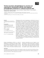
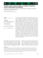
system for fungal inocul ation, up-regulat ion of OsCPK18
gene expression occurred as early as 24 h post-inoculation
with G. intraradices (Figure 1A, upper panel). The level of
OsCPK18 transcripts remained higher at the subsequent
time points in the G. intraradices-inoculated roots com-
pared to mock-inoculated roots. Concerning OsCPK4,its
expression was also found to be up-regulated in response
to G. intraradices inoculation during the time period of
24-72 hours. OsCPK4 expression returne d to a level similar
to that of non-inoculated roots by 96 h post-inoculation
(Figure 1A, middle panel). Finally, OsCCaMK expressio n
increased in G. intraradices-infected roots relative to
mock-inoculated roots by 72-96 hours post-inoculation
(Figure 1A, lower panel).
Campos-Soriano et al. BMC Plant Biology 2011, 11:90
/>Page 3 of 14
Overall, gene expression studies revealed up-regula-
tion of the rice OsCPK4 and OsCPK18 genes during
the presymbiotic phase of the AM symbiosis. These
results were consistently observed in all three indepen-
dent experiments. Although the expression of the
OsCPK18, OsCPK4 and OsCCaMK genes was up-regu-
lated in AM-inoculated roots compared to non-inocu-
lated roots, it is also true that the amplitude of the
differential expression for these genes was not very
high during the time period here analyzed. Concerning
OsCCaMK for which a role during AM symbiosis has
been demonstrated in rice [23], its variation in the
expression level in response to AM inoculation is also
low and appears to occur at a later time point com-
pared to the observed activation of OsCPK18 and
OsCPK4 gene expression.
Figure 1 Gene expression analysis by real-time qPCR for the OsCPK18, OsCPK4 and OsCCaMK genes. The single-sandwich (A) or the
double-sandwich (B) system was used for inoculation. Roots inoculated with G. intraradices and mock-inoculated roots were harvested at the
indicated times. Each sample consisted on a pool of at least 12 individual plants. Expression levels are shown relative to the housekeeping
OsAct1 gene. Data shown represents the means ± error. Three independent experiments were carried out with similar results.
Campos-Soriano et al. BMC Plant Biology 2011, 11:90
/>Page 4 of 14
Diffusible factors released by G. intraradices induce
OsCPK18 and OsCPK4 expression in rice roots
It is generally assumed that plants perceive AM fungi
even before physical contact between the two symbionts,
andthatrecognitionofMycfactors triggers alterations
in Ca
2+
levels and transcriptional responses in host
roots [7,9,11,41]. In this work, the double sandwich
method was used to investigate whether the observed
induction of OsCPK18 and OsCPK4 expression is attri-
butable to diffusible factors released by the fungus. This
system prevents contact between the tw o symbionts
while allowing the exchange of signal molecules [42].
OsCPK18 and OsCPK4 expression was analyzed by RT-
qPCR (Figure 1B). When using the double sandwich sys-
tem for inoculation of rice roots, OsCPK18 and OsCPK4
expression was found to be rapidly activated in response
to G. intraradices inoculation (Figure 1B, upper and
medium panel). However, induction of OsCPK4 and
OsCPK18 expression was not maintai ned with time (the
maximum induction occurred at 24 h post-inoculation
for the two genes). Similar levels of transcript accumula-
tion were observed in G. intraradices- and mock-inocu-
lated root s at the latest time point here analyzed
(96 hours post-inoculation). From these results it can be
concludedthatadiffusiblefungal factor elicits expres-
sion of the rice OsCPK18 and OsCPK4 genes, and that
this activation is transient. Most probably, contact
between the two partners is needed to maintain the
expression of these genes in an acti vated manner at
the subsequent stages of the infection process. U nder
the same exp erimental conditions, an activation of
OsCCaMK gene expression also occurred at 24 h post-
inoculation although differences in OsCCaMK gene
expression between AM-inoculated and mock inoculated
roots were lower than those observed for the CPK genes
(Figure 1B, lower panel).
OsCPK18 expression in microdissected root cells
The laser-microdissection (LMD) technology has been
successfully used for gene expression analysis in arbus-
cule-containing cells in different plant species such as
Medicago, Lotus or tomato [27-29,43,44]. A variety of
protocols have been developed for LMD of root tissues
in order to identify the most appropriate fixation and
embedding conditions that preserve cellular morphol-
ogy, while still enabling extraction of high quality RNA
for PCR amplification. In this way, laser microdissected
cells can be used for RNA extraction and expression
studies, thus avoiding the dilution effect of RNA sam-
ples extracted from whole roots. In this work, the proto-
col previously developed [43] for the isolation o f cells
from tomato roots was applied for the acquisition of
rice root cells. The use of paraffin tissue preparations
coupled to Methacarn fixation pro vided rice root tissues
that satisfactorily retain the cellular morphology. Next,
RNA samples of high quality were obtained from laser
microdissected root cells.
Sections of the epidermis and the cortex were pre-
pared from G. intraradices- and mock- inoculated rice
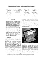
roots at four days after inoculation (F igure 2). Cells,
either epidermal or cortical cells, were collected pooled
and used for RNA extraction. The cell type-specific pat-
tern of expression of the OsCPK18 gene was examined
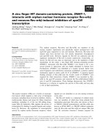
in laser microdissected cells. As it is shown in Figure 2F,
OsCPK18 transcripts were exclusively detected in cortical
cells of G. intraradices-inoculated rice roots. OsCPK18
transcripts were not detectable in epidermal cells of the
fungal-inoculated roots. The absence of PCR amplifica-
tion products in epidermal cells of the fungal-inoculated
roots was confirmed by nested PCR (results not shown).
Transcripts for the ubiquitin1 gene (Figure 2F) or the
cyclophilin gene (results not shown) were also detected
Figure 2 Laser microdissection of epidermal and cortical c ells
from rice roots. (A) Typical transverse section from the rice root.
(B and C) Representative transverse sections with targeted
epidermal cells before (B) and after (C) cutting with the laser
microdissector. (D and E) Representative transverse sections with
targeted cortical cells before (D) and after (E) cutting with the laser
microdissector. Insets in C and E show captured microdissected
cells. Two independent experiments were carried out for isolation of
epidermal and cortical rice root cells by LMD. (F) RT-PCR analysis to
detect OsCPK18 transcripts in laser microdissected cells from rice
roots. Cells were harvested from G. intraradices-inoculated (+Gi) and
mock-inoculated (-Gi) roots. Total RNA samples were obtained from
pooled microdissected cells. Expression analysis was carried out
using the one-step procedure for RT and PCR amplification. OsCPK18
transcripts were detected only in cortical cells. Bars, 100 μm (A),
50 μm (B to E).
Campos-Soriano et al. BMC Plant Biology 2011, 11:90
/>Page 5 of 14
in all the RNA samples obtained from laser microdis-
sected cells. The use of gene-specific primers that span
introns excluded the possibility of genomic DNA in total
RNA samples used for RT-PCR analyses. The absence of
an amplified product in RT-negative reactions also
excluded any DNA contamination in RNA samples
obtained from laser microdissected cells (results not
shown). Finally, OsCPK4 transcripts were detected in
RNA samples obtained from the two cell types ca ptured
from fungal-inoculated and control roots, this observa-
tion furt her supporting the integrity of the RNA samples
used in this study (Additional file 2: Figure S2).
When comparing the results obtained on OsCPK18
expression in laser microdissected cells (Figure 2F) and
whole roots (Figure 1), an apparent contradiction is
observed. Thus, OsCPK18 transcripts were not detected in
isolated cells from mock-inoculated roots (Figure 2F)
whereas RT-qPCR analysis revealed OsCPK18 expressi on
in whole roots (Figure 1A, upper panel). This finding
could be exp lained taking into account the plant material
and experimental approach used in these studies. In this
work, only two cell types of the root were harvested for
LMD-related analyses (epidermal and cortical cells). Thus,
the detection of OsCPK18 expression in whole mock-
inoculated roots could be due to the presence of cell types
constitutively expressing OsCPK18 that were not analyzed
with LMD (i.e. cells from the central cylinder). Addition-
ally, transversal sections were routinely made at aprox. 2
cm from the root tip. Thus the observed expression of the
OsCPK18 gene in regions of the rice root other than that
used for laser microdissection (i.e. meristems) might well
account for the o bserved OsCPK18 expression in mock-
inoculated whole roots. This observation also illustrates
the fact that results obtained in gene expression by using
entire roots might often be misinterpreted and spatial dif-
ferences in gene expression might not be perceived by
using whole roots. Clearly, a more detailed analysis of
OsCPK18 expression during growth and development of
the rice root is needed.
Subcellular localization of OsCPK18
Onion epidermal cells are widely used as a convenient sys-
tem in which to evaluate the subcellular location of GFP-
tagged proteins. Accordingly, the subcellular localization
of OsCPK18 was investigated in onion epidermal cells
transiently expressing gene fusions to the green fluores-
cent protein (GFP) (Figure 3A). Confocal microscopy of
transformed onion cells revealed that OsCPK18-GFP loca-
lizes to the cell periphery, likely the plasma membrane
(Figure 3B). As expected, onion cells expressing the GFP
gene showed fluorescence distributed throughout the cell
(Figure 3C).
Onion epidermal cells are also particularly useful for
analysis of plasma membrane proteins because the
environmental conditions can be manipulated to cause
plasmolysis and par tial separation of th e plasma mem-
brane from the cell wall. The onion epidermal cells were
plasmolyzed after being transformed with OsCPK18-
GFP. In plasmolyzed onion cells, the OsCPK18-GFP
displayed a pattern consistent with its location in
the plasma membrane of the shrunken protoplasm
(Figure3D).Undertheseconditions, protoplast pull
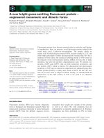
Figure 3 Plasma membrane localization of OsCPK18.Wild-type
OsCPK18 and a N-terminal myristoylation mutant were transiently
expressed as GFP fusion proteins in onion epidermal cells. Confocal
images were taken 24 h post-bombardment. (A) Diagrams of the
constructs used for particle bombardment of onion epidermal cells,
wild OsCPK18-GFP and the mutant OsCPK18-GFP fusion protein in
which the Gly
2
was mutated to Ala (OsCPK18
G2A
). (B) Localization of
wild OsCPK18-GFP fusion protein. Merged pictures of the green
fluorescence channel with the corresponding light micrographs are
shown in on the right. (C) Localization of GFP. (D) Onion cells after
plasmolysis with mannitol (15 min of treatment). Light micrographs
show the shrinkage of the protoplast (white arrow). (E) Treatment
with mannitol renders the Hechtian strands (arrows) attaching the
plasma membrane to the cell wall. (F) Onion epidermal cells
expressing a mutated version of OsCPK18 with an altered
myristoylation site (OsCPK18
G2A
-GFP). While the wild-type protein is
localized to the plasma membrane, the G
2
A mutant protein lost its
specific plasma membrane localization. Projection (B, C, D, F) and
individual (E) sections are shown. Scale bars = 20 μm (B, C, D, F),
10 μm (E).
Campos-Soriano et al. BMC Plant Biology 2011, 11:90
/>Page 6 of 14
away from the cell wall, leaving large numbers of thin
plasma membrane bridges, knownasHechtianstrands,
firmly anchored to the cell wall (Figure 3E).
Analysis of the amino acid sequence of OsCPK18 shows
that the OsCPK18 polypeptide possess a N-terminal myr-
istoylation site at the Gly residue at position 2 (Gly
2
) sug-
gestive of N-myristoylation. The need for this lipid
modification to promote and stabilize membrane associa-
tion of certain CPKs has been experimentally demon-
strated [37]. To address the role of the myristoylation site
of OsCPK18 in plasma membrane association, a mutation
at the N-termi nal myristoylation site (MGNTCVGPS) of
the OsCPK18 polypeptide was made. The Gly2 was con-
verted to Ala (G2A, referred to as OsCPK18
G2A
) and fused
to GFP (Figure 3A). Transient expression in epidermal
onion cells showed that the Gly
2
mutation abolished the
plasma membrane localization of OsCPK18 (Figure 3F).
Instead, a distribution throughout the cell was observed
for the mutated version of OsCPK18 similar to that of the
GFP alone. These findings suggest that the N-terminal
myristoylation site is required for subcellular localization
of OsCPK18 at the plasma membrane.
Phylogenetic analysis of cpk genes
In this work, the evolutionary relationships among CPKs
from rice and known CPKs from other plant species
establishing association with AM fungi was determined.
For t his analysis, the full-length CPK protein sequences
from cereal species, namely wheat and maize, as well as
CPKs so far characterized in the model symbiotic spe-
cies of Medicag o were used. As previously mentioned,
thericegenomecontains31CPK genes which classify
into four major phylogenetic groups (I-IV) [39,40].
Known CCaMK protein sequences from rice, wheat and
Medicago were also considered. In this respect, the rice
genome contains a single CCaMK gene [39]. As Arabi-
dopsis is not a host for AM fungi, this species was not
included in the phylogenetic analysis.
Phylogenetic trees of CPK and CCaMK proteins were
constructed based on the neighbor-joining method
(Figure 4) or the maximum parsimony method (Addi-
tional file 3: Figure S3). The aligment of the various pro-
teins used for construction of the phylogenetic tree is
presented in Additional file 4). Similar to what was pre-
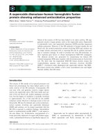
viously reported [ 40], the rice CPKs clustered into four
distinct phylogenetic groups (Figure 4). Four distinct
CPKs, OsCPK18, OsCPK4, OsCPK30 and OsCPK31,
cluster into an independent clade of CPKs, the Group
IV, which appears to have diverged significantly from
the other rice CPK sequences. Noticeably, results here
presented show that OsCPK18 and OsCPK4 are both
up-regulated by the AM fungus G. intraradices ,these
particular CPKs belonging to Group IV of rice CPKs. As
for the other members of the Group IV of rice CPKs,
no expression could be detected in the rice roots for
Oscpk31,whereasOscpk30 exhibited a low expression
but no responsiveness to AM inoculation.
Some interesting observations came from the phyloge-
netic analysis of CPK and CCaMK proteins. Firstly,
OsCPK18 and OsCPK4 appear to be closely related to
the AM-associated MtCDPK1 (Figure 4). Secondly,
Group IV of rice CPKs and CCaMKs are closely related
each other. Indeed, Group IV of rice CPKs appears to
be more related t o CCaMKs than to the other rice
CPKs. Here, it is worthwhi le to mention that the essen-
tial function of Mt CCaMK and OsCCaMK during the
mycorrhizal symbiotic association is well documented
[18,23]. Finally, the OsCPK18 is clearly related to
TaCPK6, one of the 20 CPKs described in wheat [45].
Sequence analysis of the OsCPK18 and OsCPK4 promoters
Knowing that the OsCPK18 and OsCPK4 genes are tran-
scriptionally activated in response to inoculation with
Figure 4 Phylogenetic relationships among rice, wheat, maize
and Medicago CPKs. An unrooted phylogenetic tree was created
using the MEGA4 program based on the full length sequences of
CPK proteins from rice (Os), wheat (Ta), maize (Zm) and Medicago
(M. truncatula, Mt; M. sativa, Ms). The four groups are indicated (I-IV).
Rice CPKs are highlighted in bold. Members of each phylogenetic
group of rice CPKs which have undergone expansion by segmental
genome duplication (pairs of closely related CPKs) are indicated by
brackets. Dots denote rice CPKs and CCaMK whose expression were
analyzed in this work.
Campos-Soriano et al. BMC Plant Biology 2011, 11:90
/>Page 7 of 14
the AM fungus G. intraradices, it was of interest to
investigate whether symbiosis-related cis-elements are
present in the promoter region of these genes. The
OsCPK18 and OsCPK4 promoter analysis was carried
out using the PLACE algorithm [46] and extended to
genes that are known to be required for both AM and
rhizobial root nodule symbioses, such as the MtCPK1
and MtCCaMK genes from M. truncatula and the OsC-
CaMK from rice.
Analysis of the 2 kb promoter region of the OsCPK18
and OsCPK4 genes revealed the presence of the CTCTT
element (NODCON2GM) which is found up to five and
six times in the OsCPK18 and OsCPK4 promoter,
respectively (Figure 5 and Additional file 5: Tables S1
and S2). The NODCON2GM as well as the NOD-
CON1GM element (AAAGAT) are characteristic motifs
of promoters from genes that are regulated during root
nodule and AM symbiosi s. These motifs are also part of
the “organ-specific element” (OSE) se quence [47]. Th e
MtCPK1, OsCCaMK and MtCCaMK promoters contain
several cop ies of the NO DCON1GM and NOD-
CON2GM consensus sequences.
Interestingly, multiple copies of the ABRE-related con-
sensus motif [(C/A)ACG(T/C)G(T/G/C), ABRERAT-
CAL] were present in the proximal region of the
OsCPK18 promoter (Figure 5 and Additional file 5:
Tables S1 an d S2). The A BRE-related motif is a cis-element
identified in the upstream regio n of 162 Ca
2+
-responsive
up-regulated genes [48]. Furthermore, up to three copies of
the CGCG-BOX element (GCCGCGGC) are found
in the Oscpk18 promoter, this element being involved
in Ca
++
/calmodulin-regulated gene expression [49]
(Figure 5 and Additional file 5: Tables S1 and S2). The
OsCPK4 promoter region contains one copy of the ABRE-
related motif element. The G(G/A/C/T)ATAT(G/A/C/T)C
(P1BS element) was recognized in the OsCPK4, MtCPK1
and MtCCaMK promoters (Figure 5 and Additional file 5:
Tables S1 and S2). This element is found in the upstream
region of phosphate starvation responsive genes from sev-
eral plant species [50].
Finally, the OsCPK18 and OsCPK4 promoters harbor
multiple stress-related cis-acting elements, including ele-
ments that are known to confer responsiveness to
pathogen-regulated genes. Some of them were repre-
sented many times in these promoters, such as the
TGAC-containing W box of WRKY transcription factors
(Additional file 5: Tables S1 and S2). In line with this,
we recently reported the activation of defense-and
stress-related genes during colonization of rice ro ots by
G. intraradices [4]. Whether the expression of the
OsCPK18 and OsCPK4 genes is regulated during patho-
gen infection in roots remains to be determined.
Overall, this stu dy revealed the presence of symbiotic-
related motifs, as well as putative elements related to
Ca
2+
regulation of gene expression, in the promoter
region of the OsCPK18 and OsCPK4 genes. This obser-
vation is consistent with the observed induction for the
two CPK genes in AM-inoculated rice roots.
Discussion
In this work, the expression of CP K genes was moni-
tored during the early stages of the AM symbiosis in
rice. The OsCPK18 and OsCPK4 consistently showed
up-regulation in response to AM inoculation. Evidence
is also presented on the transcriptional activation of
OsCPK18 and OsCPK4 expression by diffusible mole-
cules produced by G. intraradices. When comparing the
expression profiles of the rice CPK and CCaMK genes,
it appears that activation of the two CPK genes
(OsCPK18 and OsCPK4) occurs earlier than that of OsC-
CaMK pointing to a role for these p articular rice CPK
genes at the early stages of the symbiotic process. The
observation that the OsCPK18, OsCPK4, OsCCaMK,
MtCPK1 and MtCCaMK genes share symbiotic-relate d
cis-elements in their promoters is also indicative of the
transcriptional regulation of these genes as part of the
signaling mechanisms involved in the AM symbiosis in
rice. An expanded view of OsCPK18 gene expression
came from expression studie s in laser microdissected
Figure 5 Structural features of the promoters from the
OsCPK18, OsCPK4, MtCPK1, OsCCaMK and MtCCaMK genes. The
location of the indicated cis-acting elements is indicated in each
promoter.
Campos-Soriano et al. BMC Plant Biology 2011, 11:90
/>Page 8 of 14
cells isolated from rice roots. At 4 days post-inoculation
with G. intraradices, OsCPK18 was detected in cortical
cells and not in epidermal cells.
Clearly, the specificity of a CPK functioning in a given
signaling pathway may be achieved not only by a differ-
ential pattern of expression but also by targeting of the
CPK protein to a particular subcellular compartm ent.
Along with this, CPK proteins appear to be widely dis-
tributed among subcellular compartments including
cytosol, peroxisome, plasma membrane, oil bodies and
nucleus, as well as in association with actin filaments,
mitochondria and the endoplasmic reticulum [33]. Our
results in transformed onion cells clearly demonstrated
that OsCPK18 localizes to the plasma membrane. More-
over, the association of O sCPK18 to the plasma mem-
brane is possibly linked to N- terminal myristoylation of
this protein.
Knowing that CPKs act as Ca
2+
sensors in plant sig-
naling, and that Ca
2+
plays an important role in the AM
symbiosis, a function of OsCPK18 as a Ca
2+
sensor dur-
ing the AM-induced host responses to AM fungi can be
envisaged. Thus, perception of the fungal-produced
symbiotic signal(s) would activate downstream signaling
events required for the establishment of the symbiotic
association, including the cytoplasmic and nuclear Ca
2+
spiking responses [9-11]. Alterations in the Ca
2+
level
would be itself a major factor in mediating up-regulation
of OsCPK18 gene expression in the nucleus, as judged
by the presence of the Ca
2+
-responsive cis-elements in
the OsCPK18 promoter region [48]. In line with this,
previous studies in Arabidopsis revealed the presence of
ABRE-related sequences in Ca
2+
-responsive genes, and
exclusively in up-regulated Ca
2+
-responsive genes [48].
Tetramers of the ABRE-cis element are sufficient to
confer this transcriptional activation in response to Ca
2+
transients. The presence of multiple Ca
2+
-responsive cis-
regulatory elements in the promoter region of the
OsCPK18 gene (e.g. ABRE-related and CGCG-box ele-
ments) favors the possibili ty of a Ca
2+
-mediated up-reg-
ulation of OsCPK18 gene expression. The identity of the
trans cription factors that respond to rapid transien t Ca
2+
signals and that subsequently activate gene expression
through ABRE-related cis-elements remains to be
determined.
In addition to its transcriptional activation, a direct
regulation of the OsCPK18 enzyme acti vity by Ca
2+
can
be expected. Thus, it is well known that the activity of
CPKs is regulated b y the binding of calcium to its
intrinsic calmodulin-like domain. At basal Ca
2+
concen-
trations, t he functional autoregulatory domain acts as a
pseudosubstrate that inhibits the kinase activity of CPKs
(autoinhibited structure). In response to transient
increases in the level of cellular Ca
2+
,CPKsundergo
conformational changes that activate their kinase activity
(calcium-bound structure) [51]. It is then reasonable to
assume that the plasma membrane-localized OsCPK18
protein sense the AM-induced increase in cytoplasmic
Ca
2+
levels and transduce this signal into phosphoryla-
tion processes. The OsCPK18-mediated signaling pro-
cesses might then be crucial for root colo nization and
accommodation of the fungal symbiont in the root cor-
tex. The identification of downstream targets of the
OsCPK18 kinase act ivity requires, however, further
investigation.
On the other hand, our phylogenetic analysis of CPKs
and CCaMK of plant species that are able to establish
mycorrhizal associations revealed that Group IV of CPKs
and CCaMK are closely related each other pointing to an
evolutionary relationship between the two families of
protein kinases. In other studies carried out in the green
alga it was proposed that CCaMK originated through
gene duplication from CPK during green alga evolution
[52] Altogether, these findings are in clear support a
functional specialization of members of the Group IV of
CPKs and their relatedness with CCaMK functioning.
Adaptation steps probably occurred in different plant
species that determined t heir functional specialization
and symbiosis-specific regulation.
The current work also provides a foundation for
further functional investigation o f the complex CPK
family in relationship to the mycorrhization ability in
another c ereal species, such as wheat. Thus, the phylo-
genetic analysis of CPKs revealed that OsCPK18 and
OsCPK4 are closely related to the wheat TaCPK6 pro-
tein as well as to the Medicago MtCDPK1 pro tein. For
MtCDPK1 a role during the establishment of the AM
symbiosis is well documented [53]. It is then tempting
to speculate that the TaCPK6 gene might exhibit an
AM-regulated expression pattern in wheat plants.
An intriguing aspect is the presence of three Arabi-
dopsis proteins in Group IV of CPKs [39], even though
Arabidopsis is not a host for AM fungi. To this point, it
has been proposed that genes r equired for other aspects
of plant development might have been recruited to
function in symbiotic pathways. In line with this, inacti-
vation of the MtCDPK1 gene is associated to a signifi-
cant reduction of rhizobial and mycorrhizal symbiosis
and also results in stunted roots and short root hairs in
M. truncatula [53]. In other studies, impairment of root
hair development results in defective symbiotic interac-
tions in L. japonicus [54]. Then, the Arabidopsis CPKs
within Group IV of CPKs might play a role in normal
processes during root growth and development. The
finding of SYM genes in species that do not associate
with AM fungi (e.g. Arabidopsis and Physcomitrella),
also supports that specific genes functioning in normal
developmental processes in roots might also regulate
mycorrhizal infection. If so, this fact, would explain the
Campos-Soriano et al. BMC Plant Biology 2011, 11:90
/>Page 9 of 14
observed expression of OsCPK18 in experiments carried
out on whole roots by RT-qPCR.
Conclusions
This study provides a new view of the molecular
mechanisms involved in the AM symbiosis in rice
while defining an OsCPK18-mediated signaling path-
way functioning during this process. The rapid activa-
tion of OsCPK18 expression in response to AM
inoculation, its expression being also induced by fun-
gal-secreted signals, together with the observed plasma
membrane localization of OsCPK18, suggest that
OsCPK18 might play a role in perception and/or
recognition of the AM fungus in rice. Compared to
legume species, less effort has been invested in the
characterization of the AM symbiotic interaction in
this important crop species. OsCPK18 might be con-
sidered as a marker for the presymbiotic phase of the
symbiotic process that might play a preferential role in
the root cortex. The identification of additional com-
ponents of the AM-induced signaling processes in
which OsCPK18 participates can be now approached.
A major challenge for the future research is to deter-
mine whether interconnections and synergistic func-
tions exist between CPKs and SYM components, this
interplay determining recognition and compatibility
between the two symbiotic partners.
Methods
Plant material and growth conditions
Rice (Oryza sativa cv Nipponbare) was used as the
experimen tal material. Seeds were surface sterilized with
70% ethanol for 1 min, sodium hypochlorite (30% v/v)
for 30 min, and extensively washed with ste rile water
(four times, 10 min each). Seeds were germinated in
agar (0,4%) prepared with minimal m edium. Seedlings
were grown at 27°C ± 2°C under 18 h/6 h light/dark on
vertical plates.
G. intraradices (DAOM197198) spores were prepared
from monoxenic cultures of carrot roots that were
grown for three months as previously described [4].
Roots and G. intraradices cultures were axenically solu-
bilized in 5 volumes of sterile 10 mM sodium-citrate,
pH 6.0 for 15 min, at 37°C and filtrated four times
through a 250 μMsieve.Ricerootswereinoculated
with the G. intraradices spore suspension using either
thesinglesandwich[29]orthedoublesandwich[42]
system.
Forthesinglesandwichmethod,thericeseedlings
were directly inoculated with the arbuscular mycorrhizal
spore suspension, or mock-inoculated (sterile water),
and placed between two sterile nitrocellulose mem-
branes (Millipore, pore diameter 0.45 μm). Fo r the dou-
blesandwichmethod,thericeseedlingswerefirst
placed between two Millipore membranes. The mem-
brane-covered seedlings were then inoculated with the
fungal spore suspension and cove red with a second
layer of membranes. In this way, the physical contact
between the fungus and the root is avoided. The
assembled sandwich containing the inoculated seedlings
was placed in Petri dishes containing 0.4% agar in mini-
mal medium. Since Millipore membranes are permeable
to diffusible molecules, the root cells can perceive fungal
signals in the double sandwich method even thought
physical contact between the two symbionts does not
occur. Control seedlings were inoculated with sterile
water.
Tissue preparation and laser microdissection
The method previously described [43] was adapted for
the isolation of cells from G. intraradices-inoculated and
mock-inoculated rice roots. Root pieces of 4 - 8 mm in
length were dissected with a razor blade and immedi-
ately transferred into freshly prepared Methacarn solu-
tion (absolute methanol/chloroform/glacial acetic acid
6:3:1). Roots were maintained in the fixative solution
overnight at 4°C, and subsequently dehydrated in a
graded series of ethanol at 4°C: 50, 70 and 90% in sterile
water and 100% ethanol, followed by isopropanol
(twice), with each step on ice for 1 h. The isopropanol
was replaced gradually with paraffin (Paraplast Plus;
Sigma Aldrich, St. Louis). Transverse root sections of
10-15 μm were made using a Reichert Jung 2050 Super-
Cut Motorized Microtome (Leica, Arnsberg, Germany).
Ribbons were arranged on RNase-free, UV-treated,
PEN-membrane 2.0 μm slides. Slides were kept in a
slide warmer at 40°C until dry and stored at 4°C and
used within two days.
The Leica LDM6000 Laser Microdissection system
(Leica, Bannockburn, IL, USA) was used for laser micro-
dissection (LMD). Just before use, the paraffin sections
were deparaffinised in a neoclear (Merck, Darmstadt,
Germany) treatmen t for 10 min followed by 100% etha-
nol for 2 min, and then air dried. The deparaffinised
slides were placed face down on the microscope. The
tissues were visualized on a computer monitor through
a video camera. Epidermal and cortical cells were
marked and then cut using a UV laser (337-nm wave-
length). Target cells were collected without any extra
forces into the cap of a microcentrifuge (RNase-free
PCR tube caps). For each cell type, we isolated at least
1500 cell sections per biological replicate, and two inde-
pendent biological replicates we re made. After collec-
tion, 50 μl of RNA extraction buffer from the PicoPure
kit (Arcturus, Sunnyvale, CA, U.S.A.) were added. Sam-
ples were incubated at 42°C for 30 min, centrifuged at
800 g for 2 min, and stored at -80°C until RNA
isolation.
Campos-Soriano et al. BMC Plant Biology 2011, 11:90
/>Page 10 of 14
RNA isolation
Total RNA was extracted from whole roots at different
times after inoculation with G. intraradices spores, as
well as from mock-inoculated roots, using the TRI-
ZOL
®
Reagent (Invitrogen, Carlsbad, CA, USA). For each
time point, roots from at least 12 individual plants were
collected. Three independent experiments were carried
out. The first cDNA was synthesized from DNase-trea-
ted total RNA (1 μg) with M-MLV (Moloney-Murine
Leukemia Virus) Reverse Transcriptase (Invitrogen,
Carlsbad, CA, USA). Aliquots of the resulting RT reac-
tion product were used as template for PCR analysis.
RNA isolations f rom laser microdissected cells were
performed using a modified PicoPure kit protocol (Arc-
turus, Sunnyvale, CA, USA). Essentially, the DNase-
treatment was not performed on the kit column but the
RNA was eluted in 25 μl of DEPC-treated H
2
Oand
then treated with RNAse-free DNase (Roche, Mannhein,
Germany). After precipitation with 0.1 vol. of 3M Na-
acetate and 2.5 vol. cold ethanol (100%), the RNA was
resuspended in 20 μl of sterile water and quantified
using the NanoDrop 1000 spectrophotometer.
Gene expression analysis
Quantitative real time PCR (RT-qPCR) analyses were
carried out in optical 96-well plates in a LightCycler
®
480 Real-Time PCR System (Roche) according to the fol-
lowing program: 10 min at 95°C, followed by 45 cycles of
95°C for 10 s, 60°C for 30 s, and an additional cycle of
dissociation curves to ensure an unique amplification.
The reaction mixture contained 10 μl2×SYBRGreen
Master mix reagent (Roche, Mannhein, Germany), 2 μl
cDNA sample, and 300 μM of each gene-specific primers,
in a final volume of 20 μl. Primers used for RT-qPCR
are indicated in Additional file 6: Table S3. Details on
RT-qPCR analysis following the MIQE guidelines [55]
are included in Additional file 7. Routinely, three repli-
cate reactions were used for eac h sample. Data were
normalized with OsAct1 as internal control. The aver-
age CT values from triplicate PCRs were normalized to
the average CT values for the OsAct1 gene from the
same RNA preparations. Three independent biological
replicates were analyzed. For each biological material,
three technical replicates were made for RT-qPCR
analysis.
In this work, semi-quantitative reverse-transcription-
polymerase chain reaction (RT-PCR) was carried out to
investigate the expression pattern of the rice CPK genes,
as well as the rice SYM genes, namely the OsSYMRK,
OsCCaMK, OsPOLLUX and OsCASTOR genes. (Addi-
tional file 8: Table S4 and Additional file 9: Supplemen-
tary Methods).
For RT-PCR analysis of RNA samples obtained from
laser microdissected cells, a one-step RT-PCR was
conducted according to the manufacturer’s instructions
(Qiagen GmbH, Hilden, Germany). A 20 μl reaction was
prepared, containing the following reagents for each
reaction: 4 μl of (5X) Qiagen one-step RT-PCR buffer,
0,5 μl of Qiagen one-step RT-PCR enzyme mix, 0,5 mM
of each dNTP, 0,25 μM of each primer, 21 ng of total
RNA and RNase free water to 20 μl.
Phylogenetic analysis
The full length protein sequences were obtained from
the NCBI database and aligned using ClustalW [56].
The alignment of the various plant CPKs was created
with GeneDoc 2.7.0 Software [57] and is shown in Addi-
tional File 4.
In addition to the 31 CPKs from rice, known full-length
CPK sequences from M. truncatula, M. sativa and Zea
mays were also used. They were (accession numbers are
from Gene Bank at the National Center for Biotechnology
Information (NCBI) database .
gov/: MtCDPK1 [AAX15706], MtCDPK3 [ABE72958],
and MsCDPK3, X96723]; ZmCDPK1 [BAA12338],
ZmCDPK2 [AAA69507], ZmCDPK7 [BAA13232],
ZmCDPK9 [BAA12715], ZmCDPK10 [CAA07481] and
ZmCDPK11 [AAP57564]. As for wheat, up to 20 CPKs
have been described from which 14 available sequences
were included in this study [45]. They were: TaCPK1
[ABY59004], TaCPK2 [ABY59005], TaCPK3 [ABY59006],
TaCPK4 [ABY59007], TaCPK5 [ABY59008], TaCPK6
[ABY59009], TaCPK7 [ABY59017], TaCPK8 [ABY59010],
TaCPK9 [ABY59011], TaCPK12 [ABY59012], TaCPK13
[ABY59018], TaCPK15 [ABY59013], TaCPK18
[ABY59014], TaCPK19 [ABY59015]. Furthermore,
CCaMK sequences from rice, wheat and Medicago were
included in this analysis, the rice OsCCaMK [Q6AVM3],
wheat TaCCaMK [ADK22086] and M. truncatula
MtCCaMK [Q6RET7] sequences. Phylogenetic trees
were created according to the neighbor-joining method
(Figure 4) or the maximum-parsimony algorithm (Addi-
tional File 3: Figure S3) using MEGA4 program [58,59].
Promoter analysis
Sequences 2000 bp upstr eam of the selected cpk’s genes
were retrieved from NCBI database. Known plant motifs
were obtained from the PLACE database http://www.
dna.affrc.go.jp/PLACE/.
Biolistic cell transformation and imaging by confocal
microscopy
To investigate the subcellular localization of OsCPK18,
the green fluorescent protein (GFP) gene was transla-
tionally fused to the C-terminal end of the OsCPK18
sequence. For thi s, a GFP gene lacking the N-terminal
signal peptide and C-terminal HDEL sequences was
initially obtained by PCR from the m-GFP5-ER sequence
Campos-Soriano et al. BMC Plant Biology 2011, 11:90
/>Page 11 of 14
[60]. Primers used were 5’ -CGGGGATCCATGAG-
TAAAGGAGAAGAACTTTTCAC-3’ (forward) and 5’-
CC
GAGCTCTTATTATTTGTATAGTTCATCCATGC-
3’ (reverse). During the PCR reaction, BamHI and SacI
sites were introduced into the PCR ampli fied DNA fr ag-
ment (underlined nucleotides in PCR primer sequences).
Next, the GFP variant was cloned between the BamHI
and SacI sites of a pUC19-derived plasmid harboring
the 35SCaMV promoter and nopaline synthetase termi-
nator (Tnos) to create the pP35S:mGFP:nos construct.
The OsCPK18 DNA fragment to be translationally
fused to the GFP gene was generated by PCR amplifica-
tion. A BglII site was introduced at the 3’end of the
PCR-amplified Oscpk18 DNA fragment. Equally, a SpeI
site was introduced at the 5’ end of the Oscpk18
sequence. The full-length OsCPK18 cDNA sequence was
PCR-amplified from clone J023148F12 obtained from
the KOME ( Knowledge-based Oryza Molecular biologi-
cal Encyclopedia) database using the following primers:
5’ -AT
ACTAGTATGGGACTCTGCTCCTCCTCC-3’
(forward) and 5’-AT
AGATCTACCTGGTGGTGGC-
GATCTGTGAACACTCCT-3 ’ (reverse) (underlined
sequences indicate the SpeI and BglII restriction s ites;
bold fonts indicate the residual tai l of one glycine and
three p rolines to ensure the fusion). To obtain the chi-
meric OsCPK18-GFP gene, the PCR-amplified OsCPK18
DNA fragment was digested with SpeI and BglII and
cloned into the XbaI/BamHI-digested pP35S:GFP:nos
plasmid resulting in plasmid pP35S:OsCPK18-GFP:nos.
A chimeric gene in which a mutated version of
OsCPK18 gene was fused to the GFP gene was also pre-
pared. For preparation of the mutated version of the
OsCPK18 gene in whi ch the Gly residue at position 2
was converted to Ala (G2A), the pP35S:OsCPK18-GFP:
nos plasmid was used as the tem plate for PCR. For this,
aforwardprimerwithasinglechange(boldfont)5’-
AT
ACTAGTATGGCACTCTGCTCCTCCTCC-3’,and
the OsCPK18 reverse primer above indicated were used.
The OsCPK18
G2A
DNA was then cloned into the XbaI/
BamHI-diges t ed pP35S:mGFP:nos plasmid. All the con-
structs were verified by nucleotide sequencing.
Onion epidermal cells were transformed by particle
bombardment. The epidermis of onion bulb scales was
prepared by peeling the inner epidermis from fresh
onion bulbs. For each DNA construct, 4 μgofDNA
was mixed with 20 μμl of 0.1 M spermidine, 50 μμlof
2.5 M CaCl
2
and 15 μl of gold microcarrier (60 μgml
-
1
in 50% glicerol), vortexed vigorously for 30 sec and
centrifuged at 800 g in a microfuge for 10 sec. The
pellet was washed on ethanol. The DNA-gold particles
were bombarded into cells at a pressure of 1100 psi
and target distance of 6 cm, with a biolistic PSD-
1000/He particle delivery system (Bio-Rad). The trans-
fected cells were kept on MS medium for 16-24 h in
the dark. The subcellular localization of the fusion
proteins was determined by confocal laser scanning
microscopy (CLSM) in a Olympus Fluoview FV1000
confocal laser scanning microscope (Tokyo, Japan).
The excitation wavelength was 488 nm. The e mission
window was set at 500-60 0 nm for GFP. To confirm
plasma membrane localization, cells were plasmolysed
with 0.75 M mannitol.
Additional material
Additional file 1: Figure S1: RT-PCR analysis of rice CPK genes in
mock-inoculated ( -) and G. intraradices-inoculated (+) rice r oots.
Inoculation with G. in traradices spores was carried out using the single-
sandwich system. Roots were harvested at diffe rent times after
inoculatio n and each RNA sample was prepar ed from a pool of roots
from 12 plants. RT-PCR was performed using specific primers for the
indicated CPK genes. The subset of genes selected for expression
analysis included members of the four phylogenetic groups of CPK
genes (I-IV) (for details see Figure 4). Expression of rice SYM marker
genes, the OsCCaMK, OsCASTOR, OsPOLLUX and OsSYMRK genes was
analyzed in the same RNA samples that were used for analysis of CPK
gene expression. Transcripts for the OsCPK2, OsCPK22, OsCP K25 and
OsCPK31 could not be detected (results not shown, similar results were
reported previously [40] by microarray analysis). The constitutively
expressed actin1 (OsAct1) g ene was used as the internal control in
these experiments. Three independent experiments were carried out
with similar results.
Additional file 2: Figure S2: Expression of the OsCPK4 gene in laser
microdissected cells from rice roots. Cells were harvested from G.
intraradices-inoculated (+Gi) and mock-inoculated (-Gi ) roots. Total RNA
samples were obtained from pooled microdissected cells. Expression
analysis was carried out using the one-step procedure for RT and PCR
amplification. Although different expression levels were observed in
epidermal and cortical cells, RT-PCR analyses from RNA samples obtained
from laser microdissected cells does not provide quantitative information
on gene expression.
Additional file 3: Figure S3: Relationship between plant CPKs. The
phylogenetic tree was constructed using the maximum parsimony
method using the full length amino acid sequences of CPKs from rice
(Os), wheat (Ta), maize (Zm) and Medicago (M. truncatula, Mt; M. sativa,
Ms) and CCaMKs. The four groups are indicated (I-IV).
Additional file 4: Alignment of the amino acid sequences of plant
CPKs and CCaMKs. Amino acid sequences from rice (Os), wheat (Ta),
maize (Zm) and Medicago (M. truncatula, Mt; M. sativa, Ms) were aligned.
Black and gray backgrounds indicate amino acids that are identical or
similar, respectively. Dashes indicate no residue present at that position.
The boundaries of the kinase (amino acids 51-317 in OsCPK18) and
calmoldulin-like (amino acids 347-493 in OsCPK18) domains are shown
(black and grey arrows, respectively).
Additional file 5: Table S1 and Table S2. Table S1. Symbiosis-related
cis-elements identified in the OsCPK18, OsCPK4, MtCPK1, OsCCAMK and
MtCCaMK promoters. The 2 kb upstream region of each promoter was
analyzed by using the PLACE motif database. Table S2. Number of
symbiosis-related cis-elements present in the OsCPK18, OsCPK4, MtCPK1,
OsCCaMK and MtCCaMK promoters. The presence (x) or absence (-) of
the indicated motifs within the 2 kb upstream region of each promoter
is shown. Multiple copies of a given cis-element
were present in a
particular promoter (2x, 3x, 4x, etc.). The PLACE motif database was used
to perform this analysis. Motifs are listed by alphabetical order.
Additional file 6: Table S3: Primer sequences used in the real-time
qPCR analysis.
Additional file 7: Information of the RT-qPCR analysis based on the
MIQE (Minimum Information for Publication of Quantitative Real-
Time PCR Experiments) checklist [55].
Campos-Soriano et al. BMC Plant Biology 2011, 11:90
/>Page 12 of 14
Additional file 8: Table S4: Primer sequences used in the RT-PCR
analysis.
Additional file 9: Supplementary Methods. Gene expression analysis
by semi-quantitative RT-PCR.
Acknowledgements
LCS was a recipient of a predoctoral fellowship from the Generalitat de
Catalunya. This work was supported by the “Acción Integrada” HI2007-0258
and grant BIO2009-08719 from the Spanish Ministry of Science and
Innovation, as well as by the Consolider-Ingenio 2010 Programme CSD2007-
00036 “Centre for Research in Agrigenomics”. We also thank the
“Departament d’Innovació, Universitats i Empresa” from the Generalitat de
Catalunya (Xarxa de Referencia en Biotecnología and SGR 09626) for
substantial support.
Author details
1
Centre for Research in Agricultural Genomics (CRAG) CSIC-IRTA-UAB.
Department of Molecular Genetics. Campus UAB, Edifici CRAG, Bellaterra
(Cerdanyola del Vallès) 08193 Barcelona, Spain.
2
Department of Plant Biology,
University of Torino and Istituto per la Protezione delle Piante - CNR. Sezione
di Torino. Viale P.A. Mattioli 25, Torino 10125, Italy.
Authors’ contributions
LCS carried out most of the experimental work and data analyses. JGA
participated in laser microdissection and gene expression studies. BSS
coordinated the design and execution of this study and wrote the
manuscript. PB also participated in the design of this study and critically
revised the manuscript for important intellectual content. All authors read
and approved the final manuscript.
Received: 18 February 2011 Accepted: 19 May 2011
Published: 19 May 2011
References
1. Smith S, Read D: Mycorrhizal Symbiosis London: Academic Press; 2008.
2. Parniske M: Arbuscular mycorrhiza: the mother of plant root
endosymbioses. Nat Rev Micro 2008, 6:763-775.
3. Liu J, Maldonado-Mendoza I, Lopez-Meyer M, Cheung F, Town CD,
Harrison MJ: Arbuscular mycorrhizal symbiosis is accompanied by local
and systemic alterations in gene expression and an increase in disease
resistance in the shoots. Plant J 2007, 50:529-544.
4. Campos-Soriano L, García-Garrido JM, SanSegundo B: Activation of basal
defense mechanisms of rice plants by Glomus intraradices does not affect
the arbuscular mycorrhizal symbiosis. New Phytol 2010, 188:597-614.
5. Sawers RJH, Gutjahr C, Paszkowski U: Cereal mycorrhiza: an ancient
symbiosis in modern agriculture. Trends Plant Sci 2008, 13:93-97.
6. Akiyama K, Matsuzaki Ki, Hayashi H: Plant sesquiterpenes induce hyphal
branching in arbuscular mycorrhizal fungi. Nature 2005, 435:824-827.
7. Maillet F, Poinsot V, Andre O, Puech-Pages V, Haouy A, Gueunier M,
Cromer L, Giraudet D, Formey D, Niebel A, Martinez EA, Driguez H,
Becard G, Denarie J: Fungal lipochitooligosaccharide symbiotic signals in
arbuscular mycorrhiza. Nature 2011, 469:58-63.
8. Genre A, Chabaud M, Timmers T, Bonfante P, Barker DG: Arbuscular
Mycorrhizal Fungi Elicit a Novel Intracellular Apparatus in Medicago
truncatula Root Epidermal Cells before Infection. Plant Cell 2005,
17:3489-3499.
9. Chabaud M, Genre A, Sieberer BJ, Faccio A, Fournier J, Novero M,
Barker DG, Bonfante P: Arbuscular mycorrhizal hyphopodia and
germinated spore exudates trigger Ca2+ spiking in the legume and
nonlegume root epidermis. New Phytol 2010, 347-355.
10. Kosuta S, Hazledine S, Sun J, Miwa H, Morris RJ, Downie JA, Oldroyd GED:
Differential and chaotic calcium signatures in the symbiosis signaling
pathway of legumes. Proc Natl Acad Sci USA 2008, 105:9823-9828.
11. Navazio L, Moscatiello R, Genre A, Novero M, Baldan B, Bonfante P,
Mariani P: A Diffusible Signal from Arbuscular Mycorrhizal Fungi Elicits a
Transient Cytosolic Calcium Elevation in Host Plant Cells. Plant Physiol
2007, 144:673-681.
12. Oldroyd GE, Downie JA: Nuclear calcium changes at the core of
symbiosis signaling. Curr Opin Plant Biol 2006, 9:351-357.
13. Bonfante P, Genre A: Mechanisms underlying beneficial plant-fungus
interactions in mycorrhizal symbiosis. Nat Commun 2010, 1:48.
14. Paszkowski U: A journey through signaling in arbuscular mycorrhizal
symbioses. New Phytol 2006, 172
:35-46.
15.
Harrison MJ: Signaling in the arbuscular mycorrhizal symbiosis. Annu Rev
Microbiol 2005, 59:19-42.
16. Charpentier M, Bredemeier R, Wanner G, Takeda N, Schleiff E, Parniske M:
Lotus japonicus CASTOR and POLLUX Are Ion Channels Essential for
Perinuclear Calcium Spiking in Legume Root Endosymbiosis. Plant Cell
2008, 20:3467-3479.
17. Kanamori N, Madsen LH, Radutoiu S, Frantescu M, Quistgaard EMH, Miwa H,
Downie JA, James EK, Felle HH, Haaning LL, Jensen TH, Sato S, Nakamura Y,
Tabata S, Sandal N, Stougaard J: A nucleoporin is required for induction
of Ca2+ spiking in legume nodule development and essential for
rhizobial and fungal symbiosis. Proc Natl Acad Sci USA 2006, 103:359-364.
18. Levy J, Bres C, Geurts R, Chalhoub B, Kulikova O, Duc G, Journet EP, Ane JM,
Lauber E, Bisseling T, Denarie J, Rosenberg C, Debelle F: A Putative Ca2+
and Calmodulin-Dependent Protein Kinase Required for Bacterial and
Fungal Symbioses. Science 2004, 303:1361-1364.
19. Saito K, Yoshikawa M, Yano K, Miwa H, Uchida H, Asamizu E, Sato S,
Tabata S, Imaizumi-Anraku H, Umehara Y, Kouchi H, Murooka Y,
Szczyglowski K, Downie JA, Parniske M, Hayashi M, Kawaguchi M:
NUCLEOPORIN85 Is Required for Calcium Spiking, Fungal and Bacterial
Symbioses, and Seed Production in Lotus japonicus. Plant Cell 2007,
19:610-624.
20. Stracke S, Kistner C, Yoshida S, Mulder L, Sato S, Kaneko T, Tabata S,
Sandal N, Stougaard J, Szczyglowski K, Parniske M: A plant receptor-like
kinase required for both bacterial and fungal symbiosis. Nature 2002,
417:959-962.
21. Yano K, Yoshida S, Müller J, Singh S, Banba M, Vickers K, Markmann K,
White C, Schuller B, Sato S, Asamizu E, Tabata S, Murooka Y, Perry J,
Wang TL, Kawaguchi M, Imaizumi-Anraku H, Hayashi M, Parniske M:
CYCLOPS, a mediator of symbiotic intracellular accommodation. Proc
Natl Acad Sci USA 2008, 105:20540-20545.
22. Messinese E, Mun JH, Yeun LH, Jayaraman D, Rougé P, Barre A, Lougnon G,
Schornack S, Bono JJ, Cook DR, Ané JM: A Novel Nuclear Protein Interacts
With the Symbiotic DMI3 Calcium- and Calmodulin-Dependent Protein
Kinase of Medicago truncatula. Mol Plant Microbe Interact 2007, 20:912-921.
23. Chen C, Gao M, Liu J, Zhu H: Fungal Symbiosis in Rice Requires an
Ortholog of a Legume Common Symbiosis Gene Encoding a Ca2
+/Calmodulin-Dependent Protein Kinase. Plant Physiol 2007,
145:1619-1628.
24. Godfroy O, Debelle F, Timmers T, Rosenberg C: A Rice Calcium- and
Calmodulin-Dependent Protein Kinase Restores Nodulation to a Legume
Mutant. Mol Plant Microbe Interact 2006, 19:495-501.
25. Gutjahr C, Banba M, Croset V, An K, Miyao A, An G, Hirochika H, Imaizumi-
Anraku H, Paszkowski U: Arbuscular Mycorrhiza-Specific Signaling in Rice
Transcends the Common Symbiosis Signaling Pathway. Plant Cell 2008,
20:2989-3005.
26. Kuhn H, Küster H, Requena N: Membrane steroid-binding protein 1
induced by a diffusible fungal signal is critical for mycorrhization in
Medicago truncatula. New Phytol 2010, 185:716-733.
27. Fiorilli V, Catoni M, Miozzi L, Novero M, Accotto GP, Lanfranco L: Global
and cell-type gene expression profiles in tomato plants colonized by an
arbuscular mycorrhizal fungus. New Phytol 2009, 184
:975-987.
28.
Gomez SK, Javot H, Deewatthanawong P, Torres-Jerez I, Tang Y,
Blancaflor E, Udvardi M, Harrison M: Medicago truncatula and Glomus
intraradices gene expression in cortical cells harboring arbuscules in the
arbuscular mycorrhizal symbiosis. BMC Plant Biol 2009, 9:10.
29. Guether M, Balestrini R, Hannah M, He J, Udvardi MK, Bonfante P: Genome-
wide reprogramming of regulatory networks, transport, cell wall and
membrane biogenesis during arbuscular mycorrhizal symbiosis in Lotus
japonicus. New Phytol 2009, 182:200-212.
30. Güimil S, Chang HS, Zhu T, Sesma A, Osbourn A, Roux C, Ioannidis V,
Oakeley EJ, Docquier M, Descombes P, Briggs SP, Paszkowski U:
Comparative transcriptomics of rice reveals an ancient pattern of
response to microbial colonization. Proc Natl Acad Sci USA 2005,
102:8066-8070.
Campos-Soriano et al. BMC Plant Biology 2011, 11:90
/>Page 13 of 14
31. Balestrini R, Lanfranco L: Fungal and plant gene expression in arbuscular
mycorrhizal symbiosis. Mycorrhiza 2006, 16:509-524.
32. Harrison MJ, Buuren ML: A phosphate transporter from the mycorrhizal
fungus Glomus versiforme. Nature 1995, 378:626-629.
33. Klimecka M, Muszynska G: Structure and functions of plant calcium-
dependent protein kinases. Acta Biochim Pol 2007, 54:219-233.
34. Harper JF, Breton G, Harmon A: Decoding ca2+ signals through plant
protein kinases. Annu Rev Plant Biol 2004, 55:263-288.
35. Cheng SH, Willmann MR, Chen HC, Sheen J: Calcium Signaling through
Protein Kinases. The Arabidopsis Calcium-Dependent Protein Kinase
Gene Family. Plant Physiol 2002, 129:469-485.
36. Harmon AC, Gribskov M, Gubrium E, Harper JF: The CDPK Superfamily of
Protein Kinases. New Phytol 2001, 151:175-183.
37. Coca M, San Segundo B: AtCPK1 calcium-dependent protein kinase
mediates pathogen resistance in Arabidopsis. Plant J 2010, 63:526-540.
38. Romeis T, Piedras P, Jones JDG: Resistance Gene-Dependent Activation of
a Calcium-Dependent Protein Kinase in the Plant Defense Response.
Plant Cell 2000, 12:803-816.
39. Asano T, Tanaka N, Yang G, Hayashi N, Komatsu S: Genome-wide
Identification of the Rice Calcium-dependent Protein Kinase and its
Closely Related Kinase Gene Families: Comprehensive Analysis of the
CDPKs Gene Family in Rice. Plant Cell Physiol 2005, 46:356-366.
40. Ray S, Agarwal P, Arora R, Kapoor S, Tyagi A: Expression analysis of
calcium-dependent protein kinase gene family during reproductive
development and abiotic stress conditions in rice Oryza sativa; L. ssp.
indica. Mol Genet Genomics 2007, 278:493-505.
41. Weidmann S, Sanchez L, Descombin J, Chatagnier O, Gianinazzi S,
Gianinazzi-Pearson V: Fungal Elicitation of Signal Transduction-Related
Plant Genes Precedes Mycorrhiza Establishment and Requires the dmi3
Gene in Medicago truncatula. Mol Plant Microbe Interact 2004,
17:1385-1393.
42. Gutjahr C, Novero M, Guether M, Montanari O, Udvardi M, Bonfante P:
Presymbiotic factors released by the arbuscular mycorrhizal fungus
Gigaspora margarita induce starch accumulation in Lotus japonicus
roots. New Phytol 2009, 183:53-61.
43. Balestrini R, Gómez-Ariza J, Lanfranco L, Bonfante P: Laser Microdissection
Reveals That Transcripts for Five Plant and One Fungal Phosphate
Transporter Genes Are Contemporaneously Present in Arbusculated
Cells. Mol Plant Microbe Interact 2007, 20:1055-1062.
44. Gómez-Ariza J, Balestrini R, Novero M, Bonfante P: Cell-specific gene
expression of phosphate transporters in mycorrhizal tomato roots. Biol
Fert Soils 2009, 45:845-853.
45. Li AL, Zhu YF, Tan XM, Wang X, Wei B, Guo HZ, Zhang ZL, Chen XB,
Zhao GY, Kong XY, Jia JZ, Mao L: Evolutionary and functional study of the
CDPK gene family in wheat Triticum aestivum. Plant Mol Biol 2008,
66:429-443.
46. Hig o K, Ugawa Y, Iwamoto M, Korenaga T: Plant cis-acting regulat ory
DNA elements (PLACE) database: 1999. Nucleic Acids Res 1999,
27:297-300.
47. Stougaard J, Sandal NN, Grøn A, Kühle A, Marcker JA: 5’ Analysis of the
soybean leghaemoglobin lbc
3
gene: regulatory elements required for
promoter activity and organ specificity. EMBO J 1987, 6:3565-3569.
48. Kaplan B, Davydov O, Knight H, Galon Y, Knight MR, Fluhr R, Fromm H:
Rapid Transcriptome Changes Induced by Cytosolic Ca2+ Transients
Reveal ABRE-Related Sequences as Ca2+-Responsive cis Elements in
Arabidopsis. Plant Cell 2006, 18:2733-2748.
49. Yang T, Poovaiah BW: A Calmodulin-binding/CGCG Box DNA-binding
Protein Family Involved in Multiple Signaling Pathways in Plants. J Biol
Chem 2002, 277:45049-45058.
50. Rubio V, Linhares F, Solano R, Martín AC, Iglesias J, Leyva A, Paz-Ares J: A
conserved MYB transcription factor involved in phosphate starvation
signaling both in vascular plants and in unicellular algae. Genes Dev
2001, 15:2122-2133.
51. Wernimont AK, Artz JD, Finerty P, Lin YH, Amani M, lali-Hassani A,
Senisterra G, Vedadi M, Tempel W, Mackenzie F, Chau I, Lourido S,
Sibley LD, Hui R: Structures of apicomplexan calcium-dependent protein
kinases reveal mechanism of activation by calcium. Nat Struct Mol Biol
2010, 17:596-601.
52. Wang B, Yeun LH, Xue JY, Liu Y, Ané JM, Qiu YL: Presence of three
mycorrhizal genes in the common ancestor of land plants suggests a
key role of mycorrhizas in the colonization of land by plants. New Phytol
2010, 186:514-525.
53. Ivashuta S, Liu J, Liu J, Lohar DP, Haridas S, Bucciarelli B, VandenBosch KA,
Vance CP, Harrison MJ, Gantt JS: RNA Interference Identifies a Calcium-
Dependent Protein Kinase Involved in Medicago truncatula Root
Development. Plant Cell 2005, 2911-2921.
54. Karas B, Amyot L, Johansen C, Sato S, Tabata S, Kawaguchi M,
Szczyglowski K: Conservation of Lotus and Arabidopsis Basic Helix-Loop-
Helix Proteins Reveals New Players in Root Hair Development. Plant
Physiol 2009, 151:1175-1185.
55. Bustin SA, Benes V, Garson JA, Hellemans J, Huggett J, Kubista M, Mueller R,
Nolan T, Pfaffl MW, Shipley GL, Vandesompele J, Wittwer CT: The MIQE
Guidelines: Minimum Information for Publication of Quantitative Real-
Time PCR Experiments. Clin Chem 2009, 55:611-622.
56. Thompson JD, Higgins DG, Gibson TJ: CLUSTAL W: improving the
sensitivity of progressive multiple sequence alignment through
sequence weighting, position-specific gap penalties and weight matrix
choice. Nucleic Acids Res 1994, 22:4673-4680.
57. Nicholas K, Nicholas H, Deerfield D: GeneDoc: analysis and visualization of
genetic variation. EMBNEW NEWS 1997, 4:14.
58. Saitou N, Nei M: The neighbor-joining method: a new method for
reconstructing phylogenetic trees. Mol Biol Evol 1987, 4:406-425.
59. Tamura K, Dudley J, Nei M, Kumar S: MEGA4: Molecular Evolutionary
Genetics Analysis (MEGA) Software Version 4.0. Mol Biol Evol 2007,
24:1596-1599.
60. Siemering KR, Golbik R, Sever R, Haseloff J: Mutations that suppress the
thermosensitivity of green fluorescent protein. Curr Biol 1996,
6:1653-1663.
doi:10.1186/1471-2229-11-90
Cite this article as: Campos-Soriano et al.: A rice calcium-dependent
protein kinase is expressed in cortical root cells during the
presymbiotic phase of the arbuscular mycorrhizal symbiosis. BMC Plant
Biology 2011 11:90.
Submit your next manuscript to BioMed Central
and take full advantage of:
• Convenient online submission
• Thorough peer review
• No space constraints or color figure charges
• Immediate publication on acceptance
• Inclusion in PubMed, CAS, Scopus and Google Scholar
• Research which is freely available for redistribution
Submit your manuscript at
www.biomedcentral.com/submit
Campos-Soriano et al. BMC Plant Biology 2011, 11:90
/>Page 14 of 14